Atrophy
Historical document, translated for reference. It reflects medical knowledge of the 1920s–30s and is not medical advice.
Summary
An article from the 1928-1936 Soviet Great Medical Encyclopedia defining atrophy as a life-acquired decrease in the volume of cells, tissues, or organs due to impaired nutrition. It categorizes atrophy into seven types, including physiological, nutritional, pressure, toxic, disuse, neurotic, and hormonal forms.
Encyclopedia article (1928–1936)
ATROPHY (from Greek a- negative particle, and trophe — nutrition), a term used to designate a decrease in the volume of cells, a tissue, or an organ due to the impairment of their nutrition. Consequently, atrophy is a life-acquired, acquired process of volume reduction in a given part of the organism, which distinguishes it from hypoplasia (see) or hypogenesis (see), which imply small dimensions of an organ or body part due to underdevelopment. At the heart of tissue or organ atrophy lies the atrophy (i.e., volume reduction) of the cells constituting them, wherein usually in a portion of the ATROPHIC cells the volume reduction reaches the complete disappearance of cells, which leads to the fact that alongside the volume reduction of cells there is also a depletion in their number. In most cases, atrophy of cells decreasing in volume is not accompanied by any qualitative changes in their constituent elements, which gives reason in such cases to speak of simple atrophy. In contrast, degenerative atrophy refers to a combination of atrophy with qualitative changes in the cell body, belonging to the manifestations of various degenerations (see) and retrogressive changes. One of the frequent examples of atrophic processes of the latter kind is the so-called brown atrophy, which is based on the appearance in the protoplasm of atrophying cells of granules of a brown pigment called wear-and-tear pigment or lipofuscin. All the above-mentioned changes underlying the atrophy of various tissues or organs exclusively or predominantly concern their parenchyma, i.e., the functional elements of the given tissue, whereas the interstitial tissue, the stroma, either does not participate in the atrophy process or even
consequently increases in volume. Atrophy can be local, i.e., concerning only a single organ or its part or some tissue, or it can be general, encompassing the entire organism or at least the majority of its organs and tissues; in cases of the latter kind, such a condition of the organism is called cachexia or marasmus. The conditions of development and causes of atrophy are diverse; in general, it is accepted according to the primary causative factor to consider seven types of atrophy: 1. Physiological atrophy, which is the prototype of pathological atrophy, is a volume reduction of certain organs occurring in the order of physiological dying-off; this includes atrophy of the thymus gland in adults, atrophy of the ovaries in the menopausal period, as well as senile atrophy (senile marasmus), affecting the entire organism and expressed in that volume reduction of the majority of organs and tissues observed in old age. 2. Atrophy from nutritional deficiency can be local or general. The former is observed in cases where a single organ or part of it suffers from nutritional deficiency, which most often occurs due to the narrowing of the lumen of the supplying artery and a weakening of blood inflow (for example, atrophy of renal tissue in arteriosclerosis of the renal artery). General atrophy from nutritional deficiency is that state of depletion and emaciation (cachexia, marasmus) which is the consequence of starvation and severe prolonged diseases; the most severe degree of atrophy in such cases is undergone by adipose tissue and musculature. 3. Pressure atrophy is observed in cases where a tissue experiences prolonged pressure; atrophy under such conditions develops depending on the reduction of blood inflow to the tissue due to the compression of its vessels, as well as due to the weakening of the assimilative capacity of the compressed cells. Examples: the cord or corset furrow of the liver in women who tightly lace their waist, the small feet of Chinese women from artificial compression by a bandage, atrophy of the bone tissue of the sternum and vertebrae due to pressure from an aortic aneurysm (see), atrophy of the kidney in hydronephrosis (see), impressions on the inner surface of the skull from so-called Pacchionian granulations. 4. Atrophy due to toxic influences occurs in various prolonged diseases accompanied by intoxication, and has a general character; here one can classify the general cachexia of patients with advanced syphilis, tuberculous patients, cachexia in
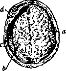
Figure 1. Atrophy of the left cerebral hemisphere: a — right hemisphere (normal); b — atrophic left hemisphere, reduced to such an extent that the cerebellum has become exposed; c — free space formed between the skull and the brain as a result of its atrophy; d — successive thickening of the cranial bones. malignant tumors, chronic malaria, etc., and it must be borne in mind that in most of these cases atrophy is the result not only of a direct toxic effect on tissues, but also of impaired general nutrition and metabolism. Close to the category of atrophy from toxic influences is atrophy resulting from the action of radiant energy, e.g., testicular atrophy from X-rays. 5. Atrophy from inactivity is a reduction in the volume of tissue and its cells observed during the inactive state of the given tissue. Activity is an important factor maintaining a sufficient blood supply to the tissue and metabolism within it; upon cessation of function, a decline of these processes sets in, and tissue atrophy occurs. An example is the atrophy of the musculature of a limb that has been in a motionless state for a long time, for example, during paralysis or in a plaster cast due to bone fracture. 6. Neurotic atrophy represents that atrophic state of tissue (e.g., musculature, bone, skin) that develops upon the disruption of this tissue's connection with the central nervous system. In former times, the existence of special trophic fibers in nerves regulating tissue nutrition was hypothesized, the elimination of the influence of which causes this type of atrophy; at present, the existence of such special nerves is not recognized by everyone, and neurotic atrophy is explained by a combination of various factors (circulatory disorders, metabolic disturbances, inactivity, etc.). Examples of neurotic atrophy can be the atrophy of muscles in lesions of the anterior horns of the spinal cord or peripheral nerves pertaining to these muscles, or atrophy of one half of the face in a lesion of the trigeminal nerve on the corresponding side. 7. Atrophy from impaired hormonal correlations implies an atrophic process developing as a result of any incorrect influences or loss of influences of hormones (see), i.e., products of endocrine glands. Examples: general cachexia in lesions of the pituitary gland (cachexia hypophysaria) or in thyroid insufficiency (cachexia hypothyreotica), atrophy of the prostate gland upon castration or destruction of the testes by a pathological process. Patho-anatomically, atrophy is expressed in the reduction of the organ or tissue in volume, wherein if the atrophy affects the organ uniformly, the usual configuration does not change; if, however, atrophy affects only certain parts of the organ while leaving others untouched, a change in the shape of the organ or only its surface occurs (e.g., granular atrophy of the kidney in arteriosclerosis). Along with volume reduction, an induration of the organ is often noted depending on the relative or even absolute increase of connective tissue in it. Sometimes this proliferation of interstitial tissue is so significant that the total volume of the tissue is not reduced and may even be increased; this is observed in muscle atrophy accompanied frequently by a significant proliferation of interstitial adipose tissue [false muscular hypertrophy (see)]. The color of organs in atrophy is somewhat paler than normal due to tissue anemia. In the liver, heart, and sympathetic ganglia, a brown color is usually observed due to the fact that these organs are characterized by "brown atrophy." Microscopically, in the atrophied organ, one finds a reduction in the size of its parenchymal elements, and this reduction occurs initially entirely at the expense of protoplasm depletion, whereas the nucleus retains its volume; only in severe degrees of atrophy does a reduction in the size of cell nuclei occur. In some types of atrophy, alongside the volume reduction of the protoplasmic
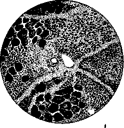
Figure 2. Muscle atrophy (in cross-section). Bundles of normal muscle fibers are visible on the left and at the bottom; in the rest of the field of view, there is severe atrophy of the muscle fibers.
part of cells, there sometimes occurs a very markedly pronounced proliferation and increase in the number of their nuclei, which is termed atrophic nuclear proliferation; this phenomenon is viewed as a manifestation of an automatic regenerative reaction. In brown atrophy (in the liver, heart muscle, sympathetic ganglia), an accumulation of brown pigment granules located near the nuclei is visible in the protoplasm of atrophic cells. Atrophying parenchymal elements are usually pushed apart from each other by the proliferated interstitial tissue. The outcome of atrophy can be expressed in the progressive escalation of the process, wherein such atrophy leads cells to death and disappearance; on the other hand, upon the elimination of conditions causing atrophy, mildly altered cells can acquire their former appearance. The significance of atrophy lies in the fact that the function of the atrophied tissue or organ is diminished. This functional decline of the atrophied organ can form the basis of a serious general disease; thus, in atrophy of the thyroid gland, myxedema (see) or cachexia (cachexia hypothyreotica) develops, and the consequence of pancreatic atrophy is frequently diabetes mellitus. A. Abrikosov.
Related articles
Mentioned in
Cite this page
“Atrophy.” Soviet Medical Encyclopedia. English translation of Bolshaya Meditsinskaya Entsiklopediya, 1st ed. (Moscow, 1928–1936), ed. N. A. Semashko. https://sovietmedicalencyclopedia.pages.dev/article/atrophy/