Nephritis
Historical document, translated for reference. It reflects medical knowledge of the 1920s–30s and is not medical advice.
Summary
Nephritis, or inflammation of the kidneys, is the most common form of Bright's disease. This article traces the historical evolution of understanding nephritis, from early observations of kidney disease to the development of classifications distinguishing between different forms of kidney inflammation and degeneration.
Encyclopedia article (1928–1936)
NEPHRITIS. Contents: I. Historical data............. 288 II. Pathological anatomy...........291 III. Classification of nephrits...........297 IV. Etiology and pathogenesis of nephrits.......299 V. Clinic and prevention of various forms of N.: A. Acute diffuse glomerulonephritis . . . 305 B. Chronic diffuse nephritis......313 C. Focal nephrits..............327 D. Interstitial and purulent nephrits . . . 329 Nephritis (from Greek nephros-kidney), inflammation of the kidneys, the most frequently observed form of Bright's disease (see). Modern pathology and clinic imply by nephritis those bilateral hematogenous diseases of the kidneys, which are accompanied by inflammatory changes in the Malpighian glomeruli (glomerulonephritis) or in the interstitial tissue of the kidneys. This disease manifests clinically with hematuria, albuminuria, and with diffuse lesions also with hypertension and edemas. I. Historical data. From a historical aspect, interstitial N. presents nothing interesting, while the doctrine of glomerulonephritis and generally of inflammations of the functional-secretory part of the kidneys has undergone a complex path of historical evolution, which on one hand coincides with the evolution of the doctrine of Bright's disease, and on the other-with the evolution of the doctrine of inflammation. In general, kidney diseases accompanied by dropsy and the appearance of protein in urine became known relatively recently. In the second half of the 17th century, the first observations appeared concerning the connection of some dropsies with kidney disease (Schenk, Bonet, Morgagni) and only in 1770 Cotugno (Co-tugno) first discovered protein in the urine in these cases. In the first half of the 18th century, Wells, Blackall, Alison already definitely indicated the connection of albuminuria and edemas with kidney disease. However, only Bright in 1827 firmly established that general dropsy with protein in the urine has a renal origin; at the same time, he accurately depicted the clinical picture of the disease and described the path.-anat. changes in the kidneys in it, which served as the reason for the subsequent designation of kidney diseases accompanied by edemas and protein in urine as Bright's disease (see). Bright himself, noting the extreme diversity of kidney changes found by him in this disease, suggested that here sometimes inflammatory changes were involved, in other cases-degenerative processes, in third cases-vascular damage. However, most researchers after Bright's work tended to believe that at the basis of the symptom complex put forward by Bright lay various types of kidney inflammation, i.e., nephritises, thanks to which for a long time the concepts of Bright's disease and nephritis became synonymous. This point of view was also supported by pathologists of that time; they were the first to attempt to give classifications of nephritises (Henle, Rokitansky), in which among other things one could find the unification under the term 'nephritis' of the most diverse kidney changes (hydronephrosis, amyloid, cysts, etc.). However, soon after this Virchow, who developed the pathological anatomy of inflammation in general, also clarified the concept of inflammations of the kidneys, excluding from the concept 'nephritis' atrophic changes and amyloid of the kidneys, which have no relation to inflammation. At the basis of Bright's disease Virchow saw two types of N.: parenchymatous and interstitial N. and viewed these forms as two different types of kidney inflammation, believing that in parenchymatous nephritis the inflammatory reaction is concentrated in the epithelial parenchyma and is expressed in the so-called degenerative changes of the latter, while in interstitial N. the inflammatory reaction proceeds in the interstitial tissue. With this view Virchow laid the beginning of the dualistic understanding of the nature of Bright's disease, which in the first time received special development among English scientists (Johnson and others). Simultaneously in Germany after the works of Cohnheim concerning inflammation, for some time the unitary view on the nature of Bright's disease became predominant. Thus, Cohnheim himself, as well as Frerichs, Traube and Klebs, put forward the position that at the basis of Bright's disease always lies inflammation of the interstitial connective tissue and that the observed change in parenchyma develops subsequently. Weigert was also a supporter of the unity of the basic anatomical process in all changes related to Bright's disease; he likewise saw the basis of all N. in inflammatory changes of the interstitial tissue and divided all N. into four types only according to the course (acute, subacute, chronic and completely chronic nephritis). Subsequently it can be seen that both clinicians and pathologists continued to adhere either to unitary or dualistic views on Bright's disease, resp. on N., with this or that variations in their opinions. From the huge amount of relevant works with corresponding classifications, one can mention the research of Bartels, who in 1871 and 1875 appeared as a defender of dualism and drew a strict distinction between acute and chronic parenchymatous N., on one hand, and interstitial N.- on the other both in clinical and path.-anat. relations. The same division with some detailing of individual forms was put at the basis of the classification of Senator, which became very popular among clinicians. Aufrecht went even further in this respect, dividing all N. into two sharply delimited forms from each other: 1) tubular N., the basis of which are degenerative changes of the tubular epithelium, and 2) vascular N. with primary damage to blood vessels, mainly vasa afferentia, and capillaries of the glomeruli. Scientists who took a completely opposite position were those who considered inflammatory changes of blood vessels, especially glomeruli (glomerulonephritis), as the only basis for all forms of Bright's disease (Charcot, Dieulafoy, Friedlander, Nauwerck, Langhans and others); this unitary doctrine subsequently received special development in the works of Löhlein. However, at the end of the 18th and beginning of the 19th centuries, partly under the influence of the clarification of path.-anatomical concepts of changes in the kidneys in general, and partly as a result of the work of clinicians on clarifying the functional activity of the kidneys in various forms of Bright's disease (Widal, Schlayer and others), the unitary point of view gradually recedes to the background, and among most pathologists and clinicians there is a tendency either to distinguish parenchymatous and interstitial forms of nephritis, or to speak of tubular and vascular (glomerular) N. The final coordination of this question however was complicated first of all by the fact that some researchers attributed the glomerular apparatus to the kidney parenchyma, others-to the vascular-interstitial tissue, and secondly by the fact that degenerative changes of kidney epithelium some scientists considered as an expression of active inflammatory reaction (Aschoff), while others excluded them from the concept of inflammation and attributed them to manifestations of pure degeneration (Ribbert, Lubarsch). In particular Ashof still adheres to Virchow's view of the existence of the so-called parenchymatous inflammation, manifested in changes of epithelium, which are called degenerative; on this basis Ashof essentially does not distinguish acute degenerative forms of Bright's disease from inflammatory ones and attributes them to manifestations of inflammation, i.e., N., distinguishing only glomerular and tubular N. Only chronic degenerative changes of the kidneys have been singled out by Ashof from inflammation and attributed to nephropathies. An attempt to coordinate all controversial points regarding N. was made in 1905 by the German path. society, which raised this question at the congress in Merano; at this congress for the first time F. Müller (F. Müller) spoke with a definite indication that those kidney changes, the basis of which are degenerative processes, do not belong to inflammation, but represent pure degeneration and therefore must be singled out from the concept of N.; for these primarily degenerative forms Müller put forward the term proposed by Ribbert- 'nephrosis'. Despite the detailed discussion of the question of N. at the congress in Merano, nothing new was developed regarding N., and both pathologists and clinicians remained with the previous division of nephritises, united by the concept of Bright's disease, into acute and chronic parenchymatous N. and chronic interstitial N. However, undoubtedly this congress gave an impetus to coordinated work on the topic of N. by clinicians and pathologists; an example of such joint work was the research of Volhard and Fahr, published in 1914 and having a tremendous influence on the entire question of nephritises and on the views on the essence of Bright's disease. Volhard and Fahr on the basis of comparison of clinical, functional and path.-anat. data came to a definite conclusion that at the basis of the symptom complex of Bright's disease there can lie three completely different kidney diseases, of which only one type (II) belongs to manifestations of inflammation: I. Primarily degenerative changes- nephroses. II. Primarily inflammatory changes of glomeruli- nephritises. III. Arteriosclerotic kidney diseases- nephroscleroses.
These three diseases differ from each other not only in the sense of pathological anatomy and pathogenesis, but also in the sense of clinical-functional manifestations both from the side of the kidneys and of the entire organism. The appearance of the classification of Volhard and Farr coincided with a period of revision of views on inflammation in general, during which purely degenerative changes as well as those representing connective tissue growth on the basis of atrophy and necrosis of the parenchyma were excluded from the concept of 'inflammation'. All this together served as the reason that this classification in recent years gradually became very widespread among clinicians as well as pathologists; at present only a small number of scientists (predominantly French, and among Germans Aschoff) do not accept it and propose their own classifications (Aschoff, Pasteur, Vallery-Radot, Oberling and others). But in any case the identification of Bright's disease with nephritis has completely fallen away and generally the term 'Bright's disease' has receded into the realm of history. II. Pathological anatomy. The description of the pathological anatomy of N. is most conveniently made separately in relation to glomerulonephritis and interstitial nephritis. I. Glomerulonephritides are divided into diffuse and focal. Diffuse glomerulonephritides in turn break down into acute and chronic. a) In acute diffuse glomerulonephritis, the macroscopic appearance of the kidney often presents nothing special: the kidney may have a normal appearance or be only more or less congested to some degree, even in very severe cases (death in the first days of the disease with symptoms of anuria and uremia); in these cases only with careful examination using side lighting can an experienced eye notice the affected glomeruli, appearing on the surface of the section in the form of the smallest grayish grains-sand particles. The picture is clearer in acute hemorrhagic forms (see below), when glomeruli are visible on the surface of the kidneys and in the section in the form of red dots. Microscopic examination of such most acute cases with little changed macroscopically normal kidneys reveals changes only in the glomeruli (acute glomerulitis): it is striking that all glomeruli are increased in volume and extremely rich in nuclei. Examination with high magnification reveals that all changes are concentrated inside the capillaries of the glomeruli (intracapillary glomerulitis) [see separate table (p. 295-96), fig. 5] and consist in the fact that inside the lumens of the capillaries, devoid of blood, among finely granular, less frequently fibrinous protein mass, leukocytes accumulate, as well as swelling and desquamating cells of the capillary endothelium; the capillaries of the glomeruli themselves are dilated, their loops fuse with each other, their contours become indistinct; sometimes individual loops undergo necrosis. Similar changes are observed in the vasa afferentia of the glomeruli, i.e., in the arteries that bring blood to the glomeruli; their wall appears swollen, having lost its structure, homogeneous; the endothelial cells are swollen, with foamy, vacuolated protoplasm, picnotic nuclei. These changes in the glomeruli, first pointed out by Lejars and clarified in detail by Fahr (Fahr and others), according to the opinion of most authors, represent an expression of inflammation of the glomerular capillaries, diffuse capillitis of the glomerular loops. In contradiction to this stands the opinion of Volhard, considering these changes not belonging to inflammation, but representing the result of ischemia of the glomerulus due to spasm of its vas afferens; such spasm of the afferent arteries Volhard considers the pathogenetic basis of glomerulonephritis. After some time, manifestations of exudation may be added to the above-described changes in the glomeruli, which consist in the secretion of exudate of one kind or another into the cavity of Bowman's capsule; sometimes this is serous fluid, which under the microscope has the appearance of a finely granular mass; in other cases it is a typical fibrinous exudate, giving a picture of a fibrous-network mass. In the exudate an admixture of leukocytes may be observed; almost always single erythrocytes are also observed; sometimes the latter accumulate in such large quantities that the cavity of Bowman's capsule is completely filled with blood (hemorrhagic glomerulonephritis). From the cavity of Bowman's capsule the exudate spreads into the tubules, which is easily noticeable under the microscope. The epithelium covering the capillary loops of the glomeruli and lining the inner surface of the capsule usually shows a picture of granular degeneration and degenerative fatty change. From the side of the tubules in the early periods of acute glomerulonephritis no changes are found. Similarly, the small blood vessels branching between the tubules, in contrast to the vessels of the glomeruli, show no noticeable changes except hyperemia. - In further periods, which represent a transition to subacute glomerulonephritis, in the tubules one or another degree of degenerative changes in the form of granular, fatty, hyaline-droplet degenerations are found, which, as is generally believed, depend on the lesion of the glomeruli and on their impassability for blood. Macroscopically at this the kidneys are more or less enlarged, their cortical layer is expanded, swollen, gray-red-brown in color, often with a mass of dark red dots and stripes, which gives the pictures of the 'large red' or 'large mottled kidney' [see separate table (vol. XX, p. 103-104), fig. 1]. The changes in the glomeruli in the subacute period of nephritis can take on different characters; sometimes the changes remain for a long time inside the capillary loops (proliferation of endothelium, clotting of fibrin, necrosis of loops), which gives the intracapillary variety of subacute glomerulonephritis; in other cases extraglomerular changes begin to predominate, which consist in the fact that the epithelium of the capsule proliferates, multiplying karyokinetically, desquamates or more often forms peculiar multilayered outgrowths from elongated cells in the form of 'half-moons' or 'half-lunes', adjacent to the glomeruli [see separate table (p. 295-296), fig. 4]; such 'productive capsulites' of the glomeruli are observed in the extracapillary variety of subacute glomerulonephritis. The extraglomerular blood vessels in the subacute period of glomerulonephritis show no special changes; sometimes in them also inflammatory-necrotic changes are observed, and later, in the subchronic period, manifestations of productive endarteritis appear; such cases Volhard distinguishes as the vascular variety of subchronic glomerulonephritis. Sometimes cases of subacute glomerulonephritis are accompanied by such severe degenerative changes in the tubular epithelium that macroscopically a picture of the 'large white kidney' is obtained (see Nephrosis); such cases of subacute glomerulonephritis with strongly expressed degenerative changes in the tubular parenchyma, simulating clinically and anatomically nephrosis, some authors denote as pseudonephrosis. b) Chronic diffuse glomerulonephritis is a transition into a chronic state of acute glomerulonephritis through a period of subacute changes. Such a course can be taken by glomerulonephritides with weakly expressed degenerative changes as well as those in which the latter are significantly expressed. The basis of chronic glomerulonephritis is the so-called secondary shrinkage, i.e., the gradual emptying of the glomerulo-tubular systems corresponding to the changed glomeruli and the growth of interstitial connective tissue. In the glomeruli a gradual disappearance of exudate and decrease in the number of nuclei takes place, collapse and hyalinization of individual capillary loops or of the entire glomerulus as a whole. In the capsule concentric growth of connective tissue and replacement of the 'half-lunes' from epithelial cells with connective tissue takes place. The emptied capillary loops fuse with the capsule, and the hyalinized glomerulus sometimes completely fuses with the proliferated capsule or between it and the capsule remain lumens, lined with epithelium and resembling glandular ducts. In the end the glomeruli turn into hyaline balls, surrounded by a layered capsule from connective tissue. At the same time collapse, atrophy, disappearance of the tubules corresponding to such glomeruli and growth of connective tissue between them takes place. In case of significant degenerative changes in the tubules in glomerulonephritis, in the shrinking kidney groups of tubules with swollen epithelium are seen, showing pictures of granular, hyaline-droplet degeneration or deposits of doubly refractive lipoids. In some better preserved tubules sometimes regenerative cell proliferations are observed. In far advanced shrinkage in the arteries of the kidneys usually a sequential sclerosis of the wall with growth of the inner intima occurs. Macroscopically chronic glomerulonephritis gives a picture of decrease in volume and densification of the kidney. The cortical layer is devoid of pattern, its boundary with the medullary substance is indistinct, the color is pale grayish-yellow or more brownish-red; the presence of degenerative changes is manifested by yellowish-gray mottling.
If the process of scarring proceeds uniformly throughout the kidney, which occurs comparatively rarely, then the surface of the organ remains smooth (cases of 'chronic glomerulonephritis without granularity' of Fara). More often, along with the overgrowth of some glomeruli with connective tissue, other portions of them return to normal; therefore the process of scarring proceeds unevenly and areas of desolation and overgrowth of connective tissue alternate with areas in which all parts are not only well preserved but are in a state of compensatory hypertrophy (glomeruli are enlarged, tubules are dilated). The result of this is either a fine or coarse granularity of the kidney surface. In view of the fact that the transition of acute forms of glomerulonephritis into subacute and chronic does not always follow the same path (see above—intracapillary, extracapillary, and vascular varieties of subacute and subchronic glomerulonephritis), the clinical manifestations of the transition into chronic glomerulonephritis until the period of clearly expressed scarring may be various (three types of Volhard). Besides changes in the kidneys in diffuse glomerulonephritis, pathological processes are also observed in other parts of the body. These include inflammatory changes in the skin vessels found by Topfer, Weiss, and Müller (Topfer, Weiss, Müller) in acute glomerulonephritis; hypertrophy of the left ventricle of the heart, especially marked in the subacute and chronic periods of the disease; hypertrophy of the muscular layer of small arteries in the subacute period and later sclerosis of their walls. (Uremic changes—see Uremia). Focal glomerulonephritises are those cases in which inflammatory changes involve only individual glomeruli and groups of them. A distinction is made between a) infectious focal glomerulonephritises and b) embolic focal glomerulonephritises. a) Infectious focal glomerulonephritises are observed in various infectious diseases, but most often in streptococcal sepsis. The kidneys may be somewhat enlarged depending on the degree of degenerative changes, usually are hyperemic; against this background numerous dark red points are visible both on the surface and on the section of the kidney, and they are scattered not regularly but in groups. Upon microscopic examination it turns out that a considerable portion of the glomeruli are completely normal, but in many of them there are changes already described above in relation to acute glomerulonephritis. Besides the fact that these changes affect individual glomeruli, even in such glomeruli they sometimes involve individual capillary loops. Necrobiotic changes in capillary loops and hemorrhagic exudate in the cavity of the capsule and in the lumens of the tubules pertaining to the glomerulus (hemorrhagic focal glomerulonephritis) are especially frequently observed. Sometimes necrotic changes in the form of swelling, dullness of tissue and disappearance of nuclei from the glomerulus spread to neighboring tubules and to the interstitial tissue of the kidneys. b) Embolic focal glomerulonephritis, first described by Leyden, differs from the preceding infectious one in that it is based on embolism of glomeruli by more or less significant thrombotic particles; usually this form of focal glomerulonephritis is observed in warty-ulcerative endocarditis of the endocarditis lenta type, most often caused by Streptococcus viridans. Thrombotic particles with microorganisms occlude individual capillary loops, more rarely vasa afferentia of some glomeruli; embolized loops undergo coagulation necrosis with subsequent transformation into a homogeneous fibrinoid mass. Along with this, exudation, often hemorrhagic (hemorrhagic embolic nephritis), into the cavity of the capsule and infiltration with leukocytes around the glomerulus (periglomerulitis) may be observed. The macroscopic appearance of the kidneys is similar to the preceding form; sometimes in addition to this ordinary infarcts are observed in the kidney. The course of focal glomerulonephritises is connected with the course of the primary disease (sepsis, endocarditis). In case of retrogression of the process in the affected glomeruli, the course of changes is observed which is characteristic of glomeruli in subacute and chronic glomerulonephritises. As a result in the kidneys there may remain individual small indentations on the surface, from which connective tissue strands, gradually narrowing, sink into the depth of the cortical layer; under the microscope in them a cellular-rich connective tissue with hyalinized glomeruli is found. II. Interstitial nephritises are based on the development of inflammatory changes in the interstitial, connective tissue where the process begins primarily; parenchyma and in general secretory parts, if they participate in the process, do so only secondarily. All interstitial nephritises according to their topography belong to focal nephritises, since they always involve only individual parts of the kidney tissue, leaving others untouched. Only with very extensive spread of the process some forms of interstitial N. may acquire a diffuse character. A distinction is made between acute non-purulent interstitial nephritis and purulent nephritis. Acute non-purulent interstitial N. is observed in various acute infectious diseases, most often in scarlet fever, streptococcal anginas, diphtheria. Anatomically the kidneys present a different picture depending on the degree of affection. Sometimes all the change amounts to the fact that in a kidney close to normal, small red specks are visible on the surface and on section. In other cases the kidneys are somewhat enlarged, soft, and after removal of the capsule on the surface indistinctly delimited, diffuse, often slightly protruding red spots are visible; on section in the cortical layer similar stripes are visible. Microscopic examination of the kidneys in such typically focal interstitial N. shows that the red spots correspond to foci of infiltrates from lymphoid and plasma cells located near small veins, sometimes with an admixture of individual leukocytes and eosinophils; blood vessels in the area of such a focus are usually dilated and engorged with blood. In more severe cases the above-mentioned infiltration diffusely involves the entire kidney, and then the kidney parenchyma usually appears to a greater or lesser degree degenerated; this gives the form of interstitial nephritis which is often called septic nephritis. The kidneys in this case are enlarged, rounded, very flaccid; the capsule is removed very easily; on section the tissue protrudes strongly, of motley gray-red appearance. It should be noted that milder degrees of such diffuse interstitial N. may not be accompanied by clearly expressed infiltration, but may be based predominantly on inflammatory edema of the interstitial tissue; this includes the so-called nephritis exsudativa serosa of Aschoff; closely related to this is also the so-called toxische Schwellniere of Kuczinski, differing only in the presence of glomerulitides. Purulent N. are characterized by leukocytic infiltration of the interstitial tissue, usually passing into purulent melting of the tissue. Purulent nephritises are caused by pus-forming bacteria, and the penetration of the latter into the kidney can occur in two ways: a) hematogenously, i.e., by embolism of bacteria by the blood current, b) urinogenously, by penetration of bacteria into the kidney from the renal pelvis from stagnant urine there. This difference in the penetration of pus-forming infection into the kidneys gives two different types of purulent nephritises. 1. Hematogenic embolic, or metastatic purulent N., also called descending N., is observed in ulcerative endocarditis and in other septicopyemic diseases. Bacterial mycotic emboli are arrested in the capillaries of the glomeruli and in the capillaries surrounding the tubules; in such cases under the microscope in the corresponding capillaries clumps of bacteria are seen, around which infiltration with leukocytes begins, which in the embolized glomeruli gives the picture of glomerulitis and periglomerulitis; in a short time in the places of embolism purulent melting of the tissue and formation of multiple abscesses occur. Macroscopically such an abscess nephritis (nephritis apostematosa) is characterized by the presence in the cortical layer of protruding on the surface yellowish-gray foci, surrounded by a red border and containing a drop of pus. The kidney tissue in the intervals between small abscesses is usually unchanged; only with a large number of abscesses it becomes hyperemic and flaccid *->
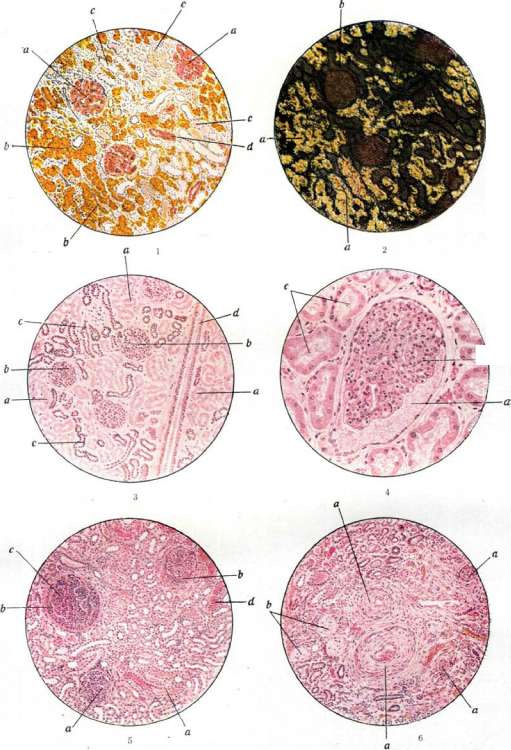
Figure ]. Acute parenchymatous nephritis with hyaline thrombi in the capillaries of the glomeruli. a-Edematous swelling of the capillary loops; b-Lipoids (with a sharp edge) outside the glomerulus; c-Swollen cells in the tubules; d-Fatty degeneration of the tubular epithelium. Fig. 2, Cortex, unstained on cut surface, in reflected light: lipoids shine with a yellowish tint (from fat); b-Shadows of the glomeruli. Fig. 3L Subacute nephritis: a-Exudation in the capsule of Bowman; b-Glomerulus; c-Exit of protein fluid into the capsule. Fig. 4. Productive stage of parenchymatous nephritis: a-Normal glomerulus; b-Proliferation of epithelial cells in the capsule of Bowman; c-Fibrous tissue; d-Erythrocyte in the lumen. Fig. 5. Embolic nephritis: a-Very erythrocyte-filled arterioles; b-Glomeruli dilated with hyaline thrombi.
1. Hematogenous, or descending, purulent N. occurs as a result of renal hyperemia, edema, and degeneration of the parenchyma. Besides the formation of emboli with subsequent development of abscesses, pyogenic bacteria, as if by secretion, can pass through the walls of capillaries and end up in the lumens of the urinary tubules, especially of the kidney medulla; as a result of this, a purulent inflammatory process develops, manifested by the appearance of yellowish-gray purulent streaks in the renal papillae; this is called papillary mycotic nephritis (nephritis papillaris mycotica). Hematogenously, larger abscesses can form, for example, when a large embolus is carried into the kidney, causing the formation of an infarct, which subsequently becomes purulent. Moreover, in the prolonged course of embolic purulent N., some abscesses may gradually increase, merge with each other, which leads to the development of large abscesses. The formation of a large purulent focus in the kidney, consisting of several adjacent abscesses, some call a renal carbuncle. 2. Urogenic, or ascending, purulent N. is observed in various inflammations of the urinary tract, accompanied by inflammation of the renal pelvis (pyelitis), from which the purulent inflammation spreads to the kidneys; on this basis, urogenic purulent nephritis coincides with what is called pyelonephritis. Macroscopically, at first the process is manifested by the appearance of grayish streaks in the parenchymal layer of the kidneys, subsequently they turn into purulent streaks extending in rays from the papillae into the cortical layer, where abscesses appear; the kidney is enlarged, flabby, and hyperemic. Under the microscope, at first, catarrhal inflammation of the tubules of the medullary layer with leukocytic infiltration of the interstitial tissue is observed, which later, as the leukocytic infiltration increases, turns into purulent melting. Purulent N. is often accompanied by involvement of the kidney capsule in the inflammation (perinephritis) and the perirenal tissue (paranephritis, see). In some cases of mild purulent nephritis with a low-virulence bacterial flora, a transition to chronic purulent N., accompanied by the proliferation of connective tissue, i.e., wrinkling, may occur; in such cases, sunken areas of dense, whitish connective tissue are visible in the kidney, among which are yellowish-gray, dry, curd-like foci. Microscopic examination reveals a picture of nephrosclerosis with encapsulated abscesses among the connective tissue from disintegrating leukocytes, and often foci of dead, amorphous detritus; in places, groups of xanthoma cells are visible. Such an outcome in wrinkling is mainly characteristic of urogenic ascending purulent Nephritis.
D. Abrikosov. III. Classification of nephritis. In the modern narrow understanding of this word, N. remains nevertheless anatomically, pathogenetically and clinically diverse suffering, and its classification presents a practically extremely important, although very difficult, task. The most important feature of nephritis, which can and must be distinguished in each individual case, is 1) the extent and nature of the anatomical process in the kidney, 2) the course of the disease, and 3) the preservation or loss of the excretory function of the kidney in N. When taking into account these three points, one can obtain a more or less clear idea of the nature, degree of kidney damage and prognosis of nephritis. According to the spread and localization, nephritis is differentiated as diffuse or focal with damage to the glomeruli or interstitial tissue, and each of these forms has a rather characteristic clinical-anatomical picture. According to the course (especially in diffuse forms of N.), acute, subacute, subchronic and chronic forms are distinguished. However, the third, predominantly clinical evaluation of individual nephritises is most important: whether the kidneys have retained or lost their main function - the removal of metabolic products and water from the body. Each of the given classifications, taking into account only one or another feature of N., still does not cover all the phenomena observed in N. It is therefore more correct in each individual case to study all the most important elements of the process, characterizing it in a multifaceted way, for example as follows: 'chronic diffuse glomerulonephritis with sufficient kidney function.' - Due to the close connection that exists in the kidneys between the tubules and glomeruli, which essentially form a single functional unit ('nephron'), strictly isolated lesions of one or another system (tubules or glomeruli) should be expected to be extremely rare. Indeed, in the presence of deep circulatory disorders in the glomeruli, for example in the extracapillary form of diffuse glomerulonephritis, degenerative changes in the corresponding tubules soon join it. Clinically, such cases, which by the way are quite common, proceed as mixed forms or nephrosis-nephritis. In some cases, the edematous syndrome of nephrosis and the corresponding degenerative changes in the kidneys are so predominant that they obscure the glomerulonephritis that was present initially. Attempts have been made to pathogenetically separate the described secondary forms of nephrosis or nephrotic overlays from true primary lipoid nephrosis. However, in pronounced cases of nephrosis-nephritis with edema, the basic, characteristic changes of nephrosis (albuminuria, hypalbuminemia, lipoidemia) are as a rule found in the urine and blood, and in the kidneys deposition of lipoids in the tubular epithelium. Nephrosis-nephritis therefore represents one of the subgroups of diffuse glomerulonephritis, in which anatomically in addition to nephritis there are also or even predominate degenerative changes in the tubular epithelium. Clinically, nephrosis-nephritis carries with it the common features of the edematous syndrome in other forms of Bright's disease (see also Nephrosis). IV. Etiology and pathogenesis of nephritis. One general regularity is decisive for the occurrence of N., and to a certain extent also for its isolation as a special anatomical-clinical unit. The common etiological factor for the occurrence of the vast majority of N., regardless of the degree and nature of their manifestation, is infection, in particular infection with cocci microbes. The most frequent causative agent of nephritis is streptococcus, less often meningococcus, pneumococcus, and in isolated cases even gonococcus. Nephritis also often occurs during diseases such as influenza, malaria, measles, and even streptotrichosis. In the second group of cases, it is extremely difficult to decide whether N. is a consequence of an additional infection that has joined or been activated due to the weakening of the patient. The primary focus of infection (its 'gateway') is rightly considered to be the lymphatic ring of the pharynx. Clinical experience teaches that in the vast majority of cases in the anamnesis of patients with N. there is tonsillitis of one or another bacterial etiology. Naturally, in cases of influenza with catarrh of the upper respiratory tract, the exclusion of such angina is hardly possible, all the more so that N. as a rule does not develop after a severe lacunar or follicular angina, but follows a relatively unnoticed, accompanied by slight fever peritonsillitis or even after a subacute superficial angina (angina superficialis). In a number of individual cases, the primary focus may be localized in other organs: gallbladder, appendix, dental granulomas, etc. Thus, a case of disappearance of N. after cholecystectomy (Bergman), elimination of N. after conducting quinine treatment in malaria, etc., has been described. However, these cases are isolated and by no means characteristic of most nephritises. Along with infection, cold has long been considered as an etiological factor, sometimes even basic, for the occurrence of N. The exceptional role of cooling in the genesis of N. is generally recognized and confirmed by a huge amount of both clinical and statistical data. Thus, 'military' N., autumn, spring outbreaks of it, different morbidity of N. in different geographical points depending on the humidity and air temperature, etc., are known. N., for example, is extremely common in northern maritime countries with a cold and humid climate. The effect of cooling can find its explanation also from the pathophysiological side. The renal vessels in terms of innervation belong to one (extraabdominal) group with the blood vessels of the skin; due to this, a constant functional correlation is created between the kidneys and the skin. Spasm of peripheral skin vessels (with cooling and chill) immediately leads to a decrease in blood supply to the kidney, and clinically reflexively to oliguria. Moreover, the latest supravital histological studies have established extremely sharp changes produced by cooling in the vascular apparatus of the kidneys (E. F. Mueller). These changes do not occur when the mentioned vegetative-nervative correlational connections are disturbed, for example in denervation of one kidney: in such a kidney bacteremia together with cooling does not lead to the occurrence of N., as is the case in the control kidney with preserved innervation. Only the possibility of development of inflammatory changes in the kidneys under the influence of cooling alone remains controversial and unproven, since in almost all cases of so-called cold N. it is extremely difficult to exclude the simultaneous presence of infectious etiology of the suffering. The role of cold is thus extremely complex and perhaps amounts to revealing or activating that hidden or dormant infection which was present or penetrated the body. Despite the undoubtedly infectious origin of N., the question of its pathogenesis - the mechanism of action of infection on the kidneys and the occurrence of inflammatory changes in them - at present cannot be considered clarified. It is quite obvious that so-called focal N. of embolic and septic nature arise as a result of the carriage (metastases) of pathogens and the corresponding direct infection of individual renal glomeruli while others remain intact and in normal condition. Another matter is diffuse glomerulonephritis, which sometimes develops with extraordinary rapidity and involves the entire glomerular system of the kidneys. Bacteriuria, observed by older authors in nephritics, as well as the detection of various bacteria on histological sections of the kidneys, have not been confirmed in recent times; and a priori it is clear that diffuse N. are not a consequence of only the excretion of pathogenic microorganisms by the kidneys, since this disease is accompanied by damage to the entire vascular system, in particular the system of the smallest vessels of the body (see below). It is also remarkable that in acute diffuse glomerulonephritis, despite the infectious etiology and the apparently maximum spread of the inflammatory process in the kidneys, a purulent process is never observed. All this gave a number of researchers to assert that the direct action of infection alone is not sufficient for the development of nephritis. The latter apparently develops under the influence of endotoxins released by the pathogen in the patient's body. An indirect confirmation of the infectious-toxic origin of glomerulonephritis can also be its manifestation usually on the third week after angina (post-infectious), in contrast to focal forms, which most often develop acutely. Such a late manifestation of N. is associated with an increased sensitivity of the renal vessels to the mentioned toxin during this interval of time. In this sense, some are inclined to consider acute glomerulonephritis as a manifestation of allergy, a 'second' disease in the post-infectious period (Schick, Pospisil). Similar, but by no means identical with infectious N., pictures are given by individual toxicoses, such as: acute poisoning with lead, a group of resin-terpenes, as well as toxicoses of pregnancy.
In particular, pregnancies ending in eclampsia are often accompanied not by tubular nephropathy—'pregnancy nephrosis,' but by a specific picture of glomerulonephritis, which would more correctly be designated as 'pregnancy kidney' (see Kidneys). It should be noted that these nephro-nephritides, arising under the influence of a special toxic etiology, also proceed in a peculiar manner, never ending until childbirth or termination of pregnancy. In acute and even subacute poisonings, e.g., lead poisoning, more or less typical manifestations of diffuse N. are also observed; in most cases, the picture of lead poisoning corresponds symptomatically to renal insufficiency, clinically resembling the final stage of chronic glomerulonephritis. As for the diffuse nephritides described in poisoning by various chemical substances, the so-called renal poisons (uranium, sublimate), or by other strongly acting agents (salvarsan, turpentine), the diffuse nature of these is more than doubtful, and the necrotic changes observed in the tubules bring these forms closer to nephroses in the form of so-called necronephroses with the syndrome of renal insufficiency (see Nephrosis). The question of the organ specificity of microorganisms causing renal lesions in N., and of the predisposition of the organ itself to this disease, is currently only being developed. Thus, Rosenow, who intensively studied the causative agents of infection in various diseases, asserts that the streptococcus that causes the development of N., when transferred to another person or animal, also gives N., just as the streptococcus of dental granuloma in dogs leads to infection of the tooth root, i.e., it shows organotropism. There are also numerous observations about the role of the genotype in the occurrence of N.; thus, out of 8,000 scarlatinal patients in the Eppendorf Hospital in Hamburg, 68 cases of nephritis occurred in 29 families, and 37 individual cases concern 186 families (Mathies). That this is not only due to the greater frequency of intra-family infection is evidenced by observations of the same author in widows' homes, where, despite epidemiologically identical conditions, such frequency of N. after scarlet fever was still not observed. A number of pedigrees with multiple cases of N. in one family or among several siblings have also been published. However, it remains questionable whether at least in some of these cases we are dealing with nephrosclerosis with hypertension, a disease whose genetic basis has been studied better. This especially concerns the cases described before 1914, when a precise differentiation between primary arteriosclerosis of the kidneys and secondary nephritic atrophy of them was not yet sufficiently precise and firmly established. Unfortunately, to this day there are no convincing observations about the occurrence of N. in monozygotic twins or about the course of the process in such patients; such observations could possibly serve as one of the proofs of predisposition to nephritis. Among the factors predisposing to the development of nephritis, perhaps the importance of tissue acidosis should be particularly emphasized. The existing observations about the prevention of scarlatinal N. by preliminary introduction of alkalis with food seem to be indirect evidence in favor of the aforementioned assumptions (Osman). In the same sense, experimental nephritides that arise under the influence of intravenous infusions of hemoglobin speak: such nephritides develop only in the presence of acidic, but not alkaline, urine. In general, it can be assumed that any disturbance in the nutrition of the parenchymal elements of the kidneys (disorders of local blood circulation, cooling, disturbance of innervation, shifts in the active reaction of tissues) creates more favorable conditions for the action of infection and toxins, or contributes to the development of an inflammatory process in the kidneys. However, the mechanism of the action of the infection and the fundamental question about the essence and origin of the main changes in the glomerulus, so-called glomerulitis, which stands at the center of the process (especially its acute form) and has given it its name, remain not fully clarified. The finding in the glomeruli of so-called intracapillary exudate, i.e., blood plasma containing leukocytes—or the usually observed typical sharp increase in nuclei in the glomerulus, gave grounds to classify glomerulitis as a form of endocapillaritis (Far). The completely obvious difficulty of explaining only an isolated diffuse glomerular capillary in the kidneys led to the necessity of seeking a generalized lesion of capillaries, which serves as the background on which the picture of nephritis is played out. Indeed, Kylin, based on clinical observations about changes in capillary circulation in N., which is manifested primarily in the narrowing of the arterial limb of capillaries and an increase in capillary pressure, transfers the entire essence of the occurrence of acute glomerulonephritis to the capillaries, designating the disease itself as capillaropathia acuta universalis. Along with this, there are numerous observations indicating that in the very earliest periods of acute glomerulonephritis, the glomeruli are not hyperemic at all, but, on the contrary, are empty. On decapsulation and biopsy of the kidneys in such cases, it could be repeatedly confirmed that the glomeruli are anemic, and the capillaries in them are dilated and open. In addition, Gülze and Ricker succeeded in injecting the entire glomerular network of an acutely nephritic kidney with a liquid mass under pressure equal to blood pressure. Obviously, during life, these kidneys were deprived of blood supply due to causes lying somewhere outside them, e.g., from a functional spasm of the afferent arterioles. Thus, the unquestionable disturbance of blood supply to the Malpighian glomeruli, which is the basis of the pathogenesis of glomerulonephritis, finds itself a twofold, almost opposite explanation. Ischemia of the glomerulus or its hyperemia—is it a spasm of the afferent vessels or an inflammatory exudation into the lumen of the capillaries that is the primary stage of the process, this is the main disagreement in the modern doctrine of the histogenesis of glomerulonephritis. Hence the further continuing disagreements on the question of the pathogenetic pathways that lead to the inflammatory changes observed in glomerulonephritis: is there a 'defensive inflammation' here due to the direct action of the toxin, or is there only an indirect reactive inflammation in response to the ischemia of the glomeruli and the emptiness of its capillaries. That angiospasm on the periphery actually occurs in diffuse glomerulonephritis can be seen by direct observation of the accessible study of the fundus of the eye, where the central artery of the retina is sometimes in a sharply narrowed state. The general functional narrowing of extensive vascular areas also lies at the basis of the cardinal symptom of acute glomerulonephritis—hypertension, which therefore has a transient and unstable character in this stage of the disease. It has been repeatedly established and confirmed by systematic two-hour measurements of blood pressure over many days that in patients with scarlet fever, or after follicular angina, hypertension, and consequently vascular spasm, precede the appearance of any objective signs of renal damage, in particular the appearance of pathological urine. Similar observations about an increase in capillary pressure 5-7 days before the appearance of clinical symptoms of N. have been made by Kylin and gave the latter occasion for the aforementioned doctrine of primary capillary disease (Kapillarschaden) as the cause of N. However, it is extremely difficult to decide this question with the same categoricity as does Volhard, that general arteriolar spasm is not only an early symptom but also the main cause of changes in the glomeruli in acute N. One thing is certain—initial hypertension in acute N. is not in itself a consequence of the pathological process in the kidneys, and at best accompanies it. Subsequently, when acute N. passes into the chronic form, hypertension acquires a more persistent character: a functional spasm of peripheral vessels, prolonged and fluctuating, leads to a series of cardiovascular disorders and to anatomical changes in the vascular system. Kidney damage that arose as a result of the action of toxic substances on them, when renal insufficiency occurs, itself becomes the cause of vascular changes on the periphery: chronic N. leads to general secondary arteriolosclerosis, clinically—to 'pale hypertension,' and the disease 'from angiospastic turns into angiopathic.' So-called local diseases of the excretory apparatus can also lead to an increase in blood pressure, this is evidenced by repeated observations about the development of hypertension in prostate hypertrophy and in other cases of difficult urination, in cystic degeneration of the kidneys, renal colic, and even in unilateral hydronephrosis. In the same sense, very convincing, although indirect, evidence of the functional origin of hypertension is the acute decrease in blood pressure after decapsulation of the kidneys in anuria.
The transient nature of hypertension in acute nephritis has led many researchers to search for some chemically vasoactive substances in the body during the described stage of 'prenephritis' that would cause general vascular spasm and, perhaps in the first place, spasms of the renal arteries. What is the chemical nature and biological properties of these substances? Are they of renal or extrarenal origin? There is every reason to assert that these substances, having a pressor effect, are contained in the blood of patients with nephritis: it has been experimentally demonstrated many times on various biological vascular preparations (according to Kravkov and Leven-Trendelenburg) that blood and extracts from organs of patients with nephritic hypertension possess an exceptionally powerful vasoconstrictive effect (Gülze and Strauss, Vovsi, Lass and Shevlyagin). These pressor substances are obviously of protein origin. Only the question of their nature remains controversial: do they represent products of higher protein breakdown-peptones, or proteinogenic amines of bacterial origin, or finally, nitrogen-free products of intestinal putrefaction-aromatic substances of the phenolic series, not excreted by the kidneys in sufficient quantity in diffuse N. The intensification and persistence of hypertension in chronic nephritis suggest that changes in the activity and anatomical process in the kidneys themselves may in turn serve as a secondary source for the formation of pressor substances and thus maintain the duration of the spastic state of the smallest vessels. Edema in acute N. is by no means an obligatory, although frequent symptom of the disease. The pathogenesis of these edemas is undoubtedly different from the usual congestive edemas in cardiac patients, and in part also from the extremely intense and characteristic edemas in lipoid-amyloid nephrosis. The former concept of nephritic edema as a consequence of oliguria and water retention in the blood in N. is completely incorrect. The presence of true hydremia (see) in N. is generally subject to doubt. Nephritic edema has a much more complex pathogenesis. Even before the onset of visible edema in N., in the stage of so-called 'pre-edema', an increase in the osmotic pressure of the blood can be noted, caused by hyperchloremia, and at the same time an accumulation of sodium chloride in the tissues. The accumulation of sodium cations and chloride anions in the tissues, however, does not proceed parallel to each other, sometimes even dissociates, and may not be accompanied by edemas, as for example is observed in the late stage of kidney insufficiency (dry retention of chlorides- retention chloree seche-Widal). In the latter case, however, there is a predominant retention of chlorine and a less intense increase in the sodium ion. In the acute stage of H. and even in nephroses, the amount of sodium in the tissues sharply increases, and this is apparently one of the factors contributing to the development of edema (Blum). It is characteristic that potassium and calcium cations in this case have the opposite, diuretic effect and are therefore even used therapeutically in renal edemas.- The second factor contributing to the development of edemas in N. is the change in the permeability of the vascular wall (membrano-genic hypo-onkia Schade). The high protein content in the edema fluid of nephritics indicates the special conditions of fluid transudation in this disease and brings nephritic edemas closer to inflammatory ones. The richness in protein can be explained according to Beckerman only by a change in the permeability of vascular walls for large protein particles.- Finally, in the edematous form of N. with a nephrotic overlay, there is always also a third factor, which is central in the pathogenesis of edemas in lipoid nephrosis. Here, as there, there is a decrease in the amount of proteins in the plasma, hypoproteinemia down to 4-5%, a shift in the albumin-globulin coefficient toward an increase in the amount of globulins and a sharp decrease in the colloid-osmotic (oncotic) pressure of the plasma to 13-16 mm Hg (Schade, Govaerts, Vovsi, Itzikson and Vagon). Despite the relatively small magnitude of oncotic pressure (25-30 mm Hg), i.e., the force with which colloids hold their water, the latter, due to their inability to osmosis, plays a decisive role in the transudation of lymph from the blood into the tissues and back. In different stages of N., edemas apparently have somewhat different pathogenesis depending on the predominant influence of one or another (of the three described) factors. In general, edemas in N. arise not so much because the water-secreting function of the kidneys is impaired, but due to extrarenal causes-'loss of the ability to retain water and salts within the vascular bed', a phenomenon caused by one or more of the above-mentioned reasons (see also Edema). U. Clinic and prevention of various forms of nephritis. A. Acute diffuse glomerulonephritis is the initial stage of the most frequently observed diffuse glomerulonephritis. In some cases it runs extremely violently, in others its symptoms are so insignificant that they are overlooked by patients and even by doctors. The most typical form of acute glomerulonephritis is scarlatinal nephritis and nephritis after angina. The disease phenomena can develop within a few hours; more often, several days before the appearance of visible objective signs of the disease, there are a number of subjective and objective precursors. Early subjective signs of acute diffuse glomerulonephritis are a feeling of brokenness, headache, pain in the lumbar region, loss of appetite and in some cases (especially those accompanied by edemas)-shortness of breath. Headache sometimes has a very persistent and tormenting character. However, the main, leading symptoms of this form of N. are hematuria, albuminuria and hypertension. These symptoms, along with the less constant edemas in acute glomerulonephritis, define the clinical picture of the acute stage of diffuse glomerulonephritis. Edemas in acute N. are very frequent, but as indicated above, by no means an obligatory symptom. The character of the edemas is to some extent peculiar: they appear first on the face, on the abdominal wall, then on the extremities and on the lumbar region. The face becomes broad, its expression is smoothed, the supraorbital arches are sharply swollen ('lion's brows'). It is necessary, however, to remember that in isolated acute N. edemas very rarely reach such degrees as in lipoid nephrosis or amyloidosis of the kidneys. The edema fluid has the same osmotic pressure as the blood, i.e., it is characterized by a high chloride content. The protein content in it is also high and usually equals 11/,-2x/a% and higher, which sharply distinguishes nephritic edema fluid from nephrotic fluid or fluid in so-called hunger (protein-free) edema (see Edematous disease). The entire skin is striking by its sharp pallor with a grayish or yellowish tint, specific for renal patients. Many, however, rightly express doubt whether this characteristic pallor is a consequence of only the edema of the skin in acute N. or whether the cause is also the general spasm of arterioles of the skin and kidneys, as well as true anemia. The sharp pallor in those nephritics who have no edemas at all or in whom the latter have already disappeared speaks in favor of vascular spasm. Edemas sometimes spread to the serous cavities-pleura, peritoneum and pericardium. In such cases, shortness of breath becomes extremely intense, and patients resemble severe cardiac patients in the stage of decompensation (cardiovascular syndrome of acute diffuse glomerulonephritis). Of the listed symptoms, hematuria is the most constant, observed in all cases of acute nephritis. Often it is so significant that the urine acquires a red or brown-red color (color of 'meaty washings') [see separate table (t. XIX, st. 99-100), fig. 5-4]. In rare cases, an admixture of blood in the urine is detected only microscopically. In such cases, there always remains doubt whether a transient hematuria, not detected in time, has occurred. Albuminuria in pure forms of acute glomerulonephritis is usually insignificant, not exceeding as a rule 1°/00- Only after the addition to nephritis of phenomena of nephrosis, which by the way is observed in a large number of cases, and thus the presence of nephroso-nephritis (see below), albuminuria can reach a significant degree. Lesion of the cardiovascular system is an integral part of the pathological phenomena that are united under the clinical concept of acute diffuse N. This includes first of all two particularly typical and constant signs-hypertension and an increase in the size of the left ventricle with accentuation of the second tone on the aorta. Already in the exposition of the pathogenesis of acute diffuse glomerulonephritis, it has been indicated that this form of N. is characterized by involving a large part of the terminal arterial system in the pathological process-be it spasm of small and smallest arterioles or an anatomical lesion of the capillary network (acute capillaritis Kilina). Both these signs, especially hypertension, are obviously a consequence of the obstacles that the blood flow encounters on its way and to which the heart and vessels adapt in the new conditions of circulation that have arisen in acute N.
In some cases, especially those accompanied by significant cavity edema, heart enlargement and cyanosis, as well as subjective symptoms, reach a considerable degree, and such patients, as indicated, resemble cardiac patients in the stage of decompensation. Insufficiency of circulation can sometimes take on such a formidable character that such patients with acute glomerulonephritis die from cardiac weakness, if timely measures are not taken to eliminate it. Changes in the composition of the blood are also extremely characteristic in N. Pallor of the skin and the usually observed decrease in the number of red blood cells, the so-called 'Bright's anemia,' were in the past usually associated with hydremia. Thus, there was a tendency to consider the oligocytopenia of N. patients not as a result of true anemia, but of pseudoanemia, i.e., a consequence of blood dilution. However, recent research (see Blood) has shown that in N., both in acute forms and in chronic renal insufficiency, there is a decrease in the total mass of blood and along with this a fall in the absolute number of red blood cells (oligocytopenic hypovolemia) (Brown, Rowntree, Tareev). In its genesis, the anemia of nephritics apparently has a hyporegenerative character, not hemolytic, since the number of reticulocytes does not increase with it, and the amount of urobilin in the excretions does not exceed the norm (see also Hemolysis, in vivo). Changes in kidney function and physicochemical shifts in the blood and urine serve as extremely important auxiliary methods, not only confirming the diagnosis but also to a large extent determining the severity of one or another case of acute glomerulonephritis or the degree and character of the anatomical damage in the kidneys. The oliguria noted above in itself serves as an indicator of the insufficient water-excreting function of the kidneys. In milder cases with a quantitatively normal diuresis at first glance, the excretion test may still prove insufficient and thus reveal a still hidden violation of the accommodative ability of the kidney. The concentrating ability of the kidneys, however, is usually not impaired, which is manifested as a rather high specific gravity of the daily or separately collected portions of urine, as well as quite satisfactory results of the concentration test. In cases where significant hematuria with oliguria is observed, such functional research (especially water loading) should be postponed, as it may lead to an exacerbation of the process. One of the characteristic features of diffuse glomerulonephritis in general, including its acute stage, is also the violation of the nitrogen-excreting function of the kidneys. The residual nitrogen of the blood (see Blood) shows a tendency to retention in the body. The amount of urea, uric acid, creatinine and other nitrogenous end products of metabolism increases both in the blood and in the tissues. It is important only to emphasize that urea, unlike other fractions of residual nitrogen, increases relatively faster in the acute stage and sometimes reaches higher figures than in chronic N. even in the pre-uremic period. Indican also appears in the blood in small amounts, however, already subject to determination. At the same time, the urine contains both absolutely and, what is especially important, relatively smaller amounts of residual nitrogen than normal. Thus, Anbar's constant begins to increase and reaches a pathological figure of 0.05 and more. The cryoscopic index of the blood sharply increases, which indicates the presence along with azotemia also hyperchloremia (see Cryoscopy). Course and complications. The course of acute diffuse glomerulonephritis can be very diverse in severity and duration. Mild forms may proceed with a small number and moreover not sharply expressed symptoms. Slight headache or lumbar pain, transient increase in blood pressure and disappearing hematuria and albuminuria may exhaust the entire picture of acute diffuse glomerulonephritis. In rapidly progressing cases, the patient may come under observation even with the presence of only hematuria, which is why such diseases are by some not entirely correctly interpreted as monosymptomatic. The functional ability of the kidneys in these cases is either not impaired at all or this impairment does not manifest itself in any way. Such a mild course is usually characteristic of acute glomerulonephritis of influenza etiology, as well as a certain part of N. after follicular angina, while most other acute N. of infectious origin proceed with more sharply expressed clinical symptoms. In these cases, hypertension is pronounced, and blood pressure slowly and with difficulty returns to normal, giving frequent surges. Examination of kidney function reveals retention of water and nitrogenous wastes, often edema, shortness of breath, signs of significant lethargy of the patient, anorexia, headaches and nausea. Sometimes symptoms of hemorrhagic diathesis are added. The clinical picture creates the impression of a severe suffering. Nevertheless, these cases, proceeding without any complications, can end very quickly and favorably, leaving behind no phenomena or only a slight 'residual' albuminuria, which is subsequently nevertheless eliminated. The third, most severe variety of acute diffuse glomerulonephritis is glomerulonephritis accompanied to a sharply expressed degree by all the basic characteristic symptoms of this disease, and the clinical phenomena proceed extremely violently and quickly and often end in uremic seizures: headaches and nausea are tormenting and very persistent, hypertension reaches high figures (maximum over 200, minimum over 120) and holds extremely persistently, oliguria is very significant and constantly threatens to turn into anuria. The weight of the patient steadily increases. Retention of nitrogen in the blood quickly reaches completely unusual for acute N. extremely high figures of 200-300 mg and more. Nevertheless, uremic seizures occurring quite unexpectedly during the severe form of acute N. are extremely rarely of the character of true azotemic uremia (see below--Clinical picture of chronic N.). For the acute form of N., convulsive uremic seizures are characteristic, resembling eclampsia and therefore designated as 'eclamptic uremia.' These seizures are accompanied by loss of consciousness, severe clonic-tonic convulsions of the body, rigidity of the neck; the designation of this condition by the term 'uremia' hardly corresponds to its pathogenetic essence, since anuria, and sometimes even retention of nitrogenous wastes, usually does not precede the onset of acute eclamptic uremia. The prerequisites for the onset of a seizure are the presence of hypertension and edema. Only in the edematous forms of diffuse glomerulonephritis does eclamptic uremia occur. Spinal fluid pressure also usually shows a significant increase and at the same time, the lumbar puncture performed often has a therapeutic effect. All this naturally leads to the conclusion that the basis of the eclamptic uremia seizure is an acute edema of the brain with an increase in intracranial pressure, as a result of which all the variety of symptoms associated with such edema occurs. Seizures usually begin abruptly, occasionally with slight precursors (headaches, nausea, vomiting); the course of the seizure is violent; in typical cases, the predominant symptom is widespread clonic-tonic convulsions of the whole body. Sometimes they have the character of focal excitation and, as in Jacksonian epilepsy, affect half of the body and even one limb. As a rule, a sharp increase in tendon reflexes and the appearance of the Babinski reflex on one or both sides are noted at this time. Consciousness is clouded or completely lost, pupils are widely dilated and do not react to light. It is extremely important to remember that in a number of cases of true eclamptic uremia, convulsive phenomena may be entirely absent and in the clinical picture, disorders of the activity of the cerebral cortex dominate: delirium, mental deafness or blindness, aphasia, etc. Along with this, the fundus of the eye shows pathological changes (retinitis) only in rare cases, more often observed in chronic N. The duration of eclamptic uremia seizures varies extremely-from several minutes to many hours and even days. In the latter case, convulsive seizures alternate with prolonged comatose or soporous states. Extremely characteristic for this type of uremia is the end of the seizure as abrupt as its beginning. The second complication or rather pathognomonic accompanying symptom of acute N., not observed, however, most often in the final stage of chronic N., is a change in the fundus of the eye-the so-called neuroretinitis (see below). Prognosis. The number of cases of acute glomerulonephritis ending in recovery is undoubtedly very much larger than the figures given in statistics, since often, especially in mild cases, this disease proceeds without subjective symptoms and is not recorded at all.
However, one should always remember that acute diffuse glomerulonephritis is a very persistent condition, difficult to treat and very prone to transition into a chronic state. In any case, the severity of the course of acute glomerulonephritis by no means determines its outcome, since a clinically extremely severe glomerulonephritis can just as often end in recovery as cases with few symptoms, and vice versa. Experience teaches us that, for example, scarlatinal nephritis, accompanied by significant albuminuria, hematuria and hypertension, nevertheless as a rule is eliminated much more quickly and with fewer relapses subsequently than other streptococcal N. Furthermore, in young individuals the outcome of acute N. is also more favorable than in the elderly. A certain influence apparently belongs here to arteriosclerotic changes in elderly subjects, which worsen the already impaired circulation in acute glomerulonephritis. The outcome of acute glomerulonephritis is influenced not a little, if not decisively, by the therapeutic measures taken from the first days of the disease. It is these measures that to a large extent determine both the course and the outcome of the condition. It is conventionally considered that if hypertension persists for more than 3 months, or if hematuria persists for six months or more, and finally if albuminuria is detected a year and a half after the disease, there are all grounds to recognize the transition of acute glomerulonephritis to a chronic stage (Rosenberg). A fatal outcome directly in the manifestations of acute glomerulonephritis is a considerable rarity. At this stage of the disease, patients most often die from concomitant or superimposed diseases on the main condition, such as: cardiac decompensation, pneumonia, erysipelas, etc. It should also be noted that eclamptic uremia, despite the severity of its course as a rule does not lead to a fatal outcome, i.e., cannot be assessed as a particularly unfavorable prognostic symptom. Diagnosis. The diagnosis of acute glomerulonephritis in pronounced cases presents no difficulties. As has been indicated, the most characteristic are changes in urine and increased blood pressure. However, it is extremely important for both therapy and prevention to recognize the condition as early as possible. In this sense, in scarlet fever, as well as after angina, it is extremely rational to perform systematic measurements of blood pressure and monitor the composition of urine, starting from the 7th-8th day from the beginning of the disease. In this way, it is possible to diagnose the initial manifestation of N., as well as those forms that occur without subjective symptoms. The diagnosis of acute N. consists of the entire set of symptoms and anamnestic data characterizing it, each of which individually, for example, hypertension, albuminuria and hematuria, is also observed in a number of other varieties of Bright's disease. The most characteristic among subjective symptoms are: headache, weakness, pain in the kidney area, shortness of breath, sometimes frequent urination and nausea; among objective signs - increased blood pressure, oliguria, change in urine composition (hematuria, albuminuria, cylindruria) and often edema (especially of the face). It is necessary to emphasize that of all the changes in urine, cylindruria perhaps has the least diagnostic significance. Depending on the severity of the case and the moment of examination, individual symptoms, including cardinal ones, may be absent (for example, increased blood pressure or albuminuria). Differential diagnostic difficulties may arise when distinguishing from relatively rare forms of acute cardiovascular decompensation with paradoxical increase in blood pressure and with stagnant kidney (Hochdruckstauung Sahli). The latter, however, occur with marked cyanosis and can therefore be relatively easily recognized. When cardiovascular weakness occurs in patients with essential hypertension, the clinical symptomatology is very close to acute diffuse glomerulonephritis. In the first case, however, there are always persistent changes in the left heart (marked hypertrophy of the left ventricle), and the increase in blood pressure itself is less labile. Great difficulty lies in distinguishing acute diffuse from focal glomerulonephritis in cases where the main differential diagnostic sign - increased blood pressure - is absent or has already disappeared. The decisive factor here can be the nature of the general infection. Thus, a past history of scarlet fever speaks with great probability in favor of diffuse glomerulonephritis. Treatment and prevention. Treatment of the acute stage of diffuse glomerulonephritis, especially in the first days of this disease, is the most responsible and at the same time most gratifying task among all forms of Bright's disease. From the pathogenesis and clinical picture outlined above, the possibility of complete clinical recovery and anatomical restitutio ad integrum in this form of condition is obvious. Properly constructed therapy, along with accurate diagnosis, thus decides not only the outcome of the disease but also the fate of the patient, because after transition to the chronic stage the disease relentlessly progresses. There is no single method for treating nephritis in general, but there is an extremely flexible and varied therapy in different periods (even days) of the course of acute glomerulonephritis. The guiding principles of treatment in this stage are: complete and unconditional rest, horizontal position, refusal of any drug therapy and extremely strict, up to complete starvation, sparing diet. Horizontal position and staying in bed carry a number of favorable therapeutic factors: uniform heat leads to dilation of skin and reflexively renal vessels. Thus, the conditions of circulation are improved for the extremely sensitive to oxygen starvation kidneys. Restriction of fluids to minimal amounts and even complete refusal of water, in other words treatment by dry diet, is the second generally recognized and most fruitful principle of therapy for acute diffuse glomerulonephritis. Complete starvation of the patient or dry diet with fruit-flour food ensures minimal excretory load on the kidneys, creates the best conditions for the excretion of retained slags and water from the body, and is therefore the best type of diet in the initial stage of diffuse glomerulonephritis. Usually, after 2-3 days on such a diet, patients note a marked improvement in well-being (headaches, shortness of breath, lethargy disappear, albuminuria and hematuria decrease, diuresis increases and edema quickly subside). The degree of hematuria, however, cannot and should not serve as a guiding criterion in prescribing as well as in evaluating the treatment carried out in the first days. Unfortunately, most patients come under observation not in the first days of the disease, but much later, when the said dietary measures do not give such quick and effective results. Such cases give rise to incorrect evaluation and sometimes unjustified refusal of these most valuable methods of treatment. The third guiding principle of dietary treatment for patients with acute diffuse glomerulonephritis is the refusal of protein nutrients. In this sense, not only meat but also the notorious salt-free milk diet, which sometimes contains up to 100 or more grams of chemically pure protein, is contraindicated, not to mention that it consists largely of liquid food, i.e., is almost incompatible with dry diet. The maximum protein content in food in the first days of strict dietary regimen in acute N. should not exceed 20-25 g, and should not later, until recovery, exceed 45-50 g. Prescription of diuretics, especially of purine group substances, appears to be a double-edged weapon, since all of them, by diluting the afferent renal vessels, can lead to hyperemia of the glomeruli. In extracapillary forms, accompanied by hemorrhages into the Bowman's capsule space, such therapy can result in direct intensification of hematuria. For the same reasons, the question of the advisability of hydrotherapeutic procedures (baths, wraps) is also extremely controversial, the effect of which varies greatly in different patients and at different stages of acute N. In general, it can be said that water procedures are rather contraindicated in acute N. V. Chronic diffuse nephritis. Chronic N., or rather, the chronic stage of diffuse glomerulonephritis is a consequence or continuation of the acute stage of this disease; although in many cases (according to Volhardt almost 2/3) of chronic nephritis, it is not possible to establish the presence of an acute form of disease in the anamnesis; however, the possibility is not excluded that these N. had some acute, perhaps even hidden, beginning. The division into acute and chronic N. is based not only on the duration of the process, but what is especially important, on the extremely different outcome of the condition. To determine the acute and chronic stage of diffuse glomerulonephritis, the criterion is thus.
"reversibility" (in acute N.) and "irreversibility" (in chronic N.) of the pathological process. In other words, the chronic stage is a progressive disease, which sooner or later ends in renal insufficiency and invariably leads to a fatal outcome. As for the duration of the process, it has already been mentioned that acute glomerulonephritis can end in recovery even 1-2 years after its onset, and at the same time, it is not uncommon for cases to be clearly outlined as early as the 2nd or 3rd month, with the transition of the process to a chronic, i.e., irreversible state. Pathologically, the subacute form is accompanied by deeper changes in the glomeruli, characterized as extracapillary changes, while the subchronic N., with a clinically longer and less severe course, corresponds to intracapillary changes of the glomeruli. In chronic N., to a greater extent than in the acute stage, it is possible to establish a certain dependence between the character of the anatomical lesions of the kidneys and the clinical variety of this suffering. Parenchymal changes in the kidney, so-called glomerular type, or "chronic parenchymatous N." of older authors, differ sharply in their clinical manifestations from the predominantly vascular form of the vascular type. In the periglomerular form of chronic N., the circulatory disturbances that have occurred in the glomeruli lead to secondary degenerative changes in the epithelium of Bowman's capsule and the corresponding tubules. The result of such a process is a slowly developing uniform, diffuse atrophy of the tubules and glomeruli. Clinically, this type corresponds to the so-called subacute or subchronic form of incurable N., in which purely renal symptoms predominate (albuminuria, hematuria, cylindruria). In the vascular form, the pathological process predominantly affects the smallest afferent renal vessels, and to a lesser extent, the glomeruli themselves and their tubular apparatus. These forms of N. usually end in granular secondary shrinkage of the kidneys. Accordingly, the course of these cases is extremely slow and persistently progressive, and their clinical manifestations are characterized by minor renal symptoms (albuminuria is transient and does not reach high values, hematuria is often absent), and at the same time, cardiovascular symptoms come to the forefront (hypertension is the leading symptom). For practical purposes, however, the anatomical substrate of each individual case and even the consideration of individual clinical symptoms are much less important than the most accurate assessment possible of the degree of loss of the kidneys' excretory function. Accordingly, chronic N., which is often a continuation of "unhealed" acute N.—its first stage—can in turn be divided into two periods—the second and third stages of N. The second stage is characterized by the duration of the course with preserved adequate kidney function. The third period, also designated as the "terminal" stage, is distinguished by the appearance and increase of renal insufficiency, which ultimately leads to a fatal outcome. Of course, various deviations are possible from the said schematic subdivision of chronic N. Thus, the rapidly progressing subacute forms of N. (Leyden) are well-known, in which a direct transition from acute N. to the phenomena of renal insufficiency is observed. Among the group of chronic nephritides, pediatricians distinguish a special form ("pedonephritis" of Heubner), characterized by significant hematuria and cylindruria, with the absence of protein in the urine and normal blood pressure. However, individual pathological-anatomical observations in this form leave open the question of whether these N. are generally diffuse. Perhaps these are special, sharply expressed focal forms of nephritis. The etiology of chronic glomerulonephritis apparently does not differ from its acute form. Here, as there, infection is the main cause of the disease. At present, there are also no proofs in favor of any etiological peculiarities of those acute forms that have passed into the chronic stage. On the contrary, there are all reasons to suppose that any acute N., under unfavorable external conditions (cold, improper or insufficient therapy, pregnancy, etc.), can acquire a prolonged course and a progressive character, i.e., pass into a chronic irreversible state (nicht ausgeheilte Nephritis of German authors). The question of the origin of chronically proceeding N. "from the very beginning" also remains completely unresolved. There are hardly any grounds to assert that prolonged or repeated infections and intoxications, by "superimposing" themselves, can sooner or later lead to the manifestation of chronic N. The above-mentioned etiological factors can either cause acute glomerulonephritis or create such pathogenetic conditions (e.g., prolonged lead hypertension) in which the existing acute form is not eliminated and consequently acquires the character of an unhealed, prolonged, progressive suffering. It should also be noted that additional infections or the presence of a constant infectious focus in the body can serve as a cause of exacerbations of N., or the addition of acute glomerulitis to a chronically proceeding process. It is difficult to say whether here we are dealing with reinfection or the intermittent course of the same process. Such flare-ups, however, extremely worsen both the course and the prognosis of chronic nephritis (see below). The clinical picture of chronic N. in its prolonged stage with adequate kidney function is extremely diverse. The main clinical signs are prolonged, usually moderate hypertension, albuminuria, and persistent changes from the heart and blood vessels. Hematuria and edema may be to a greater or lesser degree, so that the process proceeds either as a typical glomerulonephritis or as a hypalbuminemic nephrosis (edematous syndrome). Among the general symptoms, only fatigue and considerable paleness of the skin can be noted. The increase in blood pressure in the second stage of chronic nephritis with adequate kidney function usually does not reach high figures (not more than 180 mm). Hypertension does not increase, and sometimes even shows an intermittent character. The concentric hypertrophy of the left heart, caused by prolonged hypertension, is clearly expressed, especially in the vascular form, but at the same time it never reaches such a sharp degree as in primary shrunken kidney. Edemas are sometimes very considerable, especially in those glomerular forms to which marked tubular changes are added. In such cases, the clinical picture acquires more and more a nephrotic coloration (see Nephrosis). The degree of nephrotic superimposition also determines the amount of albuminuria. Usually insignificant in chronic nephritides (maximum 1-3‰), it can reach 10‰ and 15‰ or more with sharply expressed nephrotic superimposition. Hematuria (outside exacerbations) does not go beyond the limits of microhematuria, at the same time the urine is relatively rich in formed elements. It is interesting to note that, despite the frequent cardiovascular insufficiency in this stage, such patients never show urobilinuria. Kidney function in this stage is still completely preserved or shows only slight deviations from the norm. In some cases, nocturia (see) can already be noted, to be sure, of cardiac type. The test with a load usually gives quite satisfactory results, although sometimes a slowness (monotony) of water excretion or, conversely, its excessive excretion is noted. The preservation of the concentrating ability of the kidneys is particularly characteristic; it is this that makes it possible to separate the second stage from the third terminal period of renal insufficiency. The amount of residual nitrogen and sodium chloride in the blood is at the upper limit of normal or is slightly elevated, showing a significant increase only when the edemas subside or when the nephritic process exacerbates. Neuroretinitis, which usually occurs when N. passes into the terminal stage, is a symptom accompanying renal insufficiency, and therefore is relatively rarely observed in the described second stage. However, cases have been observed where the initial phenomena of retinitis were the earliest and moreover the only manifestation of a hitherto latent nephritis. Complications such as azotemic uremia are as a rule not observed in the second stage. Cases of eclamptic uremia, so characteristic of the edematous form of the acute stage, are also extremely rare. In women suffering from chronic N., during menstruation, individual symptoms of an eclamptic equivalent character (vomiting and headaches) are sometimes noted. The third (terminal) stage of diffuse glomerulonephritis can be characterized as a period of renal insufficiency. The relentlessly progressive decline in the excretory function of the kidneys is what determines the entire clinical picture of the suffering. It is hardly correct the generally accepted opinion that in these cases there is always present the so-called secondarily shrunken kidney."
Renal insufficiency is, of course, caused by the death and atrophy of the majority of glomeruli and the associated renal parenchyma in the final stage. However, the process of multiple atrophy of the excretory elements of the kidneys may find its anatomical expression not only in a reduced and shrunken, but also in a smooth, even enlarged kidney (see above pathological anatomy). Ultimately, everything depends on the pathways (vascular or glomerular type) and with what rapidity N. from the acute form passed to the final stage of renal insufficiency. Thus, cases are sometimes observed of a direct transition of acute diffuse N. to a phase of renal insufficiency ending rapidly in death (extracapillary forms of N.). In such cases, no shrunken kidney can, of course, be found. Renal insufficiency itself does not give precise indications of the nature of the anatomical changes in the kidney, but merely indicates the loss of function of a large number of excretory elements, excluded from the circulation, resp. proper functioning due to prolonged or intense glomerulitis. Being a natural continuation and final completion of the second stage, renal insufficiency is clinically characterized by the main symptoms of the previous period, sometimes more pronounced and complicated by a number of additional phenomena. Hypertension reaches maximum figures in N.—200 mm and more, moreover it acquires a very persistent and steadily increasing character. Hypertrophy of the heart, extremely sharply expressed, is accompanied by secondary insufficiency of the left ventricle or the entire heart, and consequently also by phenomena of decompensation (severe shortness of breath, sometimes swelling and even cirrhosis of the liver and serous transudates). In severe cases, disturbance of conductivity and contractility of the cardiac muscle is also observed, which manifests as a systolic gallop rhythm or pulse alternation. Incidentally, these phenomena serve as a poor prognostic sign. The edemas in the stage described, as indicated, are predominantly of cardiac character. As for the renal edemas, caused by nephrotic deposits, they show a tendency to disappear in this period. For those cases where there was a pronounced nephrotic deposit, this stage should be characterized as the phase of disappearance of nephrosis. The latter is explained by the circumstance that the main pathogenetic factor of nephrotic edema—hypoalbuminemia—is never observed in the third stage, on the contrary, in this period the blood shows a tendency to a high protein content, albuminuria disappears, and with the onset of renal insufficiency the patient 'dries up,' becoming a typical emaciated preuremic. The urine and urination show the most characteristic changes in the final stage. The previously observed hematuria and albuminuria tend to decrease sharply, resp. to almost complete disappearance. Bloody urine is almost never observed in such patients and only microscopic examination reveals a greater or lesser amount of erythrocytes in the urinary sediment. Cylindruria and renal epithelium are also found in moderate amounts. The urine is barely colored, its specific gravity is low (1.007-1.012) and, what is especially important, its value is fixed in very narrow limits (hypo- and isosthenuria). The almost complete colorlessness of such urine is explained not only by its low specific gravity but also by the loss of the kidney's ability to excrete urinary pigments. Of the disorders of diuresis, the earliest symptom is nocturia, and then polyuria. The latter, however, may not occur at all in cases of rapidly developing insufficiency or may give way to oliguria in the preuremic stage. The functional capacity of the kidneys is extremely deeply disturbed and never shows a tendency to improvement. The main symptom of insufficiency—the decreased concentrating capacity of the kidney, loss of its accommodative flexibility—is in severe and far-advanced cases so significant that patients, even on an ordinary diet, excrete throughout the day and night approximately equal, large amounts of urine of fixed specific gravity. In such cases, of course, any functional examination becomes superfluous. A lesser degree of insufficiency is found only when conducting a concentration test. The latter already in the early stages of insufficiency, with apparently still normally functioning kidneys, reveals a limitation of their concentrating capacity. The amount of solid constituents of the urine is in a related and complete dependence on the quantity of water excreted. The kidneys begin to lose that variability of function (excretion of a large amount of solid residue in a small amount of urine and vice versa), which is so characteristic of their normal activity. Despite the sometimes present polyuria, the water-excreting function is paradoxically also disturbed to a greater or lesser degree. The load test can therefore unexpectedly reveal delayed water excretion, and in the final stage the load sometimes has no effect on diuresis: despite the load the patient continues to excrete urine of constant specific gravity in large, but for him uniform, portions. Obviously the kidneys, even without a water load, function with maximum tension. It would be incorrect, however, to interpret polyuria in this period as a compensatory phenomenon arising under the influence of hypertension and an alleged adaptation of the cardiovascular system to the new conditions of urination. Polyuria persists even with normal blood pressure, it is therefore 'forced' and serves as an expression of renal insufficiency, since the kidney, having lost most of its parenchyma, has lost its main function as an organ forming urine: it has almost turned into an animal membrane filtering a protein-free filtrate of plasma.—Similar to water exchange, mineral exchange is also deeply disturbed, which is manifested primarily by insufficient excretion of sodium chloride during a salt load. Nevertheless, the chloride content in the blood is often below normal, obviously due to the retention of the chloride ion in the tissues in this stage (see above etiology and pathogenesis of nephritis). Extremely strong, constantly increasing retention of nitrogen in the blood and tissues should be recognized as typical and pathogenetically important. Of the individual fractions of residual nitrogen, the increase of indoxyl, which does not diffuse into tissues, is practically especially important: the sharp increase of indoxyl in the blood usually precedes uremia. The absolute amount of residual nitrogen can vary depending e.g. on the preceding diet, and therefore this value e.g. up to 150 mg or higher can serve only as a relative criterion of the severity of the case and the degree of retention of nitrogenous wastes. The amount and percentage content of nitrogen in the urine can long remain within limits close to normal; moreover, a urea load sometimes leads to increased excretion of nitrogen, however it will always be delayed and incomplete, and is also accompanied by a sharp increase in the residual nitrogen of the blood. Anbar's constant in the final stage sometimes reaches very high figures (sometimes up to 1.0). The non-urea part of residual nitrogen (creatinine, uric acid, amino acids and especially indoxyl) intensively increases in the blood. Many are inclined to attribute to these substances the role of a causal factor in the development of uremia. However, at present it can be considered indisputable that only the fact is certain that the increase in the amount of residual nitrogen in the blood and its non-urea fractions is a good indicator of renal insufficiency in N. In the blood, extremely close to indoxyl nitrogen-free substances of the aromatic series (phenols and their derivatives), which are products of intestinal putrefaction, also accumulate. They give the serum of preuremics an offensive odor, especially sharply felt when it is precipitated with trichloroacetic acid, and in addition they give a typical xanthoprotein reaction (see Proteins).—The insufficiency of the kidneys is also manifested by their loss of the ability to excrete excess acid valences from the body, loss of the function of ammonia formation and consequently regulation of acid-base equilibrium. Similar to specific gravity, the pH of urine is fixed and does not shift under the influence of food loads with alkalis and acids (see Kidneys, functional diagnosis). Accordingly, in the blood there is an accumulation of organic and inorganic acids and a decrease in the alkaline reserve. At the same time there is also tissue acidosis (see) and a number of clinical symptoms caused by it (shortness of breath, comatose phenomena). At present there are indications that the increase in acidosis occurs here not only due to the insufficient excretory function of the kidneys but is also caused by the occurring breakdown of tissue proteins and the decrease in the detoxifying effect of the liver. The degree of acidosis in the final stage of N. can reach no lesser degrees than in diabetic coma. The completion of the described renal insufficiency is the picture of true uremia, which is usually the final stage of the course of chronic N. The general condition of such patients is quite characteristic.
They are severely exhausted, earthy- or yellowish-pale, apathic. Headaches are usually not sharply expressed, vomiting, sometimes together with diarrhea, is persistent and agonizing, consciousness is clouded, there are no convulsions or focal symptoms, unlike in eclampsia. At times only insignificant twitching of individual muscle groups is noted, which gave reason to designate this type of uremia as 'quiet' uremia. At the height of the attack, the patient's condition becomes comatose, consciousness is darkened, breathing is deep (large Kussmaul breathing), the patient resembles a sleeper, the exhaled air has a urinous odor, the pupils are constricted, the reflexes are sharply increased. Among the accompanying phenomena deserving special attention are a number of toxic-inflammatory phenomena caused by uremia. The most frequently observed is the extremely characteristic dry pericarditis, diphtheritic colitis, gastritis, pleurisy, and finally a number of skin (dermatitis) and vascular phenomena (hemorrhages on the skin and mucous membranes, nosebleeds, etc.). An attack of uremia usually appears acutely, although upon careful analysis a number of precursors can be detected (for example, skin itching, anorexia, nausea). Folgard quite rightly asserts that true uremia in nephritis never occurs without preceding renal insufficiency and without an increase in the amount of indoxan and aromatic substances (phenolic groups) in the blood. Therefore, regardless of whether to recognize or not recognize the role of these substances as a pathogenetic factor in the occurrence of uremia, their determination has great diagnostic significance (see also Uremia). The course of chronic N., despite the noted regular progressive nature of the process, is still extremely varied and diverse. Here the greater, than in acute nephritis, duration of the disease depends on the depth and character of the anatomical lesion of the kidneys. Extracapillary glomerulonephritises and their corresponding subacute clinical forms quickly lead to renal insufficiency and death, sometimes bypassing the second stage. In the intracapillary form the disease lasts no less than 3 years and, which is extremely characteristic, to diffuse glomerulonephritis are added nephrotic overlays in the form of persistent, difficult to eliminate edemas. More benign and quietly run those vascular forms in which for a number of years or even decades residual albuminuria or moderate hypertension serve as the only symptom of the disease. Such patients consider themselves practically healthy for many years, and renal insufficiency or even uremia may be the first manifestation of their secretly suffering disease. In some cases retinitis albuminurica, accidentally discovered by a physician, may serve as an indication of the presence of chronic N. However, even these cases sharply differ from other similar sufferings, e.g. focal N. or cardiovascular diseases, by the persistence and progression of any symptom observed in chronic N. - be it hypertension, visual disturbances or decrease in the specific gravity of urine. Extremely worsens the course of any form of chronic N. the so-called 'exacerbation' of it (Nachschube of German authors), manifesting as acute hematuria, the appearance in the urine of a significant number of formed elements, and sometimes acquiring the character of acute glomerulonephritis. Diagnosis. Chronic N. is easily diagnosed in cases where there is a number of typical symptoms and there is also an acute glomerulonephritis in the anamnesis. Depending on the duration and severity of the case, as well as on the stage of the process, the symptomatology may be either very scanty or expressed by all the characteristic signs of acute N., having acquired persistent constancy. The greatest difficulties are presented by the solution of the question of the transition of acute N. into the chronic stage, starting from the second half of the year from the beginning of the disease. In such a case the presence of only 'residual' albuminuria does not yet give the right to consider glomerulonephritis chronic. Hypertension in this stage is not prone to fluctuations and is already accompanied by hypertrophy of the left heart. Hematuria, although not as sharply expressed as in the acute form, but often gives acute flashes (recurrences or exacerbations?). Albuminuria in the absence of nephrotic overlays may be insignificant, but persistent. The function of the kidneys in this stage is preserved. However, this can be judged only by the functional examination of the kidneys (see Kidneys - functional examination). It was pointed out above that with apparently normal amount of urine and even preserved specific gravity of it, the water-secreting function may already be disturbed. In such cases the residual nitrogen of the blood and Anbar's constant also begin to exceed the norm, although they do not reach such large figures and do not increase with that rapidity as is the case in the preuremic period. The diagnosis of the final stage is established by the leading symptom - the fall in the concentrating ability of the kidneys, manifested by sharply lowered and fixed specific gravity of urine in 2-hour portions with simultaneous forced polyuria. Extremely characteristic is also the persistent shift of the active reaction of urine: its pH approaching sharply acid figures (4.8 - 5.3). The existing edemas show a tendency to disappear. Characteristic is the noticeable progressive nature of the process that has entered the third stage, which until then proceeded unnoticed and lasted sometimes for years and even decades. Diagnostically very important and prognostically ominous sign is neuroretinitis. Changes in the blood are extremely characteristic for any renal insufficiency, regardless of its pathogenesis, therefore in clinically undoubtedly diagnosed diffuse nephritis an increase in residual nitrogen of the blood, an increase in Anbar's constant, an increase in the amount of aromatic substances, indoxan, creatinine, uric acid indicate retention, a fall in the excretory ability of the kidneys and threatening uremia. Differential diagnostic difficulties are presented by those cases where phenomena of nephrosis or cardiovascular insufficiency predominate and finally the primary shrunken kidney (see also Nephrosclerosis). In the first case the distinguishing sign is the presence in glomerulonephritis of at least transient hypertension, never observed in patients with lipoid nephrosis, as well as the tendency in N. to disturbance of renal function. As for the doubly refracting substances characteristic of lipoid nephrosis in the urine, as indicated, they are sometimes found in the sediment and also in the edematous forms of nephrosis-nephritis. Hypertension with arteriolosclerosis and with decompensation of the heart, resp. arteriolosclerosis in the stage of primary shrunken kidney can also occur with phenomena very similar to the final stage of diffuse glomerulonephritis. In particular the vascular forms of diffuse glomerulonephritis are so close in their symptomatology, course and outcome to primary general arteriolosclerosis, up to the typical death from stroke, and not from renal insufficiency, that their demarcation presents extraordinary difficulties. The main identifying symptom of nephritis in such cases is the presence of acute glomerulonephritis in the anamnesis. In the primarily shrunken kidney extrarenal phenomena predominate. Vascular symptoms are sharply expressed and in the first place. And here the anamnesis is of no small importance, while the renal insufficiency itself, as well as the final picture of azotemic uremia, does not present any differential diagnostic peculiarities in both forms of shrunken kidney. The clinical manifestation of individual anatomical varieties of chronic glomerulonephritis is determined not so much by the symptomatology of the disease as by its course. It has already been pointed out that intracapillary forms of N. are characterized by a long and benign course, while the extracapillary type corresponds to the so-called subacute glomerulonephritis with its typical rapid transition to the third final stage. Vascular forms, according to their anatomical substrate, show small changes in the urine and most often run under the guise of apparently long-standing cardiovascular diseases. - Of other kidney diseases having a symptomatology similar to nephritides, it is necessary to keep in mind cystic degeneration of the kidneys, hydronephroses and pyelonephritises with shrinkage, as well as hypernephromas and other tumors of the kidneys. All of them can be accompanied by renal insufficiency. Cystic kidney - usually a bilateral disease, often diagnosed by palpation and characterized by the presence of often similar changes in the liver. Hypernephromas (see) are usually unilateral and, if they are not palpable, their localization can be determined by means of catheterization of the ureters. Differential diagnostic significance also has the character of hematuria, more profuse in hypernephroma than in nephritis and not accompanied by other changes in the urine; besides in hypernephroma metastases to the lung and on the neck are extremely frequent. Pyelonephritises and pyelocystitises with secondary insufficiency of the kidneys are usually preceded by a long, sometimes multi-year disease of the urinary tract with typical disturbances of urination, which is of no small importance also for the differential diagnosis.
Amyloidosis of the kidneys presents great difficulties when it comes under observation in the stage of shrinkage (shrunken amyloid kidney) (see Kidneys). Treatment of chronic N. can only be symptomatic and to a certain extent preventive. The main therapeutic measures apply to the acute stage, when correctly prescribed dietary measures and removal of the primary focus, if found, can prevent the process from passing into a chronic state. Causal or etiological treatment of chronic N. is essentially impossible, since the disease has already entered the stage of irreversibility. Numerous observations, however, indicate that surgical removal of chronically inflamed tonsils, dental granulomas, and other foci of infection has a beneficial effect on the course of chronic N., leading to practical recovery in a large percentage of cases (according to Wiechert, even up to 54%). In a number of such patients, a marked improvement in the general condition is observed after tonsillectomy, and along with this, a sharp decrease or even disappearance of albuminuria. The surgical intervention itself, it is true, sometimes leads to a sharp exacerbation of symptoms (increased hematuria, fever, etc.). However, this artificially induced exacerbation is in most cases transient and cannot serve as a contraindication to the mentioned interventions. It is also undoubtedly true that removal of infectious foci contributes to a more benign course of nephritis in the future, preventing in some cases the occurrence of the described exacerbations, which have an extremely unfavorable effect on the course and outcome of N. Symptomatic treatment should aim to eliminate the most threatening individual symptoms, i.e., to convert the progressive chronic process into the most benign and prolonged form. One of the main points in the treatment of chronic nephritides is preventive measures in the sense of protecting the patient from systematic cooling and contracting various other infections. Both of these factors can play an exacerbating, and sometimes even fatal, role in the course of the disease. Pregnancy also represents a certain danger for patients with chronic N. There are numerous observations indicating an aggravation of the nephritic process after pregnancy. Nevertheless, in cases of fully compensated renal function, there is probably no a priori basis to prohibit pregnancy to all patients with chronic N. When signs of exacerbation of N. are found in pregnant women, only particularly careful observation is required, and when uremic phenomena occur, energetic intervention is necessary, up to artificial interruption of pregnancy (see also Kidneys-pregnant kidney). The basis of symptomatic treatment is dietary measures. However, in chronic nephritis, less than anywhere else, it is possible to recommend a universal schematic dietary regimen. Guided by the principle of sparing the kidneys, one should at the same time constantly be guided by the degree of impairment of the excretory function. The second stage with well-compensated renal function therefore allows for a much more complete and varied diet than is usually applied. Taking into account the duration of the suffering and the relative ineffectiveness of the dietary measures taken, one must be extremely restrained in prescribing uniform and severely restricted food intake. Fluid intake should be moderate (up to 1 liter), and only in particularly rare (mainly edematous) cases should one resort to a dry diet. The prohibition of table salt can be considered justified only in cases with sufficient kidney function where there are sharply expressed concomitant symptoms (hypertension, edema). Its reduced intake has significance as a dehydrating factor, mainly in nephrosis-nephritis. Allen recently proposed using an almost absolutely salt-free diet (0.2-0.5 g NaCl per day), mainly for the treatment of hypertonic forms of chronic N., and allegedly obtained brilliant results. To improve the taste of food, table salt is successfully replaced by other salts not containing chlorine and especially sodium ions. Among these salts, ammonium formate takes first place. The food of chronic nephritics should consist mainly of carbohydrates and fats, however, the exclusion of proteins while maintaining nitrogen balance should be considered poorly justified. It must be remembered that for such patients, who often continue their work activity, a diet containing a minimal amount of protein is insufficient and leads to a decrease in their work capacity. Among protein-containing foods, milk has an advantage over meat and its derivatives only in cases with signs of cardiovascular insufficiency. In such patients, milk given in the form of the Karelevsky treatment (see Karelya method) sometimes has an excellent therapeutic effect. In any case, the amount of protein in the diet of a chronic nephritic can reach 70-80 g. The practically important question of the contraindication of various seasonings (spices) in the food of patients with chronic N. has not yet been scientifically substantiated, and therefore should be considered open. The dietary treatment, or rather the dietary regimen, of patients with renal insufficiency, especially in the terminal stage of N., is completely different. Creating a minimal excretory load for the kidneys along with preventing the retention of metabolic products in the body is the main therapeutic task here. In this sense, when patients have forced polyuria and severe thirst, restriction of fluid intake is not only unjustified but even contraindicated. At the same time, the sharp decrease in the nitrogen-excretory function of the kidneys requires extreme caution in prescribing protein food. The latter can sometimes provoke the onset of true uremia. In the pre-uremic stage, a carbohydrate-vegetable diet is therefore indicated, in particular the intensive treatment with raw vegetables and fruits recommended for this purpose. The latter also have a special therapeutic role as alkalizing food, which is of no small importance for neutralizing the tissue acidosis that is usually observed in this stage. The introduction of table salt should also be limited, if only to reduce the amount of solid substances in the urine. This creates the possibility of excreting large amounts of nitrogen-containing substances, since the total amount of chlorides and nitrogenous derivatives contained in the dry residue of urine is more or less fixed in this stage: both values (N and Cl in urine) are therefore in a mutually exclusive relationship. A salt-free diet is extremely indicated also in the presence of cardiovascular symptoms (shortness of breath, edema after movement, cardiac nocturia), since only with such a diet is it possible to simultaneously partially restrict fluid intake. Of medications, only the prescription of cardiac agents (digitalis, adonis) along with diet is indicated when cardiovascular decompensation occurs. The question of the use of alkalies (NaHCO3, Na2CO3, Na3C6H5O7) stands apart. They are especially recommended in the presence of edema (M. Fischer). In individual cases, systematic treatment with alkalines gave brilliant results, but due to the observed exacerbations, many researchers recommend abstaining from using this method (Volhard, Koranyi). Extremely valuable, but unfortunately not widely enough used, is abundant bloodletting (see). In chronic N., as in acute, it prevents or at least delays the impending threat of uremia. Depending on the nature and degree of hypertension, renal insufficiency, and progressiveness of the process, it should be repeated from 1 to 2 times a month. Climatic treatment in a dry, warm climate (Geluan-Egypt), in our country in the USSR - Central Asia (Bairam-Ali, Firuza), partly the southern coast of Crimea and Transcaucasia is indicated mainly in those chronic cases of N. that are accompanied by sharply expressed nephrotic deposits, resp. edema. Uniform warmth and enhanced skin perspiration along with a dry diet have a favorable effect also in compensated N. of the second stage with persistent hypertension. It is less indicated, and sometimes even contraindicated, in cases of cardiovascular or renal insufficiency. When true azotemic uremia occurs in patients with chronic N., the condition is considered hopeless. Nevertheless, numerous attempts have been and are being made to save the patient's life. Decapsulation of the kidneys (see) is the most radical of this group of interventions. It must be taken into account, however, that in the presence of true shrinkage, no effect can be expected from this surgical treatment. In those cases where it was possible to save the lives of patients, uremia was apparently due not so much to the death of all renal parenchyma as to the loss of function of most glomeruli, including those anatomically not yet completely atrophied.
Some, attributing to decapsulation the significance not so much of a locally acting factor as of a reflex one that reduces peripheral vascular spasm, have proposed the use of kidney radiotherapy (Stephan) and diathermy of its area (Eppinger). The few favorable observations still speak in favor of these methods. Among symptomatic measures, the use of which is advisable mainly in the pre-uremic stage, systematic administration of laxatives (salts) with simultaneous subcutaneous or intravenous introduction of large amounts of easily assimilated and kidney-excretable glucose (300-500 cc of isotonic solution) should be noted; these infusions are especially indicated after abundant and repeated bloodletting, to which one also usually resorts in azotemic uremia. Nephritis and working capacity. The question of restoring working capacity in nephritis at the present time is still far from resolved. The absence of any regulated provisions on this issue is apparently explained in part by the fact that the clinical picture of diffuse nephritis does not allow one to confidently conclude what the subsequent course of the disease will be. This circumstance leads to the need to determine working capacity in each individual nephritis patient in accordance with the individual characteristics of the case being examined. Moreover, even with other equal conditions of the clinical picture and course, the decision on working capacity will obviously be completely different for a worker performing heavy physical labor in the open air or with sharply fluctuating temperatures, or for those working in an office setting. In acute diffuse nephritis, after the disappearance of hematuria, hypertension, and edema, the patient can be considered practically healthy even with the presence of a small residual albuminuria. However, considering that it is extremely difficult to exclude the possibility of relapses or subsequent exacerbations, one should always keep in mind the need to change professions if it is associated with constant cooling or staying in a damp room. In the second stage of chronic nephritis with sufficient kidney function, the patient, while observing dietary precautions (see above), can retain working capacity for many years and even decades. Factors that help preserve working capacity in these cases include the presence of appropriate domestic and working conditions (as far as possible, a sedentary lifestyle, dry and warm housing, change of climatic conditions, etc.). However, it must be taken into account that in cases accompanied by a large number of symptoms (persistent hypertension, tendency to frequent exacerbations of hematuria, severe nephrotic complications), working capacity must be recognized as unquestionably lost to a greater or lesser degree. In a considerable part of chronic nephritis, especially in its extracapillary and vascular forms, characterized by a progressive course and predominance of pathological symptoms from the organ NEPHRITIS
disorders of circulation (hypertension, heart enlargement, shortness of breath, edema), working capacity is extremely limited already at a comparatively early stage of the disease, since such patients very often after short intervals are forced to return to bed rest and to a strict diet. Finally, when symptoms of persistent renal insufficiency appear, which inevitably leads to a fatal outcome, the patient's working capacity should be considered completely lost or else partially preserved for a very limited range of activities (e.g., work as a registrar, watchman, librarian, etc.). V. Focal N. differ from its diffuse forms both anatomically and especially clinically. The essential feature of focal glomerulonephritis is that they arise during the infection itself, most often a septic disease, as a local manifestation of it in the kidney. Anatomically they manifest as individual glomerulitis, resp. miliary septic foci, usually not prone to suppuration. Focal nephritis can therefore arise in the very first days of the disease (intra-infectiously), whereas diffuse forms as a rule appear after a certain period following the infection (post-infectiously). In focal forms arising on the basis of mycotic embolism, the causative agent of the disease (e.g., streptococcus) can be found in individual inflamed glomeruli. One can therefore consider that focal glomerulonephritis is one of the localizations of infection, and not an independent disease form, similar to the diffuse nephrites described. Anatomically and functionally focal glomerulitis leaves a huge number of normally functioning glomeruli untouched, thanks to which almost all symptoms characteristic of diffuse forms are absent here or are weakly expressed. The main and sometimes only symptom in focal glomerulonephritis is hematuria, which is never accompanied by an increase in blood pressure. Obviously, for the development of hypertension there are no corresponding pathogenetic prerequisites in this form of nephritis: there is no functional spasm of all the smallest arterioles, just as there are no diffuse vascular changes in the kidneys. In some types of focal N., especially those running acutely, hematuria can be significant: the urine acquires the color of meat slops. More often however, persistent and recurrent microhematuria is observed. Albuminuria is usually insignificant and does not exceed tenths of a percent, and it can be prolonged, remaining even after the disappearance of hematuria. In such cases, in the absence of anamnestic indications, it is extremely difficult to distinguish chronic focal N. from the second stage of diffuse N., running without hypertension and with residual albuminuria (see above). The presence of focal glomerulonephritis can be undoubtedly confirmed by finding in the dried sediment of urine, stained by Gram, the causative agent of the existing general infection. Kidney function as a rule is not impaired or almost unchanged in focal N. In the acute stage, a transient decrease in concentrating ability is sometimes observed: sometimes it is also possible to find water retention in the excretion test. It is necessary however to take into account the presence of general infection, resp. fever in these cases. The course of focal glomerulonephritis depends mainly on the underlying disease. With the exception of the embolic focal nephrites described below in chronic sepsis, all other N. as a rule represent a benign disease. - T r e a t m e n t of these forms of N. should be as far as possible etiological, i.e., be directed against the causes underlying the basic infectious disease. In these forms, for example, tonsillectomy undertaken for proper indications sometimes gives maximum effect in the form of persistent cure of N. (Pesler). Dietary measures in such cases are unnecessary, since all those pathogenetic moments (hypertension, edema, circulatory disorders) against which the nephritic regime is usually directed are absent here. In significant and persistent hematuria, sometimes the prescription of calcium chloride or some alkali is beneficial. Sometimes a hemostatic effect is produced by the injection of protein (horse serum, milk, autogenous blood therapy). From the group of focal glomerulonephritis, a sharply defined variety, rather the most typical form, has been distinguished in recent years, the so-called embolic non-suppurative focal N. in sepsis lenta. The feature of this form is the finding in it of miliary and mycotic emboli, caused by the carriage of coccal causative agents of the disease into the kidney, which most often occurs in endocarditis lenta. In individual cases, where pathological changes in the kidneys predominate, some are even inclined to isolate a special variety - nephritis lenta. However, essentially we are always talking about the same symptom accompanying the basic disease, expressed to a greater or lesser degree. The course of focal embolic glomerulonephritis is always chronic, and for it the intermittent manifestation of symptoms ('impulsiveness') is extremely characteristic, in particular hematuria. The latter appears suddenly in the form of microhematuria and quickly disappears, showing a tendency to recur many times. In rare cases, in the presence of extensive embolic infarcts or involvement of a large number of glomeruli by mycotic emboli, shrinkage with signs of renal insufficiency may develop. In the majority however, the outcome of focal nephritis depends on the course of the basic disease and therefore is unfavorable. Only in rare cases, after removal of the primary infectious focus, is it possible to achieve improvement of the process. No special treatment, including dietary, is required for focal embolic N., its therapy is reduced to the general therapeutic measures undertaken in endocarditis lenta. G. Interstitial and suppurative nephritides. Another variety of focal N., fundamentally different from the first group, is the so-called interstitial N. The latter have nothing in common with the disease of the same name of older authors, who denoted by the concept of interstitial N. any kidney lesion in which participation of the interstitial tissue in the inflammatory process was found. With such an understanding, all vascular forms of N., as well as all shrunken kidneys, including nephrosclerosis (see), should be classified as interstitial. The form described, however, represents a comparatively rare focal kidney lesion, in which foci of round-cell (lymphocytic) infiltration are found in the interstitial tissue. Focal interstitial N. arise in childhood infections - scarlet fever, measles, as well as usually after necrotic anginas, in sepsis, in the presence of wounds, disintegrating tumors. The main, sometimes only clinical manifestation of this almost asymptomatic form of N. is the appearance in the urine of significant amounts of leukocytes. Edema, hypertension, hematuria and even albuminuria are usually absent in pure forms of interstitial N. In the urinary sediment upon careful examination, lymphocytes and plasma cells can sometimes be found. Naturally, the majority of interstitial N. are discovered only on the autopsy table. Apart stand the true suppurative N. and abscesses of the kidneys close to them, representing metastatic lesions of them in septic diseases (strepto- and staphycomycoses, typhoids, etc.). Most often suppurative N. is accompanied or accompanied by a picture of severe sepsis. Sometimes the phenomena of N. predominate in the entire clinical picture of the disease. However, even in these cases their symptomatology is extremely meager. The most characteristic signs are severe pain in the kidney area, chills, high temperature, and from the side of the urine, bacteriuria, marked pyuria, hematuria with a relatively small amount of other elements. The changes described in the urine occur only when the abscesses open into the pelvis, while in closed and limited suppurative nephritides the urine may remain completely normal. Often a secondary paranephritis (see) develops, and in such cases the clinical picture bears the features of this severe disease. The course is usually acute and even violent, although individual cases lasting up to half a year have been described, running with subfebrile temperature. The diagnosis in the absence of changes in the urine is very difficult and is facilitated only by the presence in the anamnesis of some purulent or septic disease (furunculosis, osteomyelitis, pyemia, etc.). Suppurative N. can mimic appendicitis, cholecystitis, infarction of the spleen and even pleurisy. In the development of paranephritis the differential diagnosis is almost impossible. - T r e a t m e n t of suppurative nephritis is always surgical. The outcome depends on the severity of the disease and on the nature of the causative agent. Thus, streptococcal suppurative N. usually give a poor prognosis.
Related articles
Cite this page
“Nephritis.” Soviet Medical Encyclopedia. English translation of Bolshaya Meditsinskaya Entsiklopediya, 1st ed. (Moscow, 1928–1936), ed. N. A. Semashko. https://sovietmedicalencyclopedia.pages.dev/article/nephritis/