Nephelometry
Historical document, translated for reference. It reflects medical knowledge of the 1920s–30s and is not medical advice.
Summary
Nephelometry is a method of analysis that measures the concentration of a substance causing cloudiness in a solution by the degree of turbidity. It can be performed by measuring light absorption or light scattered by suspended particles, with the latter being more precise for quantitative determinations.
Encyclopedia article (1928–1936)
NEPHELOMETRY (from the Greek nephe-los-fog), a method of analysis that measures the concentration of a substance causing cloudiness in a solution (colloidal suspension) by the degree of turbidity. Measurement of turbidity can in principle be performed in two ways: first, one can measure the t* absorption of light as it passes through a turbid liquid, similar to how it is done with a colored solution in colorimetry (see); this type of measurement could be called measuring the "opacity" of a turbid liquid; second, one can measure the intensity of light scattered or reflected by suspended particles; in a colloidal solution, this would be measurement of the so-called Tyndall light (see Tyndall phenomenon). Methods based on the first principle are very inaccurate and are only significant for rough determinations; in this case, the dilution or thickness of the liquid layer is usually established at which an image (for example, printed type or a line drawing) is still visible or disappears when viewed through the turbid liquid. Stas was the first to use this principle in 1894; an example is the determination of fat content in milk using a lactoscope (see) by Feser. For precise determinations, only the second principle has found application—measuring (more precisely, comparing) the brightness of light scattered by particles of a turbid liquid in a direction perpendicular to the direction of the incident light ray from the source. The application of this principle for quantitative purposes requires the premise that there is a certain relationship between the intensity of light scattered by turbidity and the number of turbid particles, i.e., the concentration of the substance causing cloudiness. The absolute brightness of light scattered by a turbid liquid depends on several factors: the number of particles, their size, the wavelength of incident light, and the refractive index of the liquid and suspended particles. This relationship is expressed by Rayleigh with the following formula: J== 7>~-k, where J is the brightness of the Tyndall light, n is the number of particles, v is the volume of an individual particle, γ is the wavelength of light, and k is a constant. Substituting the concentration of the substance c=n.v.s into this expression, where s is the specific gravity of the substance, we obtain another expression: J=-^^-fc, expressing the relationship between the concentration of the substance causing cloudiness and the brightness of scattered light. However, this expression is only applicable for the ideal case; in practice, the brightness of scattered light is influenced by phenomena of absorption and reflection of light by surrounding particles; a complete mathematical analysis of such conditions is very complex and cannot yet be considered complete. For practical purposes, this circumstance is of secondary importance, since not absolute brightness is measured, but the brightness of light scattered by two solutions is compared: the test solution and the standard solution; thus all variables drop out, and it is only important that within the concentrations used for measurement, Beer's law remains valid, i.e., that the amount of light emanating from the illuminated column of liquid is directly proportional to the height of this column. Mathematical analysis (Lednicky), confirmed by practice, shows that this condition can be satisfied if the concentrations of the compared liquids are in a ratio not exceeding 1:4. In the simplest form, comparison of turbidities can be performed simply in test tubes, where the turbidity of the test solution is compared with a series of dilutions of the standard solution of the substance in question. In this way, only very rough results can be obtained, since comparison is complicated by several extraneous reasons, the main of which is that the intensity of light scattered by a turbid liquid (brightness of turbidity) changes greatly depending on the angle at which the observer's viewing ray is to the incident light ray on the turbid liquid. Indeed, accurate results can only be obtained with the help of special instruments—n e p h e l o-meters. Nephelometry was first applied for precise analytical purposes by Richards in 1894, who built the first nephelometer. The design of this instrument was significantly improved in 1914 by Kober and in 1919 by Kleinmann. From this time, rapid development of nephelometric techniques begins. The simplest device for N. can be a blackened box with a horizontal side slit and holes in the top, into which test tubes with the test and standard liquids are inserted. Light enters it from the side through the slit, and the test tubes are viewed from above, perpendicular to the direction of the light ray illuminating them. In Richards' first nephelometer, the test tubes with turbid liquid could be partially covered with an opaque case, so that a column of liquid of arbitrary height was illuminated; from the ratio of these heights in the standard and test liquids with equal brightness, one can judge the concentration of the substance in the test solution. In modern colorimeters, the harmful effect of the upper meniscus is first eliminated by immersing hollow or solid cylinders of optical glass with a strictly horizontal bottom surface. In the latest universal colorimeter-nephelometer by Leitz, the light scattered by turbid particles goes in the opposite direction to the usual one—downward, through the flat bottom of the vessel, and then through a reflecting prism to the observer's eye. In Figure 1, the path of rays in Kleinmann's nephelometer is shown schematically, the general appearance of which is presented in Figure 2, and the details of the device in Figure 3. The change in the thickness of the illuminated layer of turbid liquid is achieved in most nephelometers by changing the width
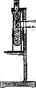
tc Figure 1. see- of the slit (window) through which light falls; the rays of the latter should be as parallel as possible, which is achieved either by sufficient distance of the light source from the instrument (Kleinman) or by the use of special lighting equipment [Hellige (Figure 4), Kober, Leitz]. Sometimes instead of changing the width of the slit, the position of the vessel containing the liquid is changed, for example in Kober's model. In this author's instrument, movement is not performed with the help of a rack and pinion as usual, but by means of a hydraulic device ensuring especially precise adjustment. To replace standard solutions, permanent standards have been proposed, in which light is scattered by the turbid walls of the test tube (they are coated with a layer of colloid
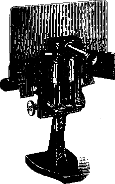
Figure 2.
with a fine powder suspended in it, such as talc or barium sulfate, or are simply lined with thin paper) and is reflected from a powder of a certain shade poured into the bottom of the test tube or scattered by prisms of varying degrees of turbidity. Such a standard is pre-calibrated precisely with solutions containing a certain amount of the substance in question. For N. to give truly accurate results, a number of conditions must be observed. First of all, this includes the cleanliness of the vessels, on which no traces of fingers or dust particles should remain; the solutions should not contain any suspended particles except those of the substance in question; large errors can be caused by filter paper fibers getting into the solutions and reagents during filtration; it is necessary to be sure of the uniformity of illumination of both test tubes, for which
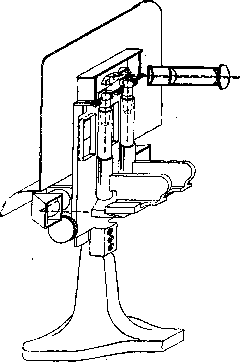
Fig. 3.
the same turbid liquid is placed in both vessels and it is verified that with the same slit width, the brightness of the two fields of view in the instrument is the same. If this is not the case, the light source is moved until this is achieved. When the compared solutions are interchanged, the ratio of slit widths with equal brightness should remain constant. The ratio of concentrations of the compared solutions should not
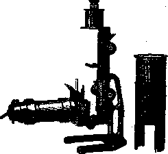
Figure 4.
should not exceed 1:4; the best results from N. are obtained with solutions of similar concentration. The size of particles in the liquids being compared must be the same, and the suspension of these particles must be sufficiently stable, at least for the duration of the study, since coagulation causes sharp changes in the brightness of light scattered by the turbid liquid. To increase stability, especially when working with inorganic substances, protective colloids sometimes have to be added. Only suspensions that appear completely homogeneous to the naked eye are suitable for N.; suspensions of coarser particles cannot be nephelometrically measured. N. must be performed in a dark room, and it is necessary to allow the eye to adapt to the darkness for 5-10 minutes beforehand. A series of readings must be taken and the average used. Provided all the stated conditions are met, N. can give precise results, quite sufficient for all kinds of biological research and often only slightly inferior to the best conventional analytical methods {on average the error does not exceed 0.5% and can be reduced to 0.3%). The advantages of N. lie in its extreme sensitivity, which allows for the analysis of minute quantities of substance. Typically, the quantities involved are on the order of several mg per 1 liter, and in individual cases even hundredths of a gram per 1 liter (for example, nephelometric determination of acetone). Moreover, micromodels of nephelometers constructed at the present time allow for the analysis of only 1.5-3 cm3 of liquid. The speed of nephelometric determinations makes them particularly valuable for serial experiments when it is necessary to perform a series of determinations within a very limited time. During its relatively very short existence of time, N. has found extremely wide application and spread in the most diverse fields. As just a few examples, it is used for determining poisonous substances (mustard gas), arsenic, quinine in blood and urine, albumins and globulins in serum, phosphoric acid, fats and lipoids, sulfates, chlorides, calcium, water hardness, for studying the action of enzymes: amylase, lipase, pepsin, trypsin and many others.
Related articles
Mentioned in
Cite this page
“Nephelometry.” Soviet Medical Encyclopedia. English translation of Bolshaya Meditsinskaya Entsiklopediya, 1st ed. (Moscow, 1928–1936), ed. N. A. Semashko. https://sovietmedicalencyclopedia.pages.dev/article/nephelometry/