Colorimetry
Historical document, translated for reference. It reflects medical knowledge of the 1920s–30s and is not medical advice.
Summary
Colorimetry is a quantitative analytical method based on determining the amount of substance by the intensity of color inherent to the substance itself or to a reaction product between the substance and a specific reagent. It is closely related to spectrophotometry and employs various techniques including standard solutions and specialized instruments called colorimeters.
Encyclopedia article (1928–1936)
Colorimetry, COLORIMETERS (from Latin color - color and Greek metron - measure). Colorimetry is a method of quantitative analysis based on determining the amount of substance by the intensity of color inherent to the substance itself or to a reaction product occurring between the given substance and a specific reagent. Accordingly, one can distinguish between direct and indirect colorimetry. In practice, the latter has the greatest significance, since direct C. is applicable only for determining substances possessing a sufficiently intense inherent color. Such substances in technology are various dyes, in biology - various pigments (chlorophyll, Hb, bile pigments, etc.). Essentially, C. is closely related to spectrophotometry. In the latter case, the absolute amount of light rays of a certain part of the spectrum absorbed in passing through a layer of the tested colored substance of a certain thickness is determined. If the absorption coefficient (Bunsen-Roscoe) for the given substance is known, the amount of the latter can be calculated. In C., instead of measuring absolute brightness, resp. light absorption, the light absorption of the tested solution is compared with that of a colored standard. The latter can be either a solution of the same substance of a certain concentration or an artificially prepared standard (colored solution, glass, gelatin film, etc.). All methods of C. are based on the premise that there is a certain relationship between the amount of colored substance and the intensity of color. In a number of cases and within certain limits of concentration, this relationship has the character of simple proportionality, i.e., when the concentration of the substance causing the color doubles, the intensity of color also doubles, and so on. For other cases, this condition does not hold, and regularities of a different order are observed. In the latter case, to judge the amount of substance based on comparing solutions of different color intensities, one can only use a formula specially established for the given case, expressing the relationship between color intensity and concentration. In such cases, it is simpler and more reliable not to compare the color intensity of solutions of different concentrations, but to find the concentration of the standard solution that gives the same color intensity as the tested solution. The comparison is made with a constant thickness of the liquid layer. Practically this is carried out as follows: a series of test tubes of exactly the same diameter are selected (this is established either by means of a gauge or by pouring the same volume of water into all test tubes and selecting those in which the water will be at the same level). In these test tubes, a series of dilutions of the standard solution of different concentrations is prepared with such calculation that the limits of the taken concentrations lie on one side above and on the other side below the presumed concentration of the tested solution. The series of dilutions is prepared according to the principle of a geometric series, i.e., so that the content of the standard substance from one test tube to another changes a certain number of times (series indicator), and not by the same amount. The table below gives examples of such series. Series indicator Number of members Concentration limits Series of dilutions 3 2 1.5 1.25 /« 4 5 7 11 9 7 1 : 27 1 : 16 1 : 11.4 1 : 9.3 1 : 0.1 1 : 0.09 1; 3; 9; 27; 1; 2; 4; 8; 16; 1; 1.5; 2.25; 3.3S; 5.1; 7.6; 11.4; 1; 1.25; 1.56; 1.95; 2.44; 3.05; 3.81; 4.76; 5.95; 7.44; 9.3; U 0.75; 0.56; 0.42; 0.32; 0.24; 0.18; 0.13; 0.1; 1; 0.67; 0.45; 0.3; 0.2; 0.13; 0.09. First, a series with a high indicator is set up, two test tubes are found between which lies the color of the tested solution, and for more accurate reading, a new series of dilutions is placed between these two test tubes, taking the concentrations of the solutions in these test tubes as the extreme members of the series and placing between them a series with a smaller indicator. Thus, that dilution is found whose color coincides as much as possible with the color of the tested liquid. The concentration of the desired substance in the latter will be equal to its concentration in the corresponding standard test tube. The volume of liquid and the amount of reagent must be the same in all standard test tubes as well as in the test tube with the tested solution. If the tested liquid is colored in itself, then in order to exclude the influence of its own color, the principle of Walpole's compensating comparator is used. This comparator is a wooden block in which two (or three) pairs of vertical recesses and three through horizontal holes intersecting the paired vertical recesses are drilled (Fig. 1a and 16). In the 1st hole, a test tube with the standard solution is placed, in the 2nd - with the tested liquid, in the 3rd - with the tested liquid, but without the addition of the color-causing reagent (instead of it, the same volume of water is added), and in the 4th - a test tube with water. Thus, when viewed through the horizontal holes (Fig. 16), the color of the standard solution is summed with the inherent color of the tested liquid, just as it happens in test tube 2; the shades are equalized, and the influence of the inherent color is compensated. For colorless liquids, when there is no need to use Walpole's principle, instead of preparing a series of standard solutions of different concentrations, one can simply gradually increase the concentration of the standard solution by adding it to the diluted reagent. Knowing the concentration of the standard solution, the content of the desired substance can be calculated by the formula x = p -^-, where x is the amount of the desired substance in the volume of liquid taken for determination, p is the amount of substance contained in the added volume of the standard solution, vx is the final volume of liquid in the test tube with the tested solution, vcm is the same in the test tube with the standard solution. Colorimetry can also be carried out in such a way that the reagent is added to both the tested and standard solutions, and then water is added to the darker solution until the colors are equalized. Based on the ratio of the final volumes, the content of the substance in the tested solution is calculated. Both of the latter methods are not very reliable, since the method of mixing reagents often affects the intensity of color, and upon dilution, the color can change not only depending on the volumes, but also due to secondary processes. Indeed, accurate results of C. can only be obtained when using special instruments - colorimeters. The latter allow equalizing the intensity of color of solutions of different concentrations by changing the thickness of the solution layers. For most (but by no means for all and not at any concentrations) colored solutions, the rule known as "Beer's law" is valid, according to which the intensity of color of two solutions is the same when the products of concentrations by the thickness of the liquid layer are equal, i.e., when C1.H1=C2.H2, where C1 is the concentration of one solution, H1 is the thickness of its layer, C2 and H2 are the same values for the second solution. Knowing the thickness of the layers $H_1$ \ $= : i i ^ ^ =&>
colony of hydrozoan polyps consists of sessile polyps (2)
Knowing the thickness of the layers and the concentration of one of the solutions (standard), it is easy to find the concentration of the tested solution by the formula Cx= Cst- Hst/Hx, where Cx and Hx are the concentration and thickness of the layer of the unknown solution, Cst and Hst are the same for the standard. The simplest instrument built on this principle is the so-called Hehner cylinders (Fig. 2), <°>
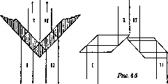
in which the change in the thickness of the liquid layer is achieved by simply draining part of the solutions through taps. The cylinders have a glass bottom; they are placed on a white lining, light passes from below, the color of the liquids is compared by looking into the cylinders from above. An important condition that greatly increases the accuracy of colorimetric research is the approximation of the compared colored fields of vision as much as possible, so that when the colors are equal, they would merge or be separated only by a very thin line. This is achieved by using various optical devices; some of them are schematically shown in Fig. 4. Fig. 2 shows a colorimeter based on the principle of Hehner cylinders.
Fig. 2. A and B - Hehner cylinders.
The double plate (Fig. 4a) is used in Autenright colorimeters;


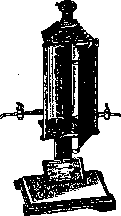
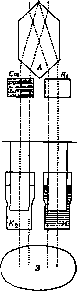
Figure 3. Diagram of the path of rays in the Bürker colorimeter: O-ocular; A-Hüfner-Albrecht body; Cm-vessel with standard solution; Я-vessel with compensating liquid; И-test solution; H%-compensating liquid; 3-mirror. In earlier Duboscq colorimeters, Fresnel prisms were usually used (Fig. 46). In the newest models, the so-called Hüfner-Albrecht body (Fig. 4b) or Lummer-Brodhun body (Fig. 4c) are more often used. Observation is made through a suitable ocular. In addition to the Autenrieth and Duboscq colorimeters (see Autenrieth colorimeter and Duboscq colorimeter), a new modification of the latter by Bürker should be mentioned here, which combines the precision of a precision optical instrument with the advantages of Walpole's compensation principle. When using this colorimeter, neither the inherent color of the test liquid nor the color of the reagent interferes, moreover the path of the rays in both halves of the field of view is exactly symmetrical (Fig. 3). In 1928, the Leitz firm introduced universal colorimeters that allow the use of both the ordinary Duboscq principle and the Bürker principle, and are also adapted for comparing turbidities; their microattachments (Fig. 5) allow measurements to be made with only 1-2 cm3 of liquid. For particularly precise work, the Krüss polarizing colorimeter has a number of advantages, in which when the intensities of the color are unequal, the hue itself also changes, which significantly increases the accuracy of setting. Generally speaking, the accuracy of colorimetric measurements when using a good instrument can be very significant and lies within several tenths of a percent up to 0.5%. To achieve this accuracy, it is necessary to take a number of precautions. Both fields of view should be illuminated with exactly the same brightness; this is verified by placing the same liquid in both vessels of the colorimeter; in this case, the scale readings should be the same. For the greatest reliability of results, it is recommended to change the solutions places after the reading has been made; the ratios of the heights should remain the same. It is necessary to make a series of readings (at least five) and take their arithmetic mean. The smaller the difference in concentrations of the standard and test solutions, the higher the accuracy of the determinations; the ratio of concentrations should not exceed 1:2. In addition to uniformity, it is also necessary to maintain constant illumination intensity; therefore it is better to use an artificial source of light; new models have an illuminator with an electric bulb attached to the colorimeter stand (Fig. 6). It is best to use light of medium brightness; insufficient or too strong brightness reduces the accuracy of readings and causes rapid eye fatigue. During colorimetry, one should as much as possible protect the eye from stray light; therefore for precise work it is better to use a darkened room.
The accuracy of colorimetry increases if possible to exclude those rays of light that are weakly or not at all absorbed by the test solution, i.e., first of all, the rays corresponding to the color of the solution itself and therefore not stopped by it. This is achieved by using appropriate light filters (e.g., yellow for blue solution color and vice versa). This not only increases the accuracy of comparing the intensity of colors but also facilitates the setting, since along with the intensity the hue of the color also changes.
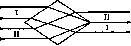
Figure 5.

The solutions to be compared should be as close as possible in temperature; the permissible difference should not exceed 3°. It is always necessary to preliminarily verify that for a given system and for given concentrations, Beer's law remains valid. Among the advantages of C., which quickly won this method wide practical application, in addition to simplicity and speed, along with sufficient accuracy for most practical purposes, belongs the extreme sensitivity of the method, which significantly expands the limits of measurement and allows one to get by with minimal amounts of substance, which is especially important in biological research, e.g., in blood analysis; on the other hand, when using colorimetric methods, it is possible to quantitatively determine such substances that are inaccessible to ordinary chemical methods for direct determination. Thus, it is possible to determine colorimetrically with an error of not more than 1-2% thousandths of a milligram of arsenic, nitrous acid, phosphorus, nitrogen, iodine, etc. These features of colorimetric methods have led to the widespread application of C. in medical and physiological laboratory techniques, so that at the present time for most substances important in clinical relations, simple, convenient and accurate colorimetric methods have been developed (as examples one can mention the determination of nitrogen by the Nessler method, the determination of uric acid, sugar, creatinine, phosphorus, cholesterol, bilirubin, lactic acid, etc.).
Methods for determining Hb are based on C. C. finds wide application in water analysis. A completely special field of application of C. is the measurement of active reaction (see Indicator method).
Related articles
Mentioned in
Cite this page
“Colorimetry.” Soviet Medical Encyclopedia. English translation of Bolshaya Meditsinskaya Entsiklopediya, 1st ed. (Moscow, 1928–1936), ed. N. A. Semashko. https://sovietmedicalencyclopedia.pages.dev/article/colorimetry/