Light
Historical document, translated for reference. It reflects medical knowledge of the 1920s–30s and is not medical advice.
Summary
Light is defined as radiant energy perceptible to the human eye, with a wavelength spectrum from 0.4 to 0.75 microns. The article discusses the nature of light, its sources (natural and artificial), and its spectral composition, emphasizing the medical importance of spectral composition rather than just intensity.
Encyclopedia article (1928–1936)
Light, a form of radiant energy perceptible to the human eye. On the scale of wavelengths of radiant energy, the visible portion of the spectrum extends from 0.4 μ to 0.75 μ (see Radiant energy, Light perception). The term light is often given a broader interpretation, referring to infrared and ultraviolet rays as light. However, when defining light units, the term L. is used in the sense indicated above (see for example the All-Union standard for light units-OST 4891, approved in 1932).-The doctrine of light is divided into several parts: 1) geometric optics, 2) physical optics, 3) colorimetry, 4) lighting engineering, 5) physiological optics. 1) Geometric optics does not consider the question of the nature of L., but deals only with the geometric problems of the propagation of light rays in various optical systems and media, and with the problems of obtaining images with the help of these systems (see Eye, dioptrics). 2) Physical optics is devoted to questions related to the nature of L., and considers phenomena in which the wave and quantum properties of L. are manifested (interference, diffraction, polarization, quantum properties). In both of these sections, L. is understood to mean the broader spectral range indicated above. 3) Colorimetry is devoted to the questions of the color of L. and methods of its measurement. 4) Lighting engineering deals with practical issues related to the production of L. and its rational use (design of L. sources and fixtures, calculation of lighting and measurement of L.). 5) Physiological optics - the doctrine of vision, i.e., the perception of L., the structure and work of the visual organs. In the last three sections, L. is understood to mean the visible portion of the spectrum. Our views on the nature of light have undergone great changes in the course of historical development (see Optics). At the present time, the electromagnetic (wave) and quantum properties of L. are firmly established (see Radiant energy, Quantum theory). Sources of L. are divided into natural and artificial. The most powerful of the natural sources is the sun. From a medical point of view, the spectral composition of the radiation of any source is extremely important, and not just its intensity. The sun as a source. The brightness of the sun is 150,000 stilbs in the center of the solar disk. In terms of its light action, the sun is equivalent to a light source with a luminous intensity of 1.7 × 1027 candles, placed at a distance equal to the average distance between the sun and the earth (1.5 × 10⁸ km). In one minute, an average of 2.1 g/cal. of solar energy falls on 1 cm2 of the earth's surface (solar constant). In the spectrum of the sun, the maximum energy falls at 0.470 μ, which corresponds to the radiation of a black body at t° 6,250°. The spectrum of the sun is crossed by a series of dark lines, explained by the absorption of solar radiation by the sun's own atmosphere and the earth's atmosphere. These lines are called Fraunhofer lines and correspond to absorption lines of various substances (sodium, hydrogen, iron, etc.). L. from the sun, passing through the earth's atmosphere, is partially absorbed and scattered by the air. At the same time, shorter waves are scattered more strongly (see below), so that the violet end of the spectrum is weakened more than the red end, and the maximum energy shifts to the red part of the spectrum. The scattering of L. in the air explains the blue color of the sky and the crimson color of the sun at sunset. In the first case, we see scattered rays, among which blue-violet rays predominate, in the second case, we, on the contrary, see what remains in the light ray after the scattering of the blue-violet part of the spectrum. Often the radiation of a source with t° equal to 5,000°, i.e. with a maximum energy at 590 μ, is taken as the standard for 'white' L. Of course, this choice is very conditional, and to avoid errors, it is always necessary to specify exactly which 'white' light is being referred to.
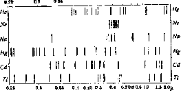
Artificial sources of L. The most common artificial sources of L.-incandescent lamps-differ greatly in their light properties from the sun. First, their filament brightness (see Illumination) ranges from several tens to several thousand stilbs, which is significantly lower than the surface brightness of the sun; secondly, the spectral composition of the radiation of these lamps differs from the solar spectrum. The latter circumstance is extremely important from a biological point of view. In the spectrum of incandescent lamps, the maximum energy lies in the infrared part, which is explained by the relatively low t° of the heated filament (1,400-3,500°). According to Wien's law, the wavelength corresponding to the maximum energy in the radiation of a heated body is inversely proportional to the t° of this body, therefore the higher the t°, the more the maximum radiation falls on a shorter wavelength. In incandescent lamps, the t° of the filament increases with the power of the lamp, so in more powerful lamps, the maximum energy is closer to the visible part of the spectrum than in less powerful lamps. Because of this, the spectral composition of L. of powerful incandescent lamps is closer to the solar spectrum than that of low-power lamps. Nevertheless, even in them, the percentage of energy falling on violet and ultraviolet rays is much lower than in solar L. L. of ordinary voltaic arcs corresponds to the radiation of a heated body at 4,000-4,500°. L. close to solar is given by a powerful projection voltaic arc of intense combustion. The temperature of the crater of an intense arc is close to the t° of the sun and therefore such an arc gives L., in terms of spectral composition, close to solar L. The brightness of the crater of an intense arc is of the order of 100,000 stilbs. In general, for temperature light sources, with an increase in t° to 7,000°, the percentage of radiant energy falling on the visible part of the spectrum increases, reaching a maximum of 14% at 7,000°, and then begins to fall.-Practically, the maximum has not yet been reached. Recently, electric discharge tubes (glow lamps) filled with rarefied gases and vapors have been widely used as sources of L. In these tubes, the glow is excited by impacts of electrons flying through the tube with the atoms of the substance filling it. Due to the fact that with this method of excitation, less energy is converted into heat than with thermal excitation, glow lamps have a higher efficiency than incandescent lamps. Glow lamps of intense combustion differ from ordinary Geissler tubes in that they have a low ignition potential and greater brightness of glow than the latter. This is achieved by the use of a heated cathode (negative electrode), which is a powerful source of electrons. The spectrum of radiation of glow tubes is not continuous, as in incandescent bodies, but consists of separate lines. A tube filled with neon gives red L. and its spectrum consists mainly of red and yellow lines. A tube with sodium vapor gives yellow L.-almost all its radiation is concentrated in one double yellow line of sodium. This tube is convenient for laboratory work, when a source of monochromatic light is needed (e.g. for a saccharimeter). In addition, it is used for lighting roads, because in its light the resolving power of the eye increases. 0.4 0.45 0.5 0.6 0.7 0.8 0.9 1.0 1.5 2.0(1", "Figure 1. Spectra of the most important gases and metal vapors; the thickness of the lines indicates intensity."
Fig. 1 shows the spectra of the most commonly used gases and vapors. The spectrum of a gas discharge tube depends on a number of parameters, even if the tube is filled with a specific gas or the vapors of a specific metal. The most significant parameters are the pressure of the gas or vapors and the current density. For example, in a mercury lamp, with an increase in the density of electric current and vapor density, the yellow line corresponding to 579 mμ grows faster than all other lines in the visible part of the spectrum, so that the light of the lamp becomes more yellow with an increase in the electrical operating conditions. The source of L., in which thermal radiation is combined with radiation from mercury vapors, is the so-called "sunlight lamp" (see Bach's mercury-quartz lamp). All artificial sources of L. are usually used "in appropriate fixtures or other lighting devices (see Lighting and Lighting Fixtures). To obtain a narrow directed beam of rays, mirrors are used, and the source must be of small size and high brightness (arc crater); to obtain a wide beam of rays, diffusing fixtures and large sources of L. can be used. On the path from the source to the illuminated object or to the observer's eye, L. usually passes through a series of media and is reflected from a series of surfaces. In this process, there is a loss of light energy and its transformation into other forms of energy, for example, into thermal or chemical energy. Reflection of L. When a beam of L. falls on the boundary between two media, part of the incident light flux is reflected back. Three types of reflection are distinguished: 1) mirror, or regular, reflection, 2) diffuse, or Lambertian, reflection, and 3) mixed reflection. 1) Mirror reflection follows the following laws: the incident ray and the reflected ray lie in the same plane with the perpendicular to the element of the reflecting surface at the point of incidence of the ray and form equal angles with it (Fig. 2). Polished surfaces of various substances exhibit mirror reflection. The reflection coefficient is the ratio of the light flux reflected by the surface to the incident flux. Polished metals, as a rule, have a high reflection coefficient. The reflection coefficient for a given metal is not a constant value, but depends on the wavelength of the incident light. The table below gives the reflection coefficients by spectrum of some metals. Wavelength in mμ Metal Silver (freshly silvered) 86.6 90.5 91.3 92.7 92.6 93.5 Silver (old) . . . 73.0 87.1 83.9 85.0 86.3 88.6 29.3 33.1 47.0 74.0 84.4 88.9 51.8 54.7 58.4 61.1 64.2 66.5 56.6 59.4 60.8 62.6 64.9 65.9 51.9 54.4 54.8 54.9 55.4 55.9 47.0 50.0 55.0 55.5 55.7 56.0 Since the reflection coefficient depends on the wavelength, the energy reflection coefficient will not be equal to the photometric reflection coefficient, because the reflected L. will have a spectral composition different from the spectral composition of the incident light (see below). Transparent media belonging to dielectrics have significantly smaller reflection coefficients. Reflection from them follows Fresnel's law, which for the case of normal incidence of a beam of L. on a polished dielectric surface takes a very simple form: R = ((n-1)/(n+1))2, where R is the reflection coefficient, n is the refractive index of the dielectric relative to the medium from which the L. falls. For example, for glass with n = 1.5, R = 4%, etc. The reflection coefficient increases with the angle of incidence. The reflection coefficient for incident scattered L. has a higher value (about 9.5%).
Fig. 2.
2) The second type of reflection—diffuse—is characterized by Lambert's law: the brightness of the reflecting surface upon incidence of a directed beam of L. is the same in all directions of observation, and the intensity of L. varies according to the cosine of the angle of observation with the perpendicular; the L. incident on such a surface is scattered uniformly in all directions (Fig. 3). To one degree or another, all matte surfaces exhibit this type of reflection. Matte metals, glass, rough paper, gypsum plates, porcelain—all can be classified as diffuse surfaces. The surface that gives the most correct diffuse reflection is considered to be a flat surface covered with magnesium oxide powder. 3) Mixed reflection. All types of reflection in which part of the light flux is reflected mirror-like and part diffusely are classified as mixed reflection. As an example, we can cite the reflection from porcelain enamel used to cover lighting fixtures (Fig. 4). The incident L. is partially reflected (according to Fresnel's law) from the surface of the enamel, while the part that passes inside the enamel is scattered there on embedded small particles. Dispersion and absorption. When L. passes through homogeneous media, two inseparably connected phenomena are observed: first, the dependence of the propagation speed on the wavelength of L. (color), and second, the absorption of the passing light flux by particles (molecules, atoms) of the medium, and the degree of absorption also usually depends on the wavelength of the passing light. The first phenomenon leads to the fact that the refractive index of the medium (see Refraction of light) also depends on the wavelength; this is called dispersion of the refractive index. The regions of the spectrum in which for a given substance the refractive index smoothly decreases with wavelength are called regions of normal dispersion; the regions in which the refractive index, after undergoing a discontinuity, begins to increase with wavelength are called regions of anomalous dispersion. The absorption of light by a medium of thickness d can be represented by Bouguer's equation: Td = td, (1) where Td is the transmittance of a medium of thickness d, and t is the transmittance of a medium of unit thickness; t, and therefore Td, are functions of wavelength. Often equation (1) is written in a slightly different form, taking as the basis not the transmittance coefficient of a layer of unit thickness, but the absorption coefficient—K—of such a layer; then the equation takes the form: Td = e-Kd. (2) K is called the specific absorption of the medium, e is the base of natural logarithms. Sometimes it is written: Td = 10-αd.
Fig. 3.
2) The second type of reflection—diffuse—is characterized by Lambert's law: the brightness of the reflecting surface upon incidence of a directed beam of L. is the same in all directions of observation, and the intensity of L. varies according to the cosine of the angle of observation with the perpendicular; the L. incident on such a surface is scattered uniformly in all directions (Fig. 3). To one degree or another, all matte surfaces exhibit this type of reflection. Matte metals, glass, rough paper, gypsum plates, porcelain—all can be classified as diffuse surfaces. The surface that gives the most correct diffuse reflection is considered to be a flat surface covered with magnesium oxide powder. 3) Mixed reflection. All types of reflection in which part of the light flux is reflected mirror-like and part diffusely are classified as mixed reflection. As an example, we can cite the reflection from porcelain enamel used to cover lighting fixtures (Fig. 4). The incident L. is partially reflected (according to Fresnel's law) from the surface of the enamel, while the part that passes inside the enamel is scattered there on embedded small particles. Dispersion and absorption. When L. passes through homogeneous media, two inseparably connected phenomena are observed: first, the dependence of the propagation speed on the wavelength of L. (color), and second, the absorption of the passing light flux by particles (molecules, atoms) of the medium, and the degree of absorption also usually depends on the wavelength of the passing light. The first phenomenon leads to the fact that the refractive index of the medium (see Refraction of light) also depends on the wavelength; this is called dispersion of the refractive index. The regions of the spectrum in which for a given substance the refractive index smoothly decreases with wavelength are called regions of normal dispersion; the regions in which the refractive index, after undergoing a discontinuity, begins to increase with wavelength are called regions of anomalous dispersion. The absorption of light by a medium of thickness d can be represented by Bouguer's equation: Td = td, (1) where Td is the transmittance of a medium of thickness d, and t is the transmittance of a medium of unit thickness; t, and therefore Td, are functions of wavelength. Often equation (1) is written in a slightly different form, taking as the basis not the transmittance coefficient of a layer of unit thickness, but the absorption coefficient—K—of such a layer; then the equation takes the form: Td = e-Kd. (2) K is called the specific absorption of the medium, e is the base of natural logarithms. Sometimes it is written: Td = 10-αd.
Fig. 4.
2) The second type of reflection—diffuse—is characterized by Lambert's law: the brightness of the reflecting surface upon incidence of a directed beam of L. is the same in all directions of observation, and the intensity of L. varies according to the cosine of the angle of observation with the perpendicular; the L. incident on such a surface is scattered uniformly in all directions (Fig. 3). To one degree or another, all matte surfaces exhibit this type of reflection. Matte metals, glass, rough paper, gypsum plates, porcelain—all can be classified as diffuse surfaces. The surface that gives the most correct diffuse reflection is considered to be a flat surface covered with magnesium oxide powder. 3) Mixed reflection. All types of reflection in which part of the light flux is reflected mirror-like and part diffusely are classified as mixed reflection. As an example, we can cite the reflection from porcelain enamel used to cover lighting fixtures (Fig. 4). The incident L. is partially reflected (according to Fresnel's law) from the surface of the enamel, while the part that passes inside the enamel is scattered there on embedded small particles. Dispersion and absorption. When L. passes through homogeneous media, two inseparably connected phenomena are observed: first, the dependence of the propagation speed on the wavelength of L. (color), and second, the absorption of the passing light flux by particles (molecules, atoms) of the medium, and the degree of absorption also usually depends on the wavelength of the passing light. The first phenomenon leads to the fact that the refractive index of the medium (see Refraction of light) also depends on the wavelength; this is called dispersion of the refractive index. The regions of the spectrum in which for a given substance the refractive index smoothly decreases with wavelength are called regions of normal dispersion; the regions in which the refractive index, after undergoing a discontinuity, begins to increase with wavelength are called regions of anomalous dispersion. The absorption of light by a medium of thickness d can be represented by Bouguer's equation: Td = td, (1) where Td is the transmittance of a medium of thickness d, and t is the transmittance of a medium of unit thickness; t, and therefore Td, are functions of wavelength. Often equation (1) is written in a slightly different form, taking as the basis not the transmittance coefficient of a layer of unit thickness, but the absorption coefficient—K—of such a layer; then the equation takes the form: Td = e-Kd. (2) K is called the specific absorption of the medium, e is the base of natural logarithms. Sometimes it is written: Td = 10-αd.
2) The second type of reflection—diffuse—is characterized by Lambert's law: the brightness of the reflecting surface upon incidence of a directed beam of L. is the same in all directions of observation, and the intensity of L. varies according to the cosine of the angle of observation with the perpendicular; the L. incident on such a surface is scattered uniformly in all directions (Fig. 3). To one degree or another, all matte surfaces exhibit this type of reflection. Matte metals, glass, rough paper, gypsum plates, porcelain—all can be classified as diffuse surfaces. The surface that gives the most correct diffuse reflection is considered to be a flat surface covered with magnesium oxide powder. 3) Mixed reflection. All types of reflection in which part of the light flux is reflected mirror-like and part diffusely are classified as mixed reflection. As an example, we can cite the reflection from porcelain enamel used to cover lighting fixtures (Fig. 4). The incident L. is partially reflected (according to Fresnel's law) from the surface of the enamel, while the part that passes inside the enamel is scattered there on embedded small particles. Dispersion and absorption. When L. passes through homogeneous media, two inseparably connected phenomena are observed: first, the dependence of the propagation speed on the wavelength of L. (color), and second, the absorption of the passing light flux by particles (molecules, atoms) of the medium, and the degree of absorption also usually depends on the wavelength of the passing light. The first phenomenon leads to the fact that the refractive index of the medium (see Refraction of light) also depends on the wavelength; this is called dispersion of the refractive index. The regions of the spectrum in which for a given substance the refractive index smoothly decreases with wavelength are called regions of normal dispersion; the regions in which the refractive index, after undergoing a discontinuity, begins to increase with wavelength are called regions of anomalous dispersion. The absorption of light by a medium of thickness d can be represented by Bouguer's equation: Td = td, (1) where Td is the transmittance of a medium of thickness d, and t is the transmittance of a medium of unit thickness; t, and therefore Td, are functions of wavelength. Often equation (1) is written in a slightly different form, taking as the basis not the transmittance coefficient of a layer of unit thickness, but the absorption coefficient—K—of such a layer; then the equation takes the form: Td = e-Kd. (2) K is called the specific absorption of the medium, e is the base of natural logarithms. Sometimes it is written: Td = 10-αd.
2) The second type of reflection—diffuse—is characterized by Lambert's law: the brightness of the reflecting surface upon incidence of a directed beam of L. is the same in all directions of observation, and the intensity of L. varies according to the cosine of the angle of observation with the perpendicular; the L. incident on such a surface is scattered uniformly in all directions (Fig. 3). To one degree or another, all matte surfaces exhibit this type of reflection. Matte metals, glass, rough paper, gypsum plates, porcelain—all can be classified as diffuse surfaces. The surface that gives the most correct diffuse reflection is considered to be a flat surface covered with magnesium oxide powder. 3) Mixed reflection. All types of reflection in which part of the light flux is reflected mirror-like and part diffusely are classified as mixed reflection. As an example, we can cite the reflection from porcelain enamel used to cover lighting fixtures (Fig. 4). The incident L. is partially reflected (according to Fresnel's law) from the surface of the enamel, while the part that passes inside the enamel is scattered there on embedded small particles. Dispersion and absorption. When L. passes through homogeneous media, two inseparably connected phenomena are observed: first, the dependence of the propagation speed on the wavelength of L. (color), and second, the absorption of the passing light flux by particles (molecules, atoms) of the medium, and the degree of absorption also usually depends on the wavelength of the passing light. The first phenomenon leads to the fact that the refractive index of the medium (see Refraction of light) also depends on the wavelength; this is called dispersion of the refractive index. The regions of the spectrum in which for a given substance the refractive index smoothly decreases with wavelength are called regions of normal dispersion; the regions in which the refractive index, after undergoing a discontinuity, begins to increase with wavelength are called regions of anomalous dispersion. The absorption of light by a medium of thickness d can be represented by Bouguer's equation: Td = td, (1) where Td is the transmittance of a medium of thickness d, and t is the transmittance of a medium of unit thickness; t, and therefore Td, are functions of wavelength. Often equation (1) is written in a slightly different form, taking as the basis not the transmittance coefficient of a layer of unit thickness, but the absorption coefficient—K—of such a layer; then the equation takes the form: Td = e-Kd. (2) K is called the specific absorption of the medium, e is the base of natural logarithms. Sometimes it is written: Td = 10-αd.


(3) The product K'd is called optical density and is denoted by the letter D (density). From (3) it is seen that lg ip- D. The quantity inverse to transmittance, ~ = 0- not- 73» is called opacity. Plotting the dependence of specific absorption K on wavelength λ, graphically we will have a characteristic of the optical properties of the medium by spectrum. It has been established that in regions of anomalous dispersion K acquires large values, forming so-called absorption bands. In the region of normal dispersion K changes little, remaining small in absolute value, so that in this region the substance is transparent up to very large thicknesses. Absorption that is especially strong is called selective; the value of the absorption coefficient K(λ) is determined by the natural frequencies of the molecules of the medium. Due to this, the medium in transmitted white L. is generally colored. This property is often used to obtain light filters—devices that separate certain regions of the spectrum from others. According to the appearance of the absorption curve, a classification of absorbing media can be given. According to Gibson, all selectively absorbing media are divided into three classes. Class 1. Strong absorption in regions λ, having some value^,,, and strong transmission in regions λ>λc. To this class belong protective light filters used to protect the eyes from ultraviolet rays when working with sources rich in these rays (quartz, mercury lamps, voltaic arc, flame during welding, etc.). Ordinary glass has a transmission limit around λc = 300-400 mμ. As protective light filters, poor green glasses with a high content of iron salts (simple bottle glass) are particularly suitable, the absorption limit of which lies around λc = 400 mμ, thus it blocks the entire erythema region. Uviol glass (vita-glass) has a transmission limit λc ^ 280-290 mμ. Water has a limit around λc = 170 mμ. The region λ < 120 mμ is not transmitted by solids and liquids. Among ordinary gases, H2 is most transparent in this region. The earth's atmosphere has a transmission limit around λc = 285 mμ, which is explained by the presence of ozone (O3) in the upper layers of the atmosphere, absorbing radiation shorter than 285 mμ. Light filters with a transmission limit of 200-350 mμ can be obtained by using certain organic liquids. In the region 300-700 mμ, solutions of many salts and organic dyes are very convenient. As media transmitting the infrared region and not transmitting the visible, various varnishes, resins, ebonite, cardboard, black paper, etc. are used. Class 2. Complete absorption in a wide region of the spectrum, where λ has a value>λC, and strong transmission in the neighboring region, where &<λe. In the region of the visible spectrum, separation of the infrared part is well achieved by aqueous solutions of copper salts (CuSO4 and others), as well as solutions of alum [K2Al2(SO4)3], which completely absorb infrared rays and have uniform and strong transmission in the visible spectrum. As solid filters for this purpose, slightly blue glasses dyed with cobalt or copper salts are used. Such glasses are used as protective goggles when working with sources rich in thermal rays (melting furnaces, etc.). Separation of the ultraviolet part from the visible is significantly more complicated. To this day, no filter is known that completely blocks the visible spectrum and transmits without noticeable weakening the ultraviolet region 400-250 mμ. For transmitting the ultraviolet spectrum closest to the visible light with a wavelength in the range 400-300 mμ, black uviol glasses, manufactured by various foreign firms and some Soviet institutes (Glass Institute in Moscow), are used. These glasses simultaneously weakly transmit the extreme red rays. For the same purpose of separating the nearest ultraviolet rays from visible light, alcoholic solutions of p-nitrosodimethylaniline in combination with blue uviol glasses can be used. For separating the region 230-310 mμ, a liquid filter of solutions of nickel sulfate (NiSO4) and cobalt sulfate (CoSO4) in a quartz cuvette is suitable. Class 3. Strong absorption everywhere except for some narrow regions of the spectrum (monochromatic light filters). The most difficult is the isolation of narrow regions in the ultraviolet part, where the best result for the region 254 mμ (isolation of the mercury resonance line) is given by Cl and Br vapors at a pressure of 6-7 atmospheres in sealed quartz vessels. For isolating the region 290-350 mμ (or narrower), a good result is given by thin layers of silver (40-100 mμ thick), applied to a quartz plate; they are usually applied by the method of cathode or thermal sputtering. The most convenient light filters for the visible spectrum are organic dyes, used in the form of solutions in glass cuvettes or in the form of gelatin films dyed with them. Due to the simplicity of manufacture, the field of application of gelatin light filters is very wide and the technique of their manufacture is not complicated and accessible to any laboratory. The gelatin used for light filters should have the greatest possible transparency, absence of turbidity, and good purification and preservation. Scattering of L. In the propagation of L. in an inhomogeneous medium (not having a uniform refractive index throughout the volume), in addition to dispersion and absorption of L., scattering of L. on inhomogeneities of the medium appears. In air, the role of such inhomogeneities is played by local densifications due to thermal fluctuations, embedded water molecules, etc. The degree of scattering depends on the wavelength λ of the incident light (Rayleigh's law). For air, inhomogeneities are small, on the order of the size of molecules; for such particles, the degree of scattering is inversely proportional to the fourth power of the wavelength (-λ-4). With an increase in the size of scattering particles, the degree at λ decreases and e.g. for milk glass, in which particles of the order of 1-2 μ are embedded, scattering is proportional to -λ-2. The law of weakening of transmitted L. for a medium with scattering does not differ from the law for pure absorption, in this case only K is not the absorption coefficient, but the sum of the absorption coefficient (K1) and the scattering coefficient (K2); K=K1+K2 is called the extinction coefficient. Beer's law is valid only for monochromatic radiation, since K is a function of wavelength. Light measurements. In light measurements, the energy of the light flux, spectral composition, polarization, and finally the effect on the eye are usually determined. (Determination of polarization—see Polarization, Polarimetry.) Light measurements are divided into two large groups. 1) Measurements of the optical characteristics of sources and the L. they emit. 2) Measurements of the optical characteristics of substances that reflect, refract, scatter, and absorb light. In this case, measurements are made in energy units or in light units (see Illumination). The latter measurements are called photometric measurements. In addition, measurements are divided into integral and spectral. In the first case, all quantities are measured for light not decomposed into a spectrum; in the second case, measurements are made for individual wavelengths.

Determination of polarization—see Polarization, Polarimetry.) Light measurements are divided into two large groups. 1) Measurements of the optical characteristics of sources and the L. they emit. 2) Measurements of the optical characteristics of substances that reflect, refract, scatter, and absorb light. In this case, measurements are made in energy units or in light units (see Illumination). The latter measurements are called photometric measurements. In addition, measurements are divided into integral and spectral. In the first case, all quantities are measured for light not decomposed into a spectrum; in the second case, measurements are made for individual wavelengths.
Measurement of light energy. To measure the amount of energy radiated by a light source, the main instruments are devices based on the thermal effect of light. The advantage of such instruments is that their sensitivity to light does not depend on its spectral composition. The simplest in design is the bolometer. The action of the bolometer is based on the change in electrical resistance of conductors when heated. Light, falling on the blackened wire or strip of the bolometer, heats it, and from the change in resistance, one can judge the intensity of the light. To obtain high sensitivity, the conductor is made extremely thin, for example in the form of a blackened platinum strip several microns wide and several tenths of a micron thick, and placed in a vacuum vessel. The change in resistance is measured either with the help of a compensation circuit of the Wheatstone bridge type (Langley's bolometer) or by the change in the strength of the current flowing through the conductor (Zeddig's bolometer). To measure currents, it is necessary to use extremely sensitive galvanometers (Fig. 5). At present, thermocouples and thermopiles are often used instead of bolometers, being more convenient in operation. The action of a thermocouple is based on the thermoelectric effect: a heated junction of two metals is a source of electromotive force. If one takes two conductors of different metals and weld their ends together, then when one of the junctions is heated, a current will flow in the conductor. The magnitude of the thermocurrent increases with temperature and depends on the combinations of welded metals. The following combinations are used: bismuth and silver, manganin and constantan, iron and bismuth, etc. Light, falling on such a blackened junction, heats it and thereby causes thermocurrents. When measuring radiant energy, the thermocouple is made of extremely thin blackened wires (several microns in diameter). The thinner the wire, the higher the sensitivity of the thermocouple per unit of energy falling on it. But since at the same time its surface decreases, and consequently the amount of absorbed energy also decreases, it is sometimes necessary to equip the thermocouple with plates that increase the absorbing surface, or to place it in the focus of a light-gathering optical instrument. To increase the sensitivity and stability of thermocouples, they are placed in a vacuum vessel. This achieves an increase in sensitivity 400 times (Fig. 6). In some cases, thermocouples are combined into a thermopile. A thermopile is a series of thermocouples connected in series (Fig. 7). To measure thermocurrents, sensitive galvanometers with low internal resistance are used. Thermocouples and thermopiles are used for both integral measurements of light energy and spectral measurements. Since the results of measurements are often of interest in calories, all these instruments are calibrated against a standard emitting a known amount of energy. In this case, the standard is placed at a certain distance, and knowing the size of the receiver, the amount of absorbed energy is calculated. By comparing the galvanometer reading with this value, the sensitivity of the instrument in calories is obtained. As mentioned, the spectral composition of the radiation is not significant in this case. The next common instrument for measuring energy is the photoelectric cell. The action of the photoelectric cell is based on the ejection of electrons from metals by light. The main disadvantage of the photoelectric cell from the point of view of measuring the energy of the light flux is its unequal sensitivity in different parts of the spectrum (see below). Due to this circumstance, with the help of a photoelectric cell, it is impossible to compare sources that give light of different spectral composition. To obtain definite results, it is necessary to know the distribution of energy across the spectrum of the sources and the spectral sensitivity curve of the photoelectric cell. The advantages of photoelectric cells are high sensitivity (especially those used in recent years with a semiconducting layer) and the convenience of amplifying photocurrents with the help of cathode lamps. Photoelectric cells must also be calibrated against a standard, and the sensitivity of some of them (gas-filled photoelectric cells) changes over time. For studying weak or instantaneous light sources, the photographic method is also used. In this case, the intensity of the energy of the light flux is judged by the blackening of the photographic plate (see Photography). The disadvantage of the photographic method is that the blackening of the plate is not proportional to the intensity of light falling on it, and that the photoemulsion has different sensitivity to different wavelengths. Due to the first circumstance, it is necessary to calibrate the plate by applying marks to it with the help of a source with intensity varying according to a known law; due to the second circumstance, the photographic method is little suitable for comparing light fluxes of different spectral composition.

Fig. 6. Scheme of a thermocouple according to Moly.
Photometric measurements. The above-mentioned objective methods are not suitable without appropriate changes for evaluating the effect of a given light flux on the eye. At the same time, the evaluation of this effect and the study of the influence of various factors on it are extremely important. As mentioned, photometry deals with the measurement of light flux not as simply an energetic quantity, but as a form of energy affecting the eye. The difference between energetic measurements and photometric measurements is extremely great, since in the latter the unequal sensitivity of the eye to different parts of the spectrum (color perception) plays a large role. As is known, the eye has maximum sensitivity in the green part of the spectrum. Therefore, if a thermocouple is used as a photometer, it is necessary to place a corresponding green light filter in front of it with maximum transmission in the green part of the spectrum. It is clear that such a combination will be equivalent to the eye, it is only necessary to calibrate the instrument against a light standard. Some photoelectric cells have a sensitivity curve in the visible part of the spectrum close to that of the eye (cesium, selenium); using light filters, such photoelectric cells can be easily and accurately equated to the eye and used as photometers and lux meters. Photometers in which the role of the measuring apparatus is played by the human eye are called visual, in contrast to the above-mentioned objective ones, in which the light energy, transforming into some other form, e.g. electrical, is measured by some instrument. The fundamental difference between the eye and all currently used energy receivers in objective photometers is that the eye reacts to brightness, while these receivers react to the magnitude of the light flux. All visual photometers are based on the null method: the field of view of the instrument is divided into two parts, one of which is illuminated by a known light source (standard), and the other by the measured source. By means of some shade, the light flux of the source whose field is brighter is shaded until the brightness of both halves is equal. At this moment, the ratio of the light fluxes falling from both sources on the photometer is equal to the degree of shading. The most common stationary photometer is the photometric bench.
A A
bench - - 1П----------------L represents two parallel horizontal bars, on which a series of carriages can move freely. On one of the carriages is mounted the photometer proper, so-called Lummer-Brodhun photometric head (Fig. 8). The two-sided white, usually porcelain plate A1A2 is illuminated by two sources L1 and L2; light from it, reflecting from mirrors S1 and S2, enters the photometric cube P, which is composed of two rectangular prisms. The hypotenuse of one of the prisms is beveled at the edges so that the prisms, being turned towards each other with hypotenuses and pressed together, form optical contact only in the middle; at this place the rays from both sources pass freely through the prisms. By placing the eyepiece O, and behind it the eye in one
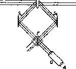
Fig. 8.
U=>J
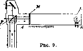
from the bundles emerging from the cube, for example in the right one, we will see the field consisting of two parts, external and internal. The external part of the field is illuminated by side A2 of the plate, and consequently by lamp L2, while the internal part is illuminated by side Ax of the plate and lamp Lx. Since the illuminance of the sides of the plate is inversely proportional to the squares of the distances to the lamps, then with different intensities of the light sources Lx and L2, one can, by moving the photometer along the bench and thereby changing the distances from plate A to the lamps, equalize the illuminance of the photometer fields; at this moment the intensities of the lamps will be inversely proportional to the squares of the distances to them. If the intensity of one of the lamps is known, then the intensity of the other is determined by simple calculation. To eliminate the error arising from different reflection coefficients of sides Ax and A2, the photometer is turned 180° and the measurement is repeated; in this case side Ax will be illuminated by lamp L2, and side A2 by lamp Lx. After performing the calculations in each case and taking the geometric mean of both values, we will obtain the true intensity of the lamp. One of the most widespread systems of portable visual photometers is the tube-photometer (fig. 9). In it, the comparison lamp L is rigidly connected to the photometer itself. The beam from the lamp falls on the ground glass M, which can be moved along the tube A-B, and thereby its brightness is changed. The light from the measured source falls on the ground glass Mx. By moving the ground glass M, equality of its brightness in the Lummer-Brodhune cube is achieved. To increase the range of the photometer, a series of attenuators can be inserted into both the arm of the measured lamp and the arm of the comparison lamp. In addition, there are a number of special photometers for studying the optical properties of bodies. Reflectometers measure the reflection coefficient of mirrors and various surfaces. Densitometers measure the transmission of exposed photographic plates and other light-absorbing bodies. For measuring the transmission of small details on photographs, visual and objective microphotometers are used. In all these instruments, the luminous flux coming directly from the source is usually compared with the luminous flux weakened by absorption in the substance (upon reflection from its surface or when passing through the substance). Finally, there are a number of universal photometers: Pulfrich's stump photometer, Bloch's unimeter, used for measuring a whole range of very diverse quantities (intensity of light, brightness, reflection coefficient, absorption, scattering, color, etc.). In phototherapy, the study of the spectral distribution in the light source, on the one hand, and the spectral characteristics of reflecting and absorbing light bodies, is extremely important. These questions are dealt with by spectrophotometry. For photometry in various parts of the spectrum, instruments called spectrophotometers are used. In them, the light beam from the investigated source and from the comparison lamp first passes through a prism and is decomposed into a spectrum, and then falls into the photometer. By moving from one region of the spectrum to another, the ratio of light energy in a given spectral region for the unknown source and the standard is measured. In the most common König-Martens spectrophotometer, the light decomposed into a spectrum from both sources falls on a Wollaston prism, where it is polarized in two mutually perpendicular directions. Behind the Wollaston prism is a Nicol prism; by rotating the Nicol prism, the intensity of both light beams passing through the latter is changed and their equality is achieved. In this case, the ratio of the intensities of both light beams will be proportional to the square of the tangent of the rotation angle of the Nicol prism: y~ = tg2 α. In objective spectrophotometers, the light from the measured source passes through a monochromator and falls on some receiver: a thermoelement, a bolometer, or a photoelectric cell. If measurements are made with the first two receivers, then the current they give is simply proportional to the energy of the source in a given spectral region, but if with a photoelectric cell, then due to its different sensitivity across the spectrum, it is necessary to measure the source with a known energy distribution across the spectrum, then the ratio of the instrument readings for each wavelength will give the ratio of the energies of the sources. By introducing a reflecting surface or an absorbing substance into the path of one of the beams in the spectrophotometer, one can determine with the spectrophotometer the reflection coefficient or transmission coefficient of the substance as a function of the wavelength of light. This method is used to study light filters and colored reflecting surfaces.
W.
Ginzburg, V. Manufacturer. The biological action of light is extremely diverse. The facts relating to this are described in both biological and agricultural and medical literature. One of the most noticeable manifestations of the biological action of L. is its influence on plant growth, particularly on seed germination; in this sense, plants can be divided into three groups: 1) those whose germination is stimulated by L. (day-growing plants), 2) plants with reduced germination under the action of L. (night-growing plants), 3) indifferent to L. For the effect in question, the law 'the product of intensity by time of exposure is a constant quantity' apparently holds, i.e., the same effect can be achieved either by brief exposure from a high-intensity source or by prolonged exposure from a weak source. The mechanism of L. action in this case is not entirely clear; the effect is due either to the development in seeds of chemical substances of the catalyst type that determine the onset of development, or, according to some, on the contrary, to the destruction of some hypothetical substances that inhibit development and determine a special anabiotic state of dormant seeds. - Undoubtedly, the influence of L. on various processes related to the life of plants can be noted, for example, red rays stimulate 'stretching growth' (Streckenwachstum); in connection with this, an increase in width is observed in plants developing under L., in contrast to the extreme elongation of etiolated plants deprived of light. Light also affects processes such as flowering, leaf formation, fruit ripening, etc. The problem of the influence of L. on plants is of considerable practical interest, in particular it is intensively developed in our Union in the special Agrophysical Institute (Leningrad). Special mention should be made of the influence of L. on the formation of chlorophyll grains; the external appearance of etiolated plants already shows that in the absence of L., their greening is impossible; however, negligible amounts of L. (exposure lasting several seconds) are sufficient to cause the appearance of chloroplasts; the mechanism of the photochemical process occurring here is largely unclear; apparently it is a matter of 'liberating' some pre-prepared reaction. It is interesting to note that the same effect can also be obtained on killed plants. L. has extremely important significance in the processes of CO2 assimilation by the plant, where, as is known, the energy base of complex and little-studied chemical processes is the use of the chemical rays of solar L., namely its violet and partly red parts. The factual material concerning the influence of L. on the growth of animal cells and tissues is significantly less abundant; most of the available data here refers to observations on the growth of tissue cultures. Apparently the most active are ultraviolet rays, and as a general rule (probably not without exceptions), it should be accepted that a short time of exposure stimulates growth, a longer duration, just like high intensity of exposure, leads to inhibition of development, and then to the death of cellular elements, which is apparently always associated with the action of L. with a change in the physicochemical state of the colloids of protoplasm—a decrease in the degree of dispersion, an increase in particle size, an irreversible precipitation of floccules, etc.—The influence of light on the course of mitosis (in particular the intermediate phases of the process—metaphase and anaphase—are most sensitive) and on development is also noted. Here too the usual sequence of stimulation and inhibition is maintained. By the classic works of Lebedev and others, the possibility of activating parthenogenetic development of eggs of marine animals under the action of ultraviolet rays, as well as obtaining deformities in embryos with excessive doses of L., has been shown. The role of short ultraviolet mitogenetic rays in the processes of division and development should be mentioned (see Mitogenetic rays). In the development of higher animals, the presence of L. (in particular small amounts of ultraviolet rays) proves to be a physiological factor of primary importance. With a deficiency of these rays, a complex of pathological phenomena arises, defined primarily by the concept of rickets and characterized by a deficiency of calcium salts in the bones, and probably also by an incorrect course of the entire histogenesis of bone tissue. These changes are associated with the absence of vitamin D. The latter arises in the body, as special studies show, under the action of ultraviolet rays on ergosterol. Along with the skeletal system, one of the most sensitive to the action of light in the adult organism is the system of hematopoietic organs, where an increase in the number of red and white blood elements is noted, as well as an increase in the amount of hemoglobin. L. has a very significant effect on the vital activity of microorganisms. Short rays (especially ultraviolet) are among the most powerful bactericidal factors, as established as early as 1878 by the works of Downes and Blunt. The action of L. consists both in inhibiting the development of microorganisms and in their death with prolonged exposure, occurring with phenomena of irreversible coagulation of protoplasmic proteins. The question of the wavelength most active in this sense is resolved differently by various researchers; apparently the maximum effect is achieved between 2,000 and 3,000 Å and 3,000-3,400 Å. Long rays have a much less pronounced effect and only under the condition of special preparation of the culture, its sensitization (see below). In addition to the bactericidal action, a number of other effects of light exposure are noted, such as a decrease in the mobility of motile forms, a change in pathogenic properties, a change in permeability under the action of ultraviolet rays (first an increase in permeability, then a decrease associated with a decrease in the degree of dispersion; the works of Academician Nadsen and his school on yeast). Ultraviolet rays affect a number of substances in the body—enzymes, toxins, and antibodies; thus, a weakening of the action of snake venom, a decrease in anaphylaxis, etc., have been described. The mechanism of the influence of L. here probably consists in changing the physicochemical state of these substances in the manner described above, i.e., by increasing the size of colloidal particles. The influence of light on metabolism in the body is manifested primarily in changes in the properties of the blood; in addition to the influence on the number of formed elements, an increase in bactericidal ability, a decrease in clotting ability, etc., are described (rays with a length of about 500 mμ). With prolonged exposure to ultraviolet rays, hemolysis occurs. The influence of L. on such processes in the body as respiration, fluctuations in the alkaline reserve, mineral metabolism, etc., is also described. The importance of light for calcium metabolism was noted above. In the field of organic metabolism, a decrease in the sugar content in the blood, an increase in fat in tissues, etc., is noted. Some influence is also noted in relation to nitrogen metabolism (a decrease in the content of urea and uric acid in the urine). There are indications of an increase in the phagocytic ability of leukocytes; perhaps in accordance with this is the influence of L. on the course of various diseases noted by many authors, as well as an increase in immunity under the action of L. Materials on the influence of light on various systems of organs do not have a systematic character. The greater part of the data concerns the influence on the endocrine, circulatory and partly nervous systems. Of the internal secretion organs, the parathyroid gland has the greatest sensitivity, in which morphological changes of the hyperplasia type can be noted when animals are raised behind colored glass. To a lesser extent, the influence of L. is manifested on the adrenal gland, pituitary gland, and thyroid gland. In the circulatory system, in addition to the observations already mentioned, the action of L. on the vessels is observed, expressed in a change in pulse and a decrease in blood pressure; the latter is probably associated with vasodilation occurring under the action of L. At the same time, the release of such large blood reservoirs as the spleen is also observed. The effect of L. on the circulatory system should be considered extremely complex and hardly primary; apparently here there is an influence on the innervation of the vessels, most likely through the formation of special substances that stimulate the work of the nervous system. It is necessary to mention the specific influence of L. on the eye, causing the act of vision (see Vision), the action on the receptor elements of the retina, and the effect on the intermediate media—the cornea, lens, vitreous body, etc. The existing hypothesis about the role of L. in the occurrence of a number of eye diseases (cataract) seems poorly founded. - The significance of light in the occurrence of various diseases is manifested primarily in the group of specific diseases associated with the action of L. on white animals in the presence of sensitization. The course of a number of skin diseases (eczema, pellagra, skin cancer) is also in close dependence on the action of L. The influence of excessive exposure to L. is well known.
(mainly ultraviolet rays), causing the phenomenon of "light shock," and when acting on small animals—"light death." One of the most important manifestations of the biological action of light is the elicitation of directed reactions in plants and animals—phototropisms and phototaxes (see Tropisms). The question of morphological changes in cells and tissues under the action of L. requires special consideration. The available data are extremely scarce and are almost entirely limited to the facts mentioned above concerning changes in the colloidal state, permeability, etc. There remains a large field for research here, since it is undoubted that proper dosing should lead to changes in the structure of the adult and growing organism. The question of the direct influence of L. on the perceiving part of the organism, i.e., on the skin (in particular human skin), was subjected to special investigation. The first effect of such exposure should be considered erythema, i.e., a local reaction of the skin associated with hyperemia, increased temperature, etc. A further result of L. exposure is the formation of pigment, located in the lower (cylindrical) cells of the epidermal layer of the skin. The formation of pigment has a protective significance, retarding the penetration of rays into the deeper tissues. It must be assumed, however, that the pigment plays some significant role in metabolism. Regarding the mechanism of pigment formation, the most widespread view is that the pigment is formed under the action of light as a result of the interaction of the protein body tyrosine and the enzyme tyrosinase. According to some authors, the question is about the action of a special enzyme. A prominent place in the doctrine of the biological action of L. is occupied by the concept of the sensitization of biological objects to L., i.e., the action of a number of substances on the organism (mainly various dyes—eosin, erythrosin, porphyrin, etc.), in the presence of which the effect of L. exposure is significantly enhanced. Among sensitizers, a distinction must be made between exogenous and endogenous substances. The latter include the hematoporphyrin of the blood. The significance of sensitizers has been most fully studied by Tappeiner and Jodlbauer, who introduced the concept of photodynamic action. In addition to dyes, sensitizers must also be attributed to certain salts, in particular salts of iron. The mechanism of photodynamic action apparently consists in the adsorption of energy by sensitizers and the transfer of this energy to biological objects. According to Neuberg, the mechanism of photodynamic action consists of complex oxidative processes. Quinine also possesses a photodynamic effect. The combined action on the organism of medicinal substances of the sensitizer type and L. should be subjected to detailed investigation in modern medicine.
L. s.
Related articles
Mentioned in
Cite this page
“Light.” Soviet Medical Encyclopedia. English translation of Bolshaya Meditsinskaya Entsiklopediya, 1st ed. (Moscow, 1928–1936), ed. N. A. Semashko. https://sovietmedicalencyclopedia.pages.dev/article/light/