Mitogenetic Rays
Historical document, translated for reference. It reflects medical knowledge of the 1920s–30s and is not medical advice.
Summary
Mitogenetic rays are ultraviolet rays with a wavelength of about 2,000 Å (200 mμ) produced by the organism itself, causing cellular divisions. Discovered by Gurvich in 1923, they are studied through mitosis induction experiments on onion roots.
Encyclopedia article (1928–1936)
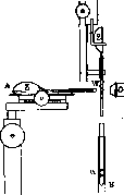
Mitogenetic rays, ultraviolet rays with a wavelength of about 2,000 Å (200 mμ), produced by the organism itself and causing cellular divisions within it. Their discovery in 1923 by Gurvich became a link in the chain of works by this scientist devoted to the study of the causes of cellular division. As a result of these studies, conducted mainly on onion roots, Gurvich concluded that 1) cellular division represents a reflex act, a response to some external (relative to the cell) irritation; 2) unlike classical reflexes, cellular division is to some extent random in the life of a given cell, and is not mandatory for all cells of any complex; 3) the surface of the cell, and in particular the submicroscopic configuration of its constituent particles, is responsible for the onset of division in each individual case. These data led Gurvich, as well as some other authors (Wassermann), to consider cellular division as a result of the interaction of the factor of the cell's readiness to divide, which in turn is an expression of certain processes inside the cell and on its surface, and the external factor of the implementation of division. The concept of the nature of this latter is based on the following reasoning. The above-mentioned role of configuration in the work of the cellular apparatus that perceives the impulse to divide allowed Gurvich to draw an analogy between it and a physical resonator apparatus, for which the same principle of configuration is particularly essential. Continuing the analogy, Gurvich suggested that the external irritation causing mitosis (karyokinesis) in cells is oscillatory in nature, i.e., to be more precise, it appears to be the action of radiant energy. This bold hypothesis found confirmation in a number of subsequent experiments by both Gurvich himself and a number of other authors. The main proof of the existence of mitogenetic rays is the induction of mitosis at a distance. The premise of the experiment is the idea that the radiant energy causing mitoses in any formation (for example, an onion root) not only 'saturates' all its cells but also exits in excess outside (especially in such a conically tapering formation as an onion root) [see separate table (pp. 71-72), figure 6] and can be detected by some sufficiently sensitive method. According to Gurvich's thinking, such a method can be an increase in the number of mitoses on one side of a symmetrical and capable of dividing object. Such a most suitable object again proved to be an onion root, naturally divided in half by the so-called central strand of large cells. The number of mitoses in both halves practically coincides, fluctuations in their number do not exceed 5%. Adding ray-A apparatus for induction through air. The horizontal bulb (Z), placed on a watch glass with water, can move in two planes using rack-and-pinion gears. The bulb (Z) of the detector root moves in one plane. The inducer root is directed at the detector root, the corresponding part of which (W) is free from the glass tube; O-objective of the horizontal microscope, with which centering is achieved; B-picture of centering the roots (inducer and detector) in the horizontal microscope. energy from the side increases by 20-30% the number of dividing cells in the half of the root facing the source of rays compared to the control. The effect of root on root (see figure), carried out through the air without direct contact, received the name of mitogenetic induction. The essence of the experiment comes down to exposing for 20 minutes the tip of one root against the productive, mitosis-rich zone of another (meristem); after 2-2.5 hours (the time needed for new mitoses to obtain sufficient morphological expression), the root is fixed, histologically processed and cut into sections in the plane of induction marked at the end of the experiment [see separate table (pp. 71-72), fig. 7]. Control experiments confirmed the basic premises of Gurvich, although critical voices denying the existence of mitogenetic induction are still heard. The induction of cell divisions is essentially the only sufficiently sensitive method for judging the presence of mitogenetic rays in one case or another. Further study of the latter went along several paths. One of the first tasks of the research was the generalization of the initial observation made on the onion root. At present, a large number of sources of mitogenetic rays are indicated, establish
ed
by the same method of induction. Among them in the world of protozoa - cultures of bacteria and yeast, in the plant world - the base of an onion bulb, the vascular-fiber bundles of a potato tuber, a rootlet, cotyledons and the first leaves of a sunflower, etc.; further - eggs of various animals (sea urchins, worms, amphibians) and the nervous system of tadpoles. In adult animals, it induces only blood (in vertebrates and invertebrates). Other tissues lack this ability. As a source of M. rays, tissue of malignant tumors and a number of organs with altered metabolism are also indicated - the epithelium of the cornea of starving animals, a liver rich in glycogen, etc. The seemingly fantastic diversity of sources of M. radiation is reduced by Gurvich and his school to three large groups, characterized by the chemistry of the underlying processes. M. rays are produced by processes 1) oxidative, 2) glycolytic, 3) proteolytic. Thus, the presence of M. rays has been established to date for all studied cases of cell division, and it is not always localized in the place of enhanced cell multiplication, which in such cases receive M. rays from outside, e.g., from the blood. The presence of M. rays has also been noted for a number of processes (muscle contraction, nerve excitation, oxidative processes in vitro), not related to cell division, but fitting into the above chemical scheme. It is necessary, however, to note that the biochemical characterization of sources of M. rays is completely preliminary and requires further research. The question is more complex regarding the detectors of M. rays, i.e., the objects on which it is possible to establish the presence of the latter. The choice of detector is connected with certain definite conditions, the main of which is the state of readiness of the irradiated cells, their ability at a given moment to divide under the influence of an external factor. This basic prerequisite is often overlooked by authors checking Gurvich's main experiments on objects incapable of division altogether. In addition, necessary conditions for the detector are 1) uniformity of the object, 2) possibility of experimental intervention, 3) existence of an ideally comparable control. At present, the universal detector, on which most of the work of Gurvich's school and a number of other authors has been done, are yeast cultures (mainly Nadsonia fulvescens) on agar-agar and on wort (liquid cultures). The criterion of the inductive effect is the increase in the number of budding cells in the experimental part of the culture compared to the control. Under certain experimental conditions (diluted cultures, prolonged exposure), it is possible to obtain a macro-effect of induction on yeast cultures (Saccharomyces and Pombe), consisting of a noticeable increase to the naked eye in the amount of yeast in the induced culture with ideal equality of the initial amounts of material in the induced and control cultures. Besides those mentioned earlier, various authors have established the following detectors: 1) bacteria - in the sense of increasing their number; this increase can be established besides simple counts also indirectly by means of fine physical methods based on the difference in light scattering in media of different turbidity; 2) the epithelium of the cornea of amphibians and mammals; 3) eggs of sea urchins and worms. Stempel's attempt to use a physicochemical detector - disturbance in the structure of Liesegang rings formed during the diffusion of silver nitrate into chrome gelatin - ended unsuccessfully, mainly due to the extraordinary lability of the ring formation process itself, making it difficult to resolve the question unequivocally (see Liesegang rings). One of the most important tasks in the study of M. rays was to clarify their physical nature. Gurvich's first preliminary experiments with the passage of rays through water and air and reflection already made their ultraviolet nature extremely probable. The next series - passage through quartz, interception by thin layers of glass and gelatin - helped to more accurately establish the wavelength, hypothetically attributed by Gurvich to the region of 200 mμ. This provisional assumption was fully confirmed by subsequent evidence: 1) obtaining a mitogenetic effect from a physical source of ultraviolet rays of the same wavelength (discharge of aluminum electrodes, arc lamp) in contrast to other nearby wavelengths; the exact boundaries of M. rays were established by Gurvich between 180 mμ and 290 mμ, and 2) direct spectrographic investigation of the radiation of a working muscle (using the usual detector - yeast), showing the same wavelength. Regarding the latter wavelength, there are disagreements in the literature between Gurvich and German authors Reiter and Gabor, indicating a different (longer) wavelength. The physical investigation of M. rays showed that we are dealing with intensities, negligibly small, lying at the threshold of sensitivity of the most delicate instruments detecting radiant energy (photoelectric cell). At present, Dessauer has proven the existence of M. rays with the help of a sensitive instrument - the so-called Geiger counter. Thus, the main criterion for the existence of M. rays is the biological method, i.e., the inductive increase in the number of cell divisions. The biological detector, according to Gurvich, is approximately 600 times more sensitive than a photographic plate. The path to the study of M. rays lies in clarifying their biological significance. The totality of data available at present forces to expand the original concept, which considered M. rays only as a necessary factor of cell division, and to assign them importance for the entire metabolism of the cell. Gurvich assigns a certain role to M. rays in the biology of malignant neoplasms, which, unlike normal tissues, are characterized by intensive production of M. rays, apparently having two sources: glycolytic (in superficial, little altered cells) and proteolytic (in foci of necrosis). Besides stimulating uncontrolled cell multiplication, M. rays in this case acquire another important significance for the biology of carcinoma. The mode of intense ultraviolet radiation inside the tumor, from this point of view, may be one of the causes of cell disintegration, which in turn becomes a source of M. rays in the future. In connection with the question under consideration, it is necessary to mention that interesting fact that the blood of carcinomatous animals early loses its ability to produce M. rays, judging by experimental data obtained on animals (mice) - several days before the appearance of a noticeable tumor. A number of data forces to assume that in this case there is a suppression of enzymatic activity, caused by the circulation in the blood of products of protein breakdown. This is probably also the explanation for the decrease in M. ray production by the blood in a number of diseases associated with the presence in the blood of significant amounts of necrotic substances - septicemia, osteomyelitis, etc. M. rays completely disappear in lesions of the blood system as such, namely - in leukemia, poisoning with nitrobenzene, etc. On the contrary, in a number of other diseases (tuberculosis, syphilis, typhus, cerebrospinal meningitis, etc.) the radiation remains in full force.
Related articles
Mentioned in
Cite this page
“Mitogenetic Rays.” Soviet Medical Encyclopedia. English translation of Bolshaya Meditsinskaya Entsiklopediya, 1st ed. (Moscow, 1928–1936), ed. N. A. Semashko. https://sovietmedicalencyclopedia.pages.dev/article/mitogenetic-rays/