Regeneration
Historical document, translated for reference. It reflects medical knowledge of the 1920s–30s and is not medical advice.
Summary
Regeneration is the process of forming new organs or tissues in place of removed parts of an organism. This article distinguishes between physiological and reparative regeneration, examining their occurrence across different species and developmental stages.
Encyclopedia article (1928–1936)
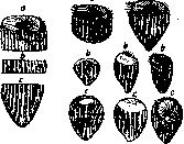
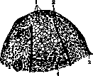
Regeneration, the process of formation of a new organ or tissue in place of a segment of the organism removed in one way or another. Very often R. is defined as the process of restoration of what has been lost, i.e., the formation of an organ similar to the one removed. Such a definition however proceeds from a false teleological point of view. First of all, the part of the organism arising during R. is never completely identical to the previously existing one, it always differs from it in one way or another (Schaxel). Then the well-known fact is sufficient that instead of the removed segment, a completely different, dissimilar one is formed. The corresponding phenomenon is also classified as R., although calling it atypical R. However, there are no data indicating that the processes occurring here differ in any way from other types of R. Thus it will be more correct to define R. in the manner indicated above. Classification of R. phenomena. Two main types of regenerative processes are distinguished: physiological and reparative R. Physiological R. takes place in the case when the process occurs without the presence of any special external influence. Such types of R. represent the phenomena of periodic molting in birds, mammals and other animals, the replacement of the desquamating epithelium of human skin, as well as the replacement of dying cells in glands and other structures by new cells. Reparative R. includes cases of new formation as a result of the organism receiving one or another damage, both as a result of artificial intervention and independently of it. Below, the phenomena of reparative R. will be mainly considered, as the most studied one. Depending on the final result of the process, reparative R. is divided into typical, when the formed organ is more or less similar to the previously existing one, and atypical, when such similarity is absent. Deviations from the typical course of R. may consist either in the formation of a completely different organ instead of the previously existing one, or in its modification. In the case when the appearance of a different organ is associated with a reversal of polarity, e.g., when instead of the cut-off tail end of a worm the head end regenerates, the phenomenon is called heteromorphosis. Modification of an organ may manifest itself in the presence of any additional parts up to the doubling or tripling of the organ, or in the absence of structures usually characteristic of it. 'It should be remembered that the division of R. into typical and atypical, based on a teleological view and oriented toward the previously existing organ, does not reflect the essence of the phenomena and is entirely conditional. The ability for R. is an extremely widespread phenomenon both among animals and plants, although individual species differ from each other both in the degree of regenerative ability and in the course of the process itself. In general, it can be considered that the higher the organization of the organism, the less its regenerative ability; however, there are a number of exceptions to this rule. Thus, many closely related species differ very strongly from each other in regenerative manifestations. On the other hand, a number of higher species are more capable of regeneration than lower ones. In amphibians, for example, even individual organs, such as the tail and limbs, can regenerate, while some worms (Nematoda) are characterized by an almost complete absence of R. As a rule however, the greatest ability for R. is found among lower animals. Unicellular organisms are characterized by a strongly expressed regenerative ability (fig. 1). In some species, pieces equal to one hundredth part of the animal are capable of restoring it completely. Among multicellular organisms, the greatest regenerative ability is found in coelenterates and worms. Some hydrozoans restore the animal from one two-hundredth of its part. Worms (especially Annelida and Turbellaria) from several segments can form all the missing parts. These species are little inferior to such a highly standing group as the tunicates, where
„
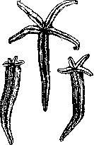
« R. of the entire animal can take place from one part of it (e.g., the basket of gills in Clavellina). The regenerative ability is also well expressed in some echinoderms; thus, starfish form a whole animal from one ray
Fig. 1. Regeneration of the infusorian Stentor, cut into three parts. (After Corsh.)
parts (After Corsh.)
Regeneration
536 of the regenerative phenomena compared to adult animals (Steinmann), the same is true for the larvae of some other animals. Already from the above it could be seen that different areas of the organism differ from each other in their regenerative ability. Weismann assumed that the ability for R. depends on how much a given part is subject to the danger of damage, and the greater the latter, the greater the regenerative ability - a property developed as a result of natural selection. However, subsequent research showed that such a regularity cannot
6,6 15 6,9 10 7,2 5 \ г°\ /i [^ 1 * .y/ ""ч> *-.„ 8 Ю 12 14 Figure 3. Solid line - change in the intensity of mitogenetic radiation of the regenerating axolotl tail. On the ordinate - conditional units of radiation intensity. Dashed line - changes in the active reaction of tissues of the regenerating axolotl limb. On the ordinate - pH values (given by Okunev). On the abscissa - days of regeneration. (From Blyakher and Noylen. according to Bromley.)
organs, not usually subject to damage during the free life of the individual and well protected, nevertheless possess a high regenerative ability (Morgan, Przibram). Ubisch associates regenerative phenomena with the differentiation of the organism; according to him, earlier developing parts more cease to regenerate with age or their R. differs in lesser intensity. Thus, in amphibians, where organs located more to the front differentiate earlier, a corresponding gradient of R. can be established - from front to back. The assertions of Ubisch, in favor of which there is a certain amount of data, still need further confirmation on a larger amount of material. In some species (mainly in worms) Child and his colleagues have also established a certain gradient of R. with respect to the longitudinal axis of the body, but its direction does not always go from front to back, but is associated with more complex regularities. Child believes that this gradient depends on the degree of physiological activity of different parts of the organism. More lowly organized animals have the ability to regenerate both parts located proximally to the amputation site, as well as
Fig 4. Regeneration of an amputated forelimb in a salamander after */4 (a) and 12 (b) hours, a: 1-blastemal cells; 2-stump of the shoulder; 3-nerve; 4-epidermis; b: 1-blastemal cells; 2-cartilage; 3-epidermis; 4-stump of the shoulder.
located distally. In higher animals only the latter regenerate. In amphibians, for example, an organ, even transplanted in an inverted position, regenerates the same formation as in the normal position.
Figure 5.:Regeneration of
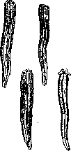
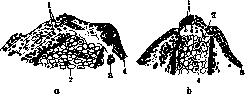
Course of the regenerative process. The regenerative process proceeds differently depending on which organism we are dealing with and which part of it is removed. As an example, we can consider the most studied object - regeneration of amphibian limbs. In this case, the following phenomena occur. After amputation of the organ, the edges of the wound come closer together due to the contraction of the severed muscles. The blood on the surface of the wound clots, releasing fibrin threads. The clotted blood, with the participation of cells from the damaged tissues, forms a scab on the surface of the wound. As a result of tissue damage and the effect of the external environment on the unprotected skin surface, processes of decay occur in the organ. The latter are manifested in a change in the acidity of the regenerate (a decrease in pH from 7.2 to 6.8, Okunev) and the appearance of mitogenetic radiation (Blyakher and Bromley). However, the wound surface does not remain unprotected for long: within the next few hours, a process of epithelial overgrowth from the edges of the wound is observed, as a result of which an epithelial film forms on the wound surface. Under this epithelial covering, all further processes take place, which come down to the destruction and rebuilding of the old and the formation of a new organ. These processes are expressed, on the one hand, in the continuing decay. The latter is revealed morphologically by pictures of tissue destruction and the arrival in the regenerate of numerous blood cells. Decay is especially strong in the period from 5 to 10 days, starting from the moment of amputation, when it apparently reaches the greatest intensity. The same is evidenced by physiological indicators. Okunev found the greatest acidity on the 5th day, when pH=6.6. At the same time, the intensity of mitogenetic radiation increases compared to previous days (Bromley). The curves of increasing acidity and intensity of mitogenetic radiation turn out to be parallel to each other throughout the entire regeneration. Both have two maximum peaks - on the 1st and 5th day of regeneration (Fig. 3). Along with this, already in the first week of regeneration, the processes of new formation are clearly outlined. They are manifested mainly in the formation under the epithelial film of an outgrowth of homogeneous cells, called a blastema. The development of the new organ goes
Figure 7. Regeneration of am-
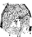
mainly at the expense of the cells of the blastema (Fig. 4-7). After a certain period of growth in the regenerate, differentiation of individual parts occurs. At this point, first the more proximal parts differentiate, and then the distal ones. In this respect, the process does not flow the same in all organisms. In some, it can even be reversed. Physiol. peculiarities of the regenerate of the limb-
but not those of a formed organ. This is manifested in particular in the fact that the regenerate possesses histolytic properties. In the case when its surface comes into contact with other tissues, e.g. when covering the regenerate of the amputated "forelimb of a salamander with a skin flap, histolysis occurs after the giant cells; 2-epi- dermis; 4-musculature; 5-shoulder ring; 6'-
regenerating organ. It also exerts its effect on the rest of the organism, which can manifest itself in various ways. Thus, a change can be detected in the blood of the animal, the mitogenetic radiation of which deviates from the normal intensity, and these fluctuations have a characteristic curve. In regeneration in hydra, decay of organs not in direct proximity to the regenerate has been noted, namely of sex cells, and predominantly male ones (Goetsch). The effect of regeneration is also manifested in the growth and other properties of the organism - a phenomenon often described under the name of regulation. Material of the regenerate. The question of the material from which the regenerate is formed must be resolved differently depending on the species of animal and the nature of the injury inflicted. If it is a matter of damage to one particular tissue, then usually the process proceeds at the expense of the outgrowth of the remainder of the corresponding tissue. The matter is more complicated in the case of regeneration of an organ or restoration of the organism from a separate part of it. However, it can be established that basically, at least in amphibians, regeneration proceeds at the expense of material directly adjacent to the wound surface, and not at the expense of cells coming from other areas of the organism. This is shown by experiments on regeneration of a haploid-nucleus limb of a triton, transplanted onto a diploid-nucleus animal. The regenerate that arises in this case consists of haploid-nucleus cells (Hertwig). The same follows from transplants of limbs from black axolotls to white ones, when the regenerating limb turns out to be black. These facts exclude the idea of regeneration at the expense of various cellular elements coming with the blood flow. When considering the material going for regeneration, one must take into account the dual possibility. Regeneration can occur either at the expense of so-called reserve, indifferent cells remaining undifferentiated during embryonic development, or there is the use of already specialized cellular elements. The importance of reserve cells was shown for a number of animals. Thus, regeneration in hydra occurs mainly at the expense of the so-called interstitial cells. The same is the case with turbellarians. In annelids, this role belongs to neoblasts, belonging to elements of a similar kind. In ascidians, indifferent cells also play an important role in regeneration. The matter is more complicated in vertebrates, where various authors attribute the main role in regeneration to different tissues. Although here too there are indications of the origin of blastema cells from unspecialized elements, however, this fact cannot be considered firmly established. Nevertheless, the positions of the previously prevailing theory of Gewebesprossung, which recognized the possibility of development of cells of any tissue only from cells of a similar tissue, have been thoroughly shaken. But if one can accept the formation of a significant mass of the regenerate at the expense of unspecialized cells, this does not exclude the possibility of development of part of the regenerate from differentiated elements. In this case, there can be a question of development of tissues - at the expense of multiplication of like elements, as well as of transition of cells of one type into another (metaplasia). In reality, in many cases it can be shown that both these processes take place. Thus, musculature usually
Figure 8. X-ray of the regeneration of a bone-
TO A CONSIDERABLE EXTENT PROCEEDS FROM THE REMAINING UNDESTROYED MUSCLE CELLS. In annelids, the formation of muscles from epithelial elements can be established. The same is the case with some crustaceans (Przibram). The formation of the nervous system from ectodermal cells has been established in ascidians (Schultze). In amphibians, it is known that regeneration of the lens can occur from the edge of the iris (Wolff, Colucci). One can also accept the formation of cartilaginous and bony skeleton without the participation of cartilaginous and bony elements of previously existing organs.
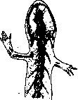
Since the regenerative process includes both development from indifferent elements and the participation of specialized elements, in each individual case special research is necessary to clarify the role of each of these processes in regeneration. If we consider as an example regeneration in amphibians, again due to its greatest studyability, then the matter here appears as follows. Nerves are always formed at the expense of the growth of endings of old nerve trunks. The matter is different with bone tissue in the case of limb regeneration. It has been shown that even when the entire bony skeleton of the limb, including the shoulder girdle, is removed, upon amputation of such a boneless limb, regeneration of an organ possessing a skeleton occurs (Fritsch, 1911; Weiss, Bischler) (Fig. 8). The matter is different in tail regeneration. In this case, bony parts are formed only when there is damage to old skeletal parts in the area of the regenerate, namely the shoulder girdle and shoulder; amputation above the elbow. The forearm with forearm bones and the hand with phalanges have regenerated. Carpus is still cartilaginous, radius and ulna are shifted into the boneless shoulder. (According to Korschelt.)

the bone elements of the latter can participate in regeneration (fig. 9). Regarding the connective tissue part of the skin, corium, we also have proof of its formation without the participation of the old corium (Weiss). As for the musculature, removal of the greater part of the musculature of a limb did not lead to any abnormalities in the development of the regenerate. Moreover, in the case of transplanting a piece of the chorda in a larva of Anura into the tail area, which was devoid of musculature, it was possible to induce the formation of a tail in this place with the corresponding direction of the tail incision. The organ formed in this way possessed musculature (Marcucci). However, histological studies show that in the usual regeneration of the tail, its muscles are formed from the corresponding elements of the old organ (Naville). Thus, a considerable part of the regenerate in amphibians may be formed not as a result of the multiplication of old tissues, but from the mass of the blastema, the origin of the elements of which, as has already been indicated, has not yet been sufficiently established. At the same time, other relationships may also occur, as we have in the regeneration of the tail, the axial organs of which regenerate only in the presence of old ones. At the same time, it should be noted that even the regeneration of the same organ may proceed from different materials depending on the conditions, as could be seen from the example of the formation of muscle elements of the tail. The experiments cited, although they indicate the possibility of the development of some tissues (e.g., bone) not from cells of similar tissue, do not yet resolve the question of how things stand under normal conditions of regeneration. Further research is necessary in this direction.
Conditions of regeneration. A. The regenerating area. The course of regeneration is of course in close dependence on what part of the organism is amputated and consequently in what area the regenerative phenomena take place. First of all, we may encounter the absence of regeneration in some parts of the organism or rather with the weak expression of the corresponding phenomena. Philippeau discovered the absence of regeneration in salamanders in the case of extirpation of a limb with the entire shoulder girdle. Schotte showed that amputation of the tail is accompanied by regeneration only in the case where the incision passes sufficiently distally (fig. 10). Vallette and Guyenot note the absence of regeneration of the nasal parts of the head when too large an area is amputated. Similarly, regeneration of the eye does not occur with complete enucleation (Shaxel). Gills do not regenerate with complete removal. Guyenot interprets these phenomena in such a way that regeneration can occur only
Figure 10. Triton cristatus after complete removal of the tail area; no signs of regeneration for 8 months.
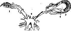
in the presence of certain cellular complexes, which can be completely removed with a sufficient degree of damage. However, reliable proof of this position has not yet been given, and it is not excluded that in some cases the absence of regeneration, discovered by the mentioned authors, is also connected with other conditions. The regenerating area also depends on the character of the formation that arises during regeneration. It is well known that when different parts of the organism are removed, different formations arise. However, this phenomenon should not be explained by the fact that the newly forming organ must be similar to the removed one. Thus, there is the experiment of Herbst, confirmed by other authors, when in the removal of an eye in a crab with the retention of the visual ganglion, an eye regenerates, and with simultaneous removal of the ganglion, regeneration of the antenna is observed (fig. 11). In extirpation in one species of insect (Dixippus morosus) of the antenna in its distal part, the formation of an antenna is observed, but when amputated at the base, a limb regenerates. The corresponding phenomena are called homoeosis. It is clear that the regenerating area also depends on the speed of regeneration, which has already been mentioned. B. Parts of the amputated organ. As could be seen from experiments on the removal of the skeleton of a limb, regeneration can take place in its absence. However, as Bishler showed, in the regeneration of a boneless organ, not the segment that is amputated regenerates, but only the more distal one, so that in regeneration, for example, of a limb, an organ arises that is shortened by one segment. Since development is observed even in the absence of bone tissue, the connection between the specificity of regeneration and the skeleton is denied. Moreover, transplanting one bone to the place of another, for example, the femur to the place of the shoulder, does not change the morphology of the regenerate. An important role in regenerative phenomena belongs to the nervous system. The necessity of the presence of nerve connections for the formation of a regenerate is proven, however not for all species. For a number of animals such a lawfulness apparently does not exist. The clearest data are available for worms, echinoderms and especially amphibians. In worms, Morgan showed the necessity of the presence of nerve endings in the area undergoing regeneration for the regenerative process to take place (fig. 12). The same has been shown for starfish (Morgulis). However, there are data contradicting the just mentioned, so that further research is necessary in this direction. For amphibians, it has been shown that the presence of the central nervous system is not a necessary condition for regeneration (Barfurth, Rubin, Godlevsky). However, in case of disruption of the peripheral innervation of the
Figure 12. Regeneration of the anterior part of the earthworm. The position of the regenerate is determined by the nerve trunk: 1 - plane of regeneration; 2 - end of the severed nerve trunk.
Figure 11. Replacement of the left eye, removed together with the eye ganglion, by an antenniform appendage (1): 2 - supraesophageal ganglion; 3 - eye; 4 - eye ganglion. (After Korschelt.) regenerating organ, the process of restoration does not exist. The relationships here were clarified as a result of detailed experiments by Schotte and Weiss. Both of them showed that in case of complete denervation, regeneration does not take place. Schotte showed that in this case only the sympathetic nervous system is important, because when the sympathetic nerves are severed and the sensory and motor innervation is left, the formation of an organ does not occur. On the contrary, regeneration is present when only sympathetic innervation is preserved. The importance of the nervous system was proven by Schotte not only for adult animals but also for larvae. Schotte's data regarding sympathetic innervation, however, are objected to by some authors who consider that the main role in the regenerative process belongs to the spinal ganglia (Locatelli). The data obtained also indicate that the role of the nervous system is not limited only to the initial stages of the process; for the continuation of regeneration, the presence of the nervous system is also necessary. A number of authors connect the specificity of the regenerate with the nervous system. According to them, there is a specific influence of the latter. Interesting data in favor of this assumption were presented by Locatelli, who obtained the formation of additional limbs in tritons by bringing the central end of the severed n. ischiadici to the surface of the body in the area of the side and hind limb (fig. 13). However, Guyenot and Schotte showed with their research that the specificity of the nerves does not play a role in this phenomenon. It is true that bringing the severed end of a nerve to this or that area of the organism causes the formation of an organ here, however the character of the organ is connected with the specificity of the area, and not of the nerve. The same nerve, when brought to the area surrounding the hind limb, causes the development of the hind leg there, and when it falls into the area located closer to the tail, causes the formation of precisely the latter organ. When the nerve is brought to intermediate areas, one can
Figure 13. Heterotopic regeneration of an organ by the method of diverting the shoulder plexus. (After Guyenot.)
obtain
Figure 14. Inhibited regeneration of the right hind limb of an axolotl due to the formation of a skin scar. (After Korschelt.)

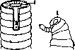

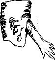
to produce chimeric formations between the tail and the limb. A number of other data in favor of the specificity of the nervous system (Wolff, Walter) also received a different explanation. In connection with this, the assumption of the specificity of nervous influence on R. must be rejected. Removal of the skin at the amputation site for a certain length of time leads to the fact that R. of the organ is delayed until the epithelium, creeping in from the edge of the skin onto the exposed surface, covers it and reaches the amputation site. Degeneration of the exposed area may also occur and then R. begins from the moment when the degeneration of the area reaches the edge of the skin and the corresponding parts fall off. Thus, the presence of skin, or rather the epithelial covering, is a necessary condition for R. of the organ. This position explains the absence of R. when the wound surface is covered with a skin flap (Fig. 14), shown by a number of authors both on amphibians (Tornier, Shaksel, Godlevsky, Efimov) and on insects (Shaksel and Adensamer). This phenomenon is due to the fact that the skin epithelium has no access to the wound surface, being separated from it by the connective tissue part of the skin, while for the occurrence of R. it is necessary to cover the wound with young epithelium. If a piece of skin is transplanted under the skin flap covering the wound surface, then R. occurs in these cases (Efimov). This fact indicates that a mechanical obstacle to the growth of the regenerate does not play a role in this phenomenon. The specificity of the skin does not affect the character of the regenerate. This is evidenced by the experiment of Taube, who transplanted a cuff of red abdominal skin onto the limb of tritons and obtained, after R., a normal black limb from the area covered with red skin. The same is confirmed by the transplantation of internal parts of the tail into a skin sleeve of the limb, when tail R. is observed (Bishler). Removal of most of the musculature affects only the speed of the process. One must also deny the specific influence of the musculature, since replacement by transplantation of musculature from one area to another does not change the character of the regenerate (Bishler). Thus, one must recognize that each of the mentioned parts of the organ (nerves, skeleton, musculature, skin), taken individually, is not a specific condition for R. Parts of the regenerate. The regenerating organ is heterogeneous not only in the sense that it consists of different tissues, but within it there are areas that differ extremely from each other in their properties. If the regenerating organ is divided into two different parts, as is usually done, the blastema and the rest of the regenerate, then their behavior turns out to be sharply different. When the blastema is removed, it is newly formed by the remaining parts, the same thing happens when a part of the organ not containing a blastema is transplanted into some other area of the organism. In this case, even very small pieces of the transplanted area can develop the corresponding organ (Fig. 15). The matter is different when transplanting another part of the regenerate—the blastema. It was discovered that up to a certain age, approximately two weeks, blastemas, when transplanted, do not develop further and dissolve (Shaksel). Blastemas in the experiments of de Giorgi, transplanted onto the back in

Figure 15. Results of transplantation of ter-
limbs in place of the tail. (According to Giéno and Pons.) They grew for up to 30 days, although they took root and increased somewhat, but did not undergo differentiation. It is difficult to say what conditions are of importance here; in any case, the conclusion from the stated facts can only be that for regeneration to occur, a connection between the blastema and the remaining parts of the regenerate is necessary. A number of authors attempted to determine which part of the regenerating organ is specific, distinguishing one organ from another. Particular attention was paid to the question of whether the material of the blastema is specific. Corresponding research involved transplanting blastemas from one organ to another in order to determine whether the specificity of the organ formed from the blastema would change. Transplantations of blastemas were performed on various species of animals. It was discovered that when a regenerate was transplanted before a certain age, it developed in the region of the tail where the shoulder was, and in the region of the front part of the organism to which it was transplanted. Thus, these experiments speak for the non-specificity of blastemas. However, all research conducted to date is not sufficiently convincing. Milosevic, when transplanting young regenerates of the hind limb to the position of the forelimb, obtained in a number of cases the formation of a forelimb at the new location, i.e., development corresponding to the site of transplantation. However, these data are not conclusive due to the lack of a reliable criterion for determining that the forming organ actually originates from the tissue of the transplant, and not from the regenerating forelimb itself. In the experiment of Giéno and Schotte, where the blastema of a limb, when transplanted to the tail, gave rise to the formation of a tail, the authors themselves doubt the origin of the material of the organ: Finally, Weiss transplanted tail regenerates into the region of the forelimb and obtained limb development in three cases. However, in these experiments as well, there can be no certainty as to whether the regeneration occurs at the expense of the tissues of the transplant. Thus, the question of the possibility of changing the developmental pathway of the regenerate in amphibians, and along with it the question of the specificity of the blastema, remains open. A similar situation exists for lower animals. The experiments of Gebhardt, who obtained head formation from the regeneratory bud of the tail in planaria in two cases, can be interpreted as the result of participation in regeneration by the tissues of the head region, where the transplantation was performed. All that has been said applies only to young regenerates, since all authors agree that newly forming tissues, taken at a relatively late age, already differ in specificity. Despite the insufficient obviousness of experiments with young regenerates, most authors consider not the blastema, but only the remaining part of the organ, to be specific. The presence of mitogenetic radiation in the regenerate has made it possible to suggest the possibility of the influence of radiation from one part of the regenerate on another, especially mitogenetic rays arising during tissue resorption, on the multiplication of blastema cells (Blyakher and Bromley). However, the significance of mitogenetic radiation in regeneration cannot yet be considered established. It is certain, however, that by acting with mitogenetic rays on the regenerate, one can cause acceleration of the process (Blyakher, Vorontsova, Irikhimovich, Lyozner). The same authors showed the presence of stimulation of regenerative processes in cases where wound surfaces have the opportunity to influence each other (for example, with a triangular excision of a section of the tail). G. Processes occurring in the organism during regeneration. Regeneration is a process that depends not only on the condition of a given organ, but also on the entire organism. Therefore, the processes occurring in the latter can have a decisive influence on the regenerative process. In the experiments of Gekh, decapitation of hydra did not lead to regeneration when the hydra possessed a kidney. Only regulatory processes occurred, as a result of which the head of the growing kidney occupied the place of the head of the polyp. If in a two-headed planaria one head is amputated, the latter does not regenerate (Steinmann). However, a change in the localization of the regenerating organ in relation to the organism may not affect the character of regeneration. Kurz transplanted the amputated limb to the back, where a normal limb regenerated. Weiss exchanged the positions of the front and hind limbs of the triton, and again, regeneration of the transplanted limbs led to the development of the organ that would have formed had they been left in place. The same occurs when a section of the tail or the front part of the head is transplanted. Thus, one or another location of the developmental process is not specific in regeneration. The influence of the organism on the regeneration of its parts can manifest not only in determining the very possibility of regeneration, but also in the character of the regenerate, its form, position, and course of the process. An example of such an influence can be, for instance, the significance of function for the regenerative process, when the use of an organ strongly affects the regenerate. The significance of other parts of the organism for regeneration of a given area is revealed in experiments with endocrine glands; removal of endocrine glands or the effect of their secretions can influence the course of regeneration. Undoubtedly, a number of processes occurring in the organism affect the regenerative process. Among them, one can mention cases of the simultaneous presence of several regenerative processes in the organism. Whether stimulation or inhibition of regeneration occurs in this case depends on the specific conditions, expressed in the size of these injuries, their location, etc. (Zeleny). The influence of existing connections in the organism on regeneration is manifested in experiments involving the excision of small sections from the bodies of hydras or planaria. This can lead to a distortion of polarity, when identical organs form on both sides of the regenerate (formation of animals with two heads or two tails, depending on the area from which the regenerating section was excised).
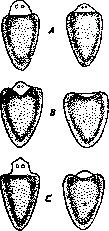
D. Environment. That regeneration can only occur in a suitable environment is sufficiently obvious. With a composition of the environment that adversely affects tissues, the regenerative process is of course impossible. For normal regeneration to occur, the environment must meet a number of conditions. These include, first of all, a certain oxygen content (Leb). Furthermore, regeneration is possible only within certain temperature limits. The optimum for amphibians, for example, is 28°C; above and below this temperature, regeneration slows down, and at 10°C it completely ceases. According to research by Moore, the speed of regeneration depending on temperature follows the van't Hoff law. For aquatic animals, the composition of the liquid surrounding them is of great importance. Regeneration is possible only with a certain concentration of seawater (Leb, Steinmann). The best regeneration is observed in diluted seawater. Certain salts (potassium, magnesium) are also necessary for the existence of the regenerative pro-
Fig. 16. Tail segments of Planaria go-nocephala with regeneration. Other factors influence the speed of it. Popov obtained significant stimulation of the regenerative process when acting with MgCl2, KJ with glycerin for 1 day; without action; C-the same through 7 days. (According to Korschelt.)
Pий и ПОЛИПОВ paCTBOpa- танином+KJ-через4
ми MgCl2, KJ c глицери- дня; C-то же через 7
ном, танином и др веще- дней.(ПоКоршельту.)
ствами (рисунок 16). Сти. мулирующее действие на регенерацию оказывают также вещества, понижающие поверхностное натяжение среды, E. Character of damage. The regenerative process depends not only on the area where amputation is performed, but also on the character of the damage. With a small incision on the body wall of an animal, rapid healing can occur with almost complete absence of tissue formation. However, when several incisions are made in the same place, preventing such healing, a pronounced regenerative process occurs, in re-
Fig. 17. Development of a hydranth from the lateral area of the polyp Corymorpha palma under the influence of radial incisions: 1-incisions; 2, 3, 4-gradual development of the hydranth-
ta. (From Child.) result of which a whole organ develops (e.g. the head of an animal; Lebed, Child) (fig. 17). The nature of the damage can determine an atypical course of R. Thus, when an amputated organ is split, double formations arise. The position of the regenerate can also depend on how the amputation is performed, since the long axis of the developing regenerate is usually perpendicular to the plane of amputation. Theories of R. The phenomenon of R. became known very long ago. Among scientists of ancient times, one can find indications of familiarity with this phenomenon. However, systematic experiments devoted to the study of R. were conducted only closer to modern times. Réaumur studied regeneration in crayfish, attributing this phenomenon to the presence of additional "organ rudiments" (1721). Data from Trembley on hydras, dating from 1744, are known, establishing the pronounced regenerative capacity of this animal. The middle and end of the 18th century count a number of other studies on R. These include the data of Bonnet and Spallanzani. These studies cover not only lower but also higher animals (vertebrates). In the following years, the study of R. progressed very slowly. Only at the end of the 19th century does the intensive study of regenerative phenomena begin, covering the most diverse types of animals. This study is characterized not only by its systematicity and detail, but also by the fact that researchers already penetrate much more deeply into the essence of the phenomenon of R. Researchers of the late 19th century devoted much attention to clarifying the connections of the regenerative process, its necessary conditions, and on this material built corresponding theories of R. The principal approach of these authors to the study of the process received its justification in the works of Roux and can be called the causal-analytical method of research. Its characteristic features are the mechanistic and formal analysis of phenomena; the moments leading to the emergence of the phenomenon under study are taken not in the process of development, but as static. By decomposing the process into individual components, the main component is isolated, which is taken as the initial one, and the phenomenon itself is considered as the result of the effect of various conditions on this basis. On the other hand, since the direction of the process is considered in isolation from its driving forces, a separate factor responsible for the direction of the process is also identified on the basis of formal analysis. Thus, the sources of development and the direction of the phenomenon turn out to be external in relation to the individual components of the process. Since the source of development appears as external in relation to the other components of the process, the inevitable question arises as to what causes the development of the source of development itself. If any factor is identified as the latter, the question of the source of development of this new factor will again arise. Thus, we must either come to a divine first push or refuse to finally resolve the question. The entire incorrectness of the causal-analytical method clearly follows from this description. However, the generality of the method does not prevent researchers of R. from differing among themselves on a number of essential issues, thus forming different camps. Part of the scientists, closer to Roux himself, held a view of a preformationist nature. The development of the regenerate itself is caused, in their opinion, by the irritation inflicted by amputation. The direction of R. is determined mainly under the influence of reserve hereditary rudiments, which thus represent the properties of the future organ and, when they come into further cell multiplication in various parts of the regenerate, stimulate them to corresponding development. Most of these researchers also held the view that each tissue of the regenerating organ is formed from similar tissue of the stump of the organ, and their development proceeds to a certain degree independently of each other (theory of R. "Teil fur Teil"). The preformationist, causal-analytical theory of R. must be decisively rejected. It excludes the idea of actual new formation, interpreting the phenomenon as the realization of something that already existed previously. Preformationist ideas are based on the assumption that we have in hidden form in the hereditary rudiments a preformed structure of the future organ. All this assumption is extremely artificial and contradicts modern data. Also, a number of observations have refuted the position on the independent development of individual tissues of the regenerate from the corresponding tissues of the stump. Along with the aforementioned view, another arises, the justification of which belongs to Driesch and is in sharp contradiction with the first view. Driesch assumes that the regenerate is not preformed in the regenerating parts, otherwise one would have to assume the presence in each part of innumerable mechanisms corresponding to different developmental possibilities. This conclusion is based on the fact that at the most different levels of amputation a normal organ develops, consequently the same area of the regenerate can develop in one case one formation, in another case another. Driesch therefore considers that the regenerate is homogeneous in the sense of the regenerative capacity of its individual parts and is devoid of any structure predetermining future development. The differences between the parts of the future organ are determined not by differences in the parts of the regenerate, but by the unequal position of these parts in the whole (regenerate). Hence Driesch's well-known position that the fate of a part depends on its position in the whole. However, the character or essence of the differences under consideration is determined not by the position in the whole, but by some immaterial factor called by Driesch entelechy. The striving of entelechy is directed toward the regenerate developing in the direction needed by the organism. To the recognition of the immateriality of the factor determining the direction of R., Driesh comes by excluding other possible explanations in his opinion, which reduce to crude mechanistic views. Thus, according to Driesch, the picture of the regenerative process is drawn in such a form. The moment causing R. is an indefinable disturbance of the organism obtained as a result of amputation and prompting the organism to correct the deficiency. The direction of R. is determined by entelechy acting purposefully and therefore depends on the final goal of R., i.e. the form of the organ that should be formed. The undeniable idealism of Driesch's concepts does not prevent him from remaining a mechanist. It is easy to see that the method used by Driesch to explain phenomena is the same causal-analytical method of Roux, but this time serving to justify vitalistic concepts. The source of development is also external in relation to the developing object in Driesch's view, and development is analyzed only in its formal determination. As a result of such an analysis, a purely formal position about the dependence of differences on the position of the part is obtained. Driesch thinks he understands the essence of the process by isolating a special factor influencing the character of the phenomenon—entelechy. If in this part of Driesch's constructions he cannot be accused of a lack of at least formal logic, this cannot be said about his reasoning regarding the activity of entelechy. Here at once the prejudice and artificiality of Driesch's theory strike the eye. Roughly breaking mechanistic views and assuming that thereby all materialistic understanding of the process is excluded, Driesch tries to explain the phenomenon of R. by introducing an immaterial principle. However, such a position in essence means only the appearance of explanation, and in reality is a refusal of the latter; the place of actual study is taken by the activity of imagination.—Very soon, a number of studies showed the unsuitability of Driesch's theory for explaining R. and its direct contradiction with observed facts. It was shown that the regenerative process occurs regardless of whether it is purposeful. Transplanted organs regenerate in an unusual place for them, giving there formations that disrupt the harmony of the organism, which cannot therefore be considered the goal toward which the regenerative process is directed. The induction of the regenerative process in an unusual place by bringing in a nerve shows that the absence of an organ is at all not the driving moment of R. and the direction of the latter is not connected with a purposeful, immaterial principle, but with the quite material properties of the regenerating area. Moreover, since the forming organ is never quite similar to the previously existing one, and sometimes not similar at all, the striving to "restore what was lost" can be disputed altogether. The unsatisfactoriness of Driesch's vitalistic constructions prompted researchers to seek another resolution of the regenerative problem. At the same time, the old preformationist doctrine had been sufficiently compromised. This explains the attempts to construct theories of R. that would go in a different direction and be free from the shortcomings of the old ones.
The most developed theories in this regard belong to Guyénot and Weiss and date to the 1920s. From the epigeneticists, these researchers borrow the concept of homogeneity in the sense of potentials of regenerative material, while at the same time they believe that the development of the blastema is determined by the tissues located directly behind the regenerate. Thus, the direction of development, in the opinion of these authors, is introduced by a factor external to the regenerate, on the other hand, such a factor turns out to be the remainder of the amputated organ, i.e., a very specific object of study, and not a mystical transcendent factor, as is the case with Driesch. The possibility of such a construction is achieved by opposing two different parts of the regenerate to each other: the newly formed tissues and the old ones lying behind them. The former are declared, on the basis of transplantation experiments, to be devoid of specificity for a certain time. On the contrary, the latter is characteristic of old tissues. The conclusion drawn from this is that the development of newly formed tissues occurs under the influence of old ones; the former do not possess an independent, inherent direction of regeneration, it is induced in them by the tissues lying behind, which impart to the blastema their characteristic structure. This basic initial premise receives this or that development and nuances depending on which view the author adheres to. Guyénot, closer to preformationism, opposes the old epigenetic point of view about the dependence of the direction of R. on the organism as a whole with the idea that the organism represents a mosaic of autonomous areas, each of which is capable of forming only a specific organ characteristic of it. Guyénot calls such isolated parts of the organism 'regeneration territories.' Accepting that the specificity of development is communicated to the regenerate by the tissues lying behind, Guyénot attempts to continue the analysis and determine which specific part of these tissues can be considered responsible for the direction of R. Since none of the tissues used in the experiment (nerves, muscles, skeleton, skin) turns out to be a specific condition for R., Guyénot concludes that either one must attribute this property by the method of exclusion to connective tissue or connect it with the territory as a whole. Either of these statements would be premature from his point of view. Weiss formulates his views differently, being more inclined toward epigenetic concepts. He also accepts that newly formed tissues contain no tendency to develop this or that organ, they are 'nullipotent,' unorganized. Any organization, according to Weiss, can arise only under the influence of already organized material. The latter are the parts lying behind the regenerate. The influence of organized material on unorganized material does not occur in such a way that its parts influence independently of each other - the organized material influences as a whole, it carries a 'field.' What the regeneration field essentially represents, Weiss does not clarify; he only points to some purely formal properties of it, for example, the possibility of merging two 'fields' into one, etc. Each area of the organism has its specific 'field,' so the organism, according to Weiss, also represents a mosaic of 'fields.' However, this mosaic is the result of embryonic development, the result of the division of once homogeneous embryo into independent parts or the division of the general 'field' of the embryo into several 'fields.' The resolution of the regeneration problem given by Guyénot and Weiss cannot be considered satisfactory at all. Their error lies again in the mechanistic analysis, in the application of the causal-analytical method. The direction of R. is studied by them not in connection with the driving forces of the regenerative process, but independently of them, only its formal determination is studied. Only formal analysis allows one to draw from the position that the regenerate is non-specific up to a certain stage the conclusion that the direction of R. is introduced from the outside, under the influence of the tissues lying behind. This is achieved by artificially opposing the parts of the regenerating area, presenting them as external to each other. - It is easy to show that the theories under consideration do not resolve the contradiction between the epigenetic and preformationist points of view. The idea of the source of development as a part of the organism external to the object under consideration is not directly discredited only as long as we are dealing with phenomena of R. But if, logically continuing the line of reasoning of the authors, we ask what determines development at the initial moment of ontogenesis, when an undifferentiated egg is present, then we must inevitably either recognize the presence of some factor external to it or return to the irresolvable contradictions of the old preformationist point of view. The difficulties arising before the theory under consideration naturally manifest themselves in the fact that we still do not get an explanation of the regenerative process. Guyénot completely refuses to judge the essence of the action of the territory, while Weiss's 'field,' despite all the author's efforts to deprive it of a mystical character, still remains no clearer a concept than Driesch's entelechy and undoubtedly indicates on the vitalistic tendencies of Weiss. The theories mentioned so far are characterized by a purely morphological approach to the object under study. The opposite of this point of view is the theory of physiological gradients by Child. Child puts at the head of his theory the differences in physiological properties of different areas of the organism. The latter can be revealed in various ways: by studying oxygen consumption, sensitivity to various reagents, etc. To the resulting quantitative differences, Child attributes decisive significance in terms of influence on development. The degree of physiological activity determines the appearance of this or that formation. Child thus replaces the one-sidedness of the morphological point of view with no less one-sided physiological, purely quantitative point of view. Such a resolution of the question is of course also unsatisfactory. Since in R. we are dealing with the formation of qualitatively different organs, a purely quantitative view is doomed to sterility. And indeed, the connection between the presence of this or that gradient and the emergence of a specific organ remains unclear in Child. Furthermore, the differences in physiological activity of various areas have, according to Child, their source in a certain area of the organism, from which the necessary influence of an energetic nature emanates. The emergence of such a 'dominant' area is the result of the reaction of protoplasm to a factor external to it. The concept under consideration essentially does not answer the inevitably arising question of why the reaction has exactly this character. Child's theory bears the same stamp of mechanistic and formal approach to the phenomenon as the previously discussed ones, and therefore cannot give a correct and consistent representation of the process. Thus all the theories of R. that we have considered cannot be considered corresponding to reality. They are unable to reveal the driving forces of the phenomenon, the moments determining it, giving a wrong idea of the process. Due to the fact that researchers of R. were guided by an erroneous method, the results obtained by them have to be interpreted quite differently than they do. One has to deny the determining role of the various factors 'isolated' as a result of the study of R., and to recognize these factors only as conditions of the process. However, one cannot limit oneself to this representation; since the isolation of these conditions in most works proceeded from an incorrect point of view, the conclusions of the authors can be disputed in a number of points. On the other hand, it is clear that one cannot be satisfied with the position of conditionalism and must reveal the determining relations that underlie the regenerative process. This leads to the necessity of developing a dialectical-materialistic theory of R., which alone can give a deep knowledge of the phenomenon. At present, we do not yet have such a theory, however, it can be pointed out that its construction presupposes considering the process in its self-movement, not a formal analysis, but the uncovering of the real driving forces of the process.
L. Liozner. Regeneration in humans, just as in all living beings, occurs in two types. A. Normological, or physiological, R. takes place in the daily normal life of humans and manifests in the continuous replacement of aging tissue elements with newly formed cells. It is observed to varying degrees in all tissues; in particular, in the bone marrow, there is continuous regenerative multiplication and maturation of erythrocytes that replace dying red blood cells; in the covering epithelium, where there is continuous shedding of keratinizing cells, there is constant replacement by multiplying cells of the deeper layers of the epithelial covering. B. Pathological R. occurs as a result of pathological death of tissue elements. The process of R. in the latter type of cases, strictly speaking, is not a pathological process; pathological R. differs from normological R. not in its essence, but in its scale and other features related to the nature of the preceding loss of tissue elements. Since the death of tissue elements as a result of various pathological factors is something that differs greatly from physiological aging of cells both quantitatively and qualitatively, pathological R. also quantitatively and qualitatively differs from normological R. The manifestations of pathological R. are most often associated with the inflammatory process and from it they are inseparable by a sharp boundary; it is often impossible to strictly delimit what belongs to inflammation and what to R.; in particular, the proliferative factor in the inflammatory reaction is very difficult to separate from regenerative multiplication of cells. In any case, every inflammation implies subsequent R., although R., as indicated, may also be unrelated to inflammation. The course of the R. process varies depending on the nature of the damage and the way tissue elements die. If there was action of a factor that caused along with damage an inflammatory reaction of the tissue, then manifestations of R. usually begin only after the acute period of inflammation, accompanied by significant disruption of the vital activity of the tissue, subsides. If in connection with damage or as a result of the developing inflammatory process tissue necrosis occurred, then R. precedes or is combined with processes of absorption of dead material; the latter often proceed with the participation of the inflammatory reaction. In contrast to this, if the death of cells is a result of degenerative and atrophic changes in them, then R. proceeds simultaneously with these necrobiotic processes and is not accompanied by inflammation; in particular, in the liver, in the kidneys, along with degeneration of some parenchymal elements, one can see phenomena of regenerative multiplication of better preserved cells; in atrophy of one lobe of the liver from pressure, e.g., by a hydatid, multiplication of cells occurs in the other lobe, often completely compensating for the occurring loss of liver tissue. The basis of R. is cell multiplication corresponding to their normal division; in this, indirect, karyokinetic (mitotic) cell division has the main significance, while direct, amitotic division is rarely observed. Besides pictures of normal karyokinesis in pathological R., pathological forms of mitotic division may occur in the form of abortive, asymmetrical, multipolar mitoses, etc. (see Karyokinesis). As a result of regenerative multiplication of cells, young, immature cellular elements are formed, which subsequently mature, differentiate, reaching the degree of maturity characteristic of normal cells of this type. If the R. process concerns individual cells, then morphologically it is expressed in the appearance among the tissue of individual young cellular forms. If, however, it is about the restoration of a more or less extensive tissue area, then as a result of regenerative multiplication of cells, the formation of immature, indifferent tissue of an embryonic type occurs; this tissue, consisting at first only of young cells and vessels, subsequently differentiates, matures. The period of immature state of the regenerating tissue, depending on the pace of the process and various external conditions, can have different duration. In some cases, the entire process of new tissue formation proceeds gradually, imperceptibly, and new tissue elements are formed and mature not simultaneously; under such conditions, as for example happens with proliferations of the interstitial tissue of parenchymal organs (liver, kidneys, heart muscle) due to atrophy of the parenchyma, the period of immature state of the tissue is morphologically indeterminate. On the contrary, in other cases, namely, when the tissue of a given area undergoes energetic regenerative proliferation, a morphologically evident immature tissue is formed, which subsequently matures at one time or another; most demonstrative in this sense is the proliferation of granulation tissue. In most regenerative processes, the rule of preservation of specific productivity of tissues is realized, i.e., the circumstance that multiplying cells during R. form the tissue from which this multiplication originates: multiplication of epithelium gives epithelial tissue, multiplication of connective tissue elements forms connective tissue. However, based on data on R. in lower vertebrates, and with respect to humans—data concerning pathological R., inflammatory proliferations and tumors, one has to admit exceptions to this rule in the form of the possibility of formation in some cases from multiplying and so to speak embryonalizing epithelium of mesenchymal nature tissues (connective tissue, muscles, vessels), and from connective tissue—the development of muscle elements, vessels, blood elements. Moreover, during regeneration in certain tissue groups (epithelium, connective tissue formations), a change in the type of tissue may occur, i.e., what is called metaplasia (see). Conventionally, a distinction is made between complete and incomplete R. Complete R., or restitution (restitutio ad integrum), is called such restoration of tissues in which in place of the dead tissue a new tissue is formed corresponding to the one that was lost, e.g., restoration of muscle tissue in violation of the integrity of the muscle, restoration of the epithelial covering in healing of a skin wound. Incomplete R., or substitution, includes those cases when the defect is not filled with tissue similar to what was here before, but is replaced by proliferation of connective tissue, which gradually turns into scar tissue; in connection with this, incomplete R. is also designated as healing by scar formation. Very often it happens that there are signs of R. of specific elements of a given tissue (e.g., in a damaged muscle formation from muscle fibers of "muscle kidneys"), however R. does not go to completion and the defect is replaced mainly by connective tissue. Incomplete R. occurs in cases of large or very significant losses of tissue substance, as well as in those cases when either due to peculiarities of the organization of the damaged tissue (see below) or due to the presence of certain unfavorable conditions for multiplication of specific elements of this tissue, it does not occur at all or proceeds too slowly; under such conditions, proliferation of connective tissue predominates. It should be noted that in reality complete R. in the sense of restoration of tissue not differing from the previous, normal tissue of this place, is never observed. The newly formed tissue, corresponding in morphological and functional sense to the previous tissue, always still differs from it to some degree. These differences are sometimes small (underdevelopment of individual elements, some irregularity of tissue architecture); in other cases they are more substantial; for example, formation of the same tissue, but of a simplified type (so-called hypotypia) or development of tissue in a smaller volume. This also includes cases of superregeneration, manifesting in lower animals in the formation of extra organs, limbs (see above), and in humans in so-called overproduction of tissues; the latter consists in the fact that regenerative proliferation of tissue goes beyond the limits of the defect and gives an excess of tissue. This is observed very often, e.g., in bone injuries, when excessively newly formed bone tissue protrudes in the form of thickenings, outgrowths, sometimes very significant; in R. in epithelial coverings and glandular organs, when the multiplying epithelium forms very significant proliferations approaching manifestations of tumor growth, e.g., atypical proliferations of epithelium in R. of ulcers and wounds of skin and mucous membranes, regenerative adenomas in liver and kidneys in diseases of these organs accompanied by death of part of their parenchyma. In most cases, such excessively proliferated tissue is devoid of functional significance; sometimes (in bones) it subsequently undergoes loss by absorption. The conditions for R. in humans are very diverse and complex.
Among them, great importance is attached to the numerous factors associated with the body's reactive abilities in general; these include hereditary-constitutional features of the organism, age, condition of the blood and blood circulation, state of nutrition and metabolism, function of the endocrine and autonomic systems, as well as the living and working conditions of the individual. Depending on the influence of these factors, R. may proceed at one pace or another, with a greater or lesser degree of perfection; in different individuals, upon damage of the same type, R. of tissue may proceed normergically, hyperergically, anergically, or may be absent altogether. Local conditions in the area where R. occurs are also important for R.: the state of blood and lymph circulation there; absence or presence of inflammation, especially suppuration. It is self-evident that the formation of new cells can occur only with sufficient delivery of nutritive material by the blood; furthermore, the multiplication and maturation of cells cannot occur in tissues in a state of acute inflammation. The nature of the regenerating tissue in terms of the degree of its organization and specific differentiation, as well as other structural and existential features of the tissue, is of very substantial importance for R. The higher the development of the tissue, the more complex its organization and differentiation, the more specialized its function, the less capable the tissue is of R.; and, conversely, the less complexly constructed and differentiated the tissue, the more it is characterized by regenerative manifestations. This rule of inverse proportionality between the ability of tissues to undergo R. and the degree of their organization is not, however, absolute; in addition to the degree of differentiation, other biological and structural features of the tissue are always of importance; e.g., cartilage cells are much less capable of R. than the more complexly organized epithelial cells. In general, however, it can be noted that, for example, poorly differentiated cells of connective tissue, cells of covering epithelium have a great capacity for R., whereas the possibility of regenerative multiplication of such highly differentiated elements as nerve cells of the brain and spinal cord, as cardiac muscle fibers, has not yet been proven and is doubtful. In the middle are the cells of the secretory epithelium of glandular organs and fibers of voluntary muscle, which are capable of R., but by no means to such a perfect degree as connective tissue and covering epithelium. The circumstance that regenerative multiplication is more characteristic of less mature and developed cells is also manifested in the fact that in every tissue R. originates from those zones where less mature elements are preserved (in covering epithelium from the basal or germinal layer, in glands from the terminal parts of the excretory ducts, in bone from the endosteum and periosteum); these zones are called proliferative centers or centers of growth. Regeneration of individual tissues. R. of blood, e.g., after blood loss, occurs in such a way that first the blood plasma is restored through diffusion and osmosis through the vascular wall, after which new red and white blood cells appear in the blood, which are regenerated in the bone marrow and in lymphoid tissue (see Hematopoiesis).---R. of blood vessels is of great importance because it accompanies R. of every tissue. There are two types of formation of new vessels.-A. Most often, budding of old vessels occurs, which consists in the fact that in the wall of a small vessel, a swelling of the endothelial cell and karyokinetic division of its nucleus occurs; an outward bulging like a bud (formation of the so-called angioblast) is formed, which, with continued division of endothelial nuclei, is drawn out into a long cord; in the latter, a lumen appears in the direction from the old vessel to the periphery, thanks to which the initially solid cord is transformed into a tube beginning to allow blood to pass. The newly formed vascular branches connect with each other, which gives the formation of vascular loops.-B-. The second type of new vessel formation is called autogenous development of vessels. It is based on the formation of vessels directly in the tissue without connection with the previous vessels; directly among cells, slits appear into which capillaries open and blood is poured out, and the adjacent cells acquire all the features of endothelial elements. This method, similar to the embryonic development of vessels, can be observed in granulation tissue, in tumors and apparently in organizing thrombi. Depending on the conditions of blood circulation, the newly formed vessels, which initially had the character of capillaries, can later acquire the character of arteries and veins; the formation of other elements of the vascular wall, in particular smooth muscle fibers, in such cases occurs through the multiplication and differentiation of the endothelium. The formation of new connective tissue occurs as a regenerative manifestation in damage to connective tissue itself and also as an expression of incomplete R. (see above) of the most diverse other tissues (muscle, nerve, etc.). In addition, the formation of new connective tissue is observed in very diverse pathological processes: in so-called productive inflammations, in the disappearance of parenchymal elements in organs due to their atrophy, degeneration and necrosis, in the healing of wounds, in processes of organization (see) and encapsulation (see). Under all these conditions, first the formation of young, immature granulation tissue (see) occurs, which matures to the degree of mature connective tissue.-R. of adipose tissue occurs from the nucleated remnants of the protoplasm of fat cells or by the transformation of ordinary connective tissue cells into fat cells. In both cases, round cells-lipoblasts are first formed, whose protoplasm is filled with a mass of small fat droplets; subsequently these droplets merge into one large droplet, pushing the nucleus to the periphery of the cell. R. of bone tissue in bone damage is based on the multiplication of osteoblasts of the endosteum and the cambial layer of the periosteum, which together with newly formed vessels form osteoblastic granulation tissue. In bone fractures (see), this osteoblastic tissue forms the so-called provisional (preliminary) callus. Subsequently, dense, homogeneous substance appears between the osteoblasts, thanks to which the newly formed tissue acquires the property of osteoid tissue; the latter, upon petrification, is transformed into bone tissue. In fractures, this coincides with the formation of the definitive (final) callus. Under functional load, a certain architecture of the newly formed bone tissue is established, which is accompanied by the resorption of excess parts and the formation of new ones (bone remodeling).- Cartilage tissue is capable of R. to a relatively weak degree, and cartilage cells do not participate in regenerative manifestations. With minor damage to cartilage, multiplication of cells of the deep layer of the perichondrium, called chondroblasts, occurs; together with newly formed vessels, these cells form chondroblastic granulation tissue. Between the cells of the latter, the ground substance of cartilage is produced; part of the cells 'atrophies, disappears, another part transforms into cartilage cells. Large defects of cartilage heal by scarring.-R. of muscle tissue-see Muscles. Epithelial tissue, especially the covering epithelium of the skin, mucous membranes, serous membranes, is highly capable of R. In defects in the multi-layered flat epithelium of the skin and mucous membranes, new epithelial tissue is formed, which is a product of the karyokinetic division of cells of the germ layer of the preserved epithelium. The resulting young epithelial cells advance over the defect and cover it first with a single layer of low cells; subsequently, with continued multiplication of these cells, a multi-layered covering is formed, in which the maturation and differentiation of cells corresponding to the structure of the ordinary multi-layered flat epithelium occurs. On mucous membranes covered with cylindrical epithelium, defects are replaced by advancing epithelial cells, which are products of the multiplication of preserved gland cells (in the intestine-Lieberkühn's, in the uterus-uterine glands); here too, the defect is first covered with low, immature cells, which subsequently mature, become high, cylindrical. In R. of the uterine and intestinal mucous membranes, from such an epithelial covering, with continued multiplication of its cells, tubular glands are formed.
The flat epithelial covering of serous membranes (peritoneum, pleura, pericardium) is restored through the karyokinetic division of remaining cells; at first, the newly formed cells have larger sizes and a cubic shape, and then they flatten. Y57 With regard to the regeneration of glandular organs, it is necessary to distinguish, on the one hand, the death and revival of only the glandular epithelium while preserving the basic structure of the organ, and on the other hand, damage followed by the regeneration of the entire tissue of the organ as a whole. The regeneration of the epithelial parenchyma of glandular organs after its partial death due to necrosis and degenerations occurs very completely. In various degenerations and necrosis, for example, of the epithelium of the liver, kidneys, the remaining cells undergo karyokinetic (less often direct) division, thanks to which the lost elements are replaced by equivalent glandular cells. The revival of parts of glandular organs as a whole is more complex and in general is rarely completely perfect. In some glands, for example, the thyroid gland and the lacrimal glands, the formation of offshoots from the remaining glandular tissue and the formation of new glandular alveoli are sometimes observed. In other organs, regeneration is much weaker; often hypertrophy and hyperplasia of the remaining epithelial elements prevail over it. In particular, in the liver, when its tissue dies, multiplication and at the same time an increase in the volume of liver cells occur only within the limits of the remaining lobules; on a section of such a liver, with the naked eye, a larger structural pattern of the lobules is often noticeable in the corresponding places. In general, such processes of multiplication and increase in cell volume in the remaining liver tissue can reach a very high degree; there are observations indicating that with the gradual removal of 2/3 of the liver, the remaining third can give an increase in volume that covers the aforementioned loss. In contrast to this, the formation of new liver tissue as a whole, i.e., new lobules with their system of capillaries, etc., is never observed. Very often there is the formation of new bile ducts, which give numerous new branches; at their ends, the cells often undergo an increase in volume and begin to resemble liver cells, but their development does not go beyond this. In the kidneys, when their tissue dies, for example, in the formation of an infarct, new kidney tissue is not formed at all; only the formation of small offshoots from the tubules is sometimes observed. At the same time, an increase in the volume of glomeruli and tubules in the preserved parts of the kidney can occur. During the regeneration of epithelial tissue, a significant restructuring of it often occurs, i.e., a change in the shape and relationships of the structural parts. Sometimes metaplasia takes place; atypical proliferations of epithelium in the form of excessive tissue production are often encountered (see above). In nervous tissue, regeneration to varying degrees affects the nerve elements themselves and the neuroglia. The revival of dead nerve cells in the formed human central nervous system apparently does not occur at all; only occasionally were not entirely convincing pictures described of what seemed to be the beginning of division of the nuclei of these cells. Ganglion cells of the sympathetic nervous system in a young organism can multiply, however this occurs very rarely. All losses of substance in the central nervous system heal through the filling of the defect by proliferating tissue of neuroglia, which is highly capable of regenerative manifestations, especially the so-called mesoglia. In addition, large defects in brain tissue can be filled with connective tissue proliferating from the meninges or from the vicinity of blood vessels. Regeneration of peripheral nerves-see Nerve fibers, regeneration of nerve fibers. a. Abricosov.
Related articles
Mentioned in
Cite this page
“Regeneration.” Soviet Medical Encyclopedia. English translation of Bolshaya Meditsinskaya Entsiklopediya, 1st ed. (Moscow, 1928–1936), ed. N. A. Semashko. https://sovietmedicalencyclopedia.pages.dev/article/regeneration/