Hypertrophy
Historical document, translated for reference. It reflects medical knowledge of the 1920s–30s and is not medical advice.
Summary
Hypertrophy refers to the excessive volume of tissue or organ, resulting from increased cell size rather than cell number. It can be caused by external factors like inflammation or mechanical stress, or internal factors related to the body's development.
Encyclopedia article (1928–1936)
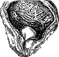
HYPERTROPHY (from Greek hyper- excessively and trophe- nutrition), excessive volume of any tissue or organ. In the narrow sense, H. should be understood (Virchow) as such an increase in tissues or organs which manifests as an increase in the volume of the cells forming them, rather than in the number of these cells (hyperplasia, see). In essence, almost all increases in tissues and organs exceeding their normal size in the organism proceed by both paths: by increasing the volume of the cells themselves and by increasing their number. The condition for distinguishing the concept of H. from other processes of 'progressive growth' is greater or complete functional identity of the excessive-sized tissue with the pre-existing normal maternal tissue. In contrast, in tissue and organ regeneration, the identity of the products of progressive growth with the original tissue, according to the latest data (Fritsch, Kurz, Weiss), manifests itself in the most limited dimensions. Likewise, in tumor processes, the products of excessive growth in the vast majority of cases lose functional identity with the original tissue. The term H. itself, i.e., 'enhanced nutrition', cannot be considered successful, since the cause of enhanced growth is not an excess of nutrition. The immediate causes of H. should be considered: 1) external factors, lying both outside the organism as a whole and inside the organism, when one of its systems is an external factor in relation to another system; 2) internal factors lying in the developmental laws of the organism itself. To the first type of H. belongs the excessive formation of tissues as a result of strong and prolonged inflammatory processes, for example, the formation of bony callus, hypertrophic granulation tissue (sago luxuriens). This is so-called inflammatory H. This also includes: chronic hypertrophic catarrhs of mucous membranes, which often acquire a polypous form; H. of the epidermis, hyperkeratoses, combining with papillary formations such as acute condylomas; inflammatory forms of elephantiasis; exostoses and hyperostoses of bones, etc. To the same type of H., on the principle of causality, should be attributed those excessive increases in tissues which arise in the organism under other external circumstances, such as: increase in muscles with excessive load from work in persons engaged in heavy physical labor, in athletes (work H.). Morgago obtained work H. of voluntary muscles experimentally, in dogs, by first keeping them for a month in a confined space, and then for 80 days making them run a large amount of work covering 3,218 km. The author found that the muscle bundles had increased by 7½ times; the number of individual fibers remained the same, while the diameter of each fiber increased 8 times. These measurements by Morgago showed that work H. of muscles is caused not by hyperplasia, but exclusively by an increase in the size of existing cellular elements. Directly related to this last type of H. are H.'s which are the result of increased functional load on an organ depending on another system of the organism or the influence of one part of an organ on another. Such H.'s include H. of the cardiac muscle, namely: of the right ventricle with increased resistance to blood flow in the branches of the pulmonary artery (in emphysema, chronic t.b.c.); H. of the left ventricle following narrowing of the aortic orifice, with increased resistance to blood flow in the arteries of the systemic circulation (see figure 1 and 2*) (hypertension, kidney diseases, general arteriosclerosis). Mention should also be made of the hypertrophy of the urinary bladder following narrowing of the urethra (see figure 3); H. of the smooth muscle of the stomach with narrowing of the pyloric and in parts of the intestine with scar strictures of its distal sections. The dimensions of hypertrophied muscles can be very large and exceed normal dimensions 5-6 times or more; such is, for example, the 'ox heart' or the urinary bladder in cystitis, urethral strictures, prostate enlargement, when the thickness of the bladder wall can reach 12 cm. All these types of H. are essentially mechanical hypertrophies.

Figure 1. Wall of a normal left ventricle.
As for H. of glandular organs, their increase is caused by the influence of chemical agents. Functional H. of these organs appears in the form of so-called vicarious, compensatory H. (see Vicarious processes). Such are H.'s of the liver, kidneys, testicles, mammary glands, thyroid gland, parathyroid glands, pancreas with Langerhans islands, etc. In those cases,
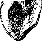
Figure.
Wall of a hypertrophied left ventricle. when part of the liver or one of the kidneys perishes or for some reason (traumatic defects, chronic inflammatory processes) is long-affected in its function, the remaining undamaged, capable of functioning part increases, taking upon itself, according to the needs of the whole organism, an increased
* Figure 1 and 2 are given at the same reduction.
Figure 3. Hypertrophy of the prostate gland, causing difficulty in urination and subsequent hypertrophy of the bladder. Trabeculae of the bladder are well expressed. In hypertrophy of the liver, not only do the cells increase in size, but also their number (mitoses are observed), so that the liver cords become thicker, more strongly curved, and more numerous. Sometimes the typical structure of the lobules is disrupted, and adenoma-like nodules form. In hypertrophy of the kidneys, the glomeruli increase, the tubules become wider and longer, and therefore curve more strongly, and then the size of the epithelium increases. New tubules and glomeruli, some believe (Boret), do not form. Compensatory hypertrophy is also observed in congenital defects of organs. The nature of vicarious hypertrophy is also possessed by increases in some endocrine glands following the loss of function of others when there is a synergistic correlation between them. Such is, for example, hypertrophy of the pituitary gland after thyroidectomy or hypertrophy of the adrenal cortex following castration. In individual links of the hematopoietic system (spleen, lymph glands, bone marrow), hypertrophic states of vicarious significance may also arise. Thus, vicarious substitution is observed not only in homogeneous but also in different organs. In other correlational hypertrophies, the matter is not about the replacement of the function of another organ, but rather about antagonistic action (antagonistic hypertrophy). To the hypertrophic processes also belong the increases in tissues and organs that, it is believed, appear when there is a decrease in the normal pressure on them from neighboring parts - so-called vacant hypertrophy (hypertrophia ex vacuo); examples of the latter can be the thickenings on the inner surface of the bones of the skull in atrophy of the brain and the overgrowth of the synovial membrane of joints after the absorption of long-existing effusions in them. Others (Boret) believe that here the matter concerns a change in metabolism of a local nature. The types of hypertrophy listed above are in one way or another, directly or indirectly, connected with the influence of external factors in the indicated sense. But there are hypertrophies of a congenital nature, belonging to the phenomena of heredity and manifesting as changes in the internal regularities of development. These hypertrophies appear already at birth or in the earliest age. Such quantitative changes in the development of tissues and organs are found in the so-called congenital giant growth and can spread either to the entire organism (general gigantism) or to individual parts. Partial giant growth affects either all limbs, or arms or legs, or individual organs, such as the brain, mammary gland, etc. In these cases, the proportions of the tissues making up the organs are preserved: in the limbs, for example, the increase concerns both bones and muscles, as well as the skin. To the congenital hypertrophic states that disrupt the form belong symmetrical and asymmetrical increases in tissues and organs, bearing the name of elephantiasis (elephantiasis congenita). This includes primarily the lesion of the skin and subcutaneous layer, connective tissue (elephant. fibrosa), fatty tissue (elephant. lipomatosa), blood vessels (elephant. angiomatosa), lymphatic spaces (elephant. lymphangiectatica), nerves (elephant. neurinomatosa). Such congenital elephantiasises are found on the tongue (macroglossia), on the cheeks (macrocheilia), on the lips (macrocheilia). On the skin, diffuse forms also occur. It should be borne in mind, however, that not all elephantiasises have a congenital origin (see Elephantiasis). To congenital hypertrophies belong excessive development of hair (hypertrichosis) and excessive development of the horny substance of the skin (hyperkeratosis). Diffuse congenital hyperkeratosis, resembling the scaly skin of fish, bears the name ichthyosis (ichthyosis congenita) and is hereditary. The pathological hypertrophies of the mammary glands (see Mammary gland), which can manifest during pregnancy and lactation, are probably based on a congenital predisposition. It should be noted that not every increase in organs and tissues is a true hypertrophy of them; false hypertrophy (pseudohypertrophy) is distinguished, when the function of the organ is not strengthened and not even preserved at its previous level, but, on the contrary, decreases; an example can be the apparent increase in muscles due to the enhanced development of intermuscular fatty tissue with atrophy of the muscular tissue itself (pseudo-hypertrophia musculorum lipomatosa).
Related articles
Mentioned in
Cite this page
“Hypertrophy.” Soviet Medical Encyclopedia. English translation of Bolshaya Meditsinskaya Entsiklopediya, 1st ed. (Moscow, 1928–1936), ed. N. A. Semashko. https://sovietmedicalencyclopedia.pages.dev/article/hypertrophy/