Pulmonary Artery
Historical document, translated for reference. It reflects medical knowledge of the 1920s–30s and is not medical advice.
Summary
This article from the first edition of the Great Medical Encyclopedia (1928–1936) details the anatomy, embryology, and histology of the pulmonary artery and veins. It also covers clinical aspects, including congenital defects, traumatic injuries, and the incidence of pulmonary embolism.
Encyclopedia article (1928–1936)
PULMONALES ARTERIA, VENAE, pulmonary artery and veins. The pulmonary artery delivers venous blood from the right ventricle to both lungs, from where oxygenated blood flows through the pulmonary veins into the left atrium. The pulmonary artery extends from the right ventricle for a distance of 50 mm alongside the aorta, covered by a common layer of the pericardium, which forms the sinus pericardii (see Pericardium). Upon exiting the sac, the pulmonary artery divides into two branches—right and left—for the respective lungs. The right branch is, as it were, a continuation of the common trunk and has a length of 50 mm; the left is 35 mm. The diameter of the right is 21 mm, and the left is 19 mm. The right pulmonary artery is directed to the right between the right and left auricles, forming a convexity posteriorly, and, curving behind the ascending aorta and the superior vena cava, it then passes to the right side, crossing the entry of the v. azygos into the superior vena cava, and enters the hilus pulmonis (Figure 1).
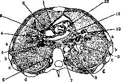
Figure 1. Course of the a. pulmonalis and its right branch. Cross-section at the level of the attachment of the II rib to the sternum and through the upper half of the VI thoracic vertebra: 1-right auricle; 2- n. phrenicus dext.; 3-v. cava sup.; 4- a. pulmonalis dext.; 5- right bronchus; 6-n. sympathicus; 7-esophagus; 8-thoracic aorta; 9-left bronchus; 10-left atrium; 11-n. phrenicus sin.; 12-a. pulmonalis communis; a, b, c and d-III-VI ribs. (After Braune.)
In the latter, the bronchus is located highest of all; anteriorly and inferiorly lie the branches of the pulmonary artery and, separately, the pulmonary veins. Before entering the right lung, the pulmonary artery divides into 3 branches corresponding to the three lobes of the lung. In the lung, the pulmonary artery branches according to the magistral or scattered type (see Lungs). The left branch of the pulmonary artery departs from it at a right angle, accompanied by the bronchus and pulmonary veins, and, forming a single bundle, enters the hilus (Figure 2). The entire bundle
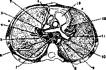
Figure 2. Origin and course of the left branch of the a. pulmonalis. Cross-section in the II intercostal space and through the upper half of the V thoracic vertebra: 1-right atrium; 2-v. cava sup.; 3-a. pulmonalis dext.; 4-right bronchus; 5-n. vagus dext.; 6-v. azygos; 7-esophagus; 8-thoracic aorta; 9-n. vagus sin.; 10-left bronchus; 11-a. pulmonalis sin.; 12-n. phrenicus sin.; 13-right ventricle; a, b and c-III-V ribs. (After Potter.)
is located between the left atrium on one side and the arch and descending part of the aorta on the other. Above lies the artery, in the middle the main bronchus and the branches of the superior pulmonary vein, and below the undivided inferior pulmonary vein. From the left pulmonary artery, 3 small branches depart to the upper lobe of the lung and one to the lower. The pulmonary veins, in the amount of 4 (2 from each lung), carry arterial blood to the left atrium, emptying either all together or in pairs, one pair above the other. The pulmonary veins have a length of 15 mm and a diameter of 13-16 mm, larger on the right than on the left.
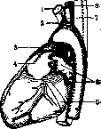
Figure 3. Relation of the a. pulmonalis to the heart and vessels. View of the heart from the left: 1- v. anonyma; 2- a. anonyma; 3- a. pulmonalis communis; 4-left auricle; 5-sinus coronarius cordis; 6-vv. pulmonales sin.; 7- a. subclavia sin.; 8- a. carotis communis sin. (After Sauerbruch.) The superior pulmonary veins run horizontally, the inferior ones obliquely, rising from the hilus upwards. Embryology. The pulmonary artery is formed from the 6th pair of embryonic arches, like the aorta, and in the embryonic period has a connection with the aortic arch in the form of the ductus arteriosus (see), which soon after birth becomes obliterated and persists as an elastic connective tissue cord, passing from the a. pulmonalis to the anterior wall of the end of the aortic arch, forming the arterial ligament (lig. arteriosum). Histology. The pulmonary artery belongs to the large arteries and is constructed according to the elastic type from three layers—intima, media, and adventitia (see Aorta). Topography. The pulmonary artery, upon exiting the heart, lies to the left of the aorta and is covered together with it by the pericardium (Figures 3 and 4). On the left, the artery borders the left auricle. Anteriorly, the pulmonary artery is covered by the left lung and pleura. When projected onto the chest wall, the pulmonary artery corresponds to the II intercostal space along the sternal line (Figure 5). Near the left semicircle of the pulmonary artery passes the n. phrenicus sin. The left branch of the pulmonary artery is located behind and below the bronchus and runs horizontally to the hilus, covered anteriorly by the left auricle and lung, crossing the n. vagus and v. hemiazygos located behind it. The right branch of the pulmonary artery at the level of the IV-VI thoracic vertebrae, being located anteriorly, crosses the ascending aorta and the superior vena cava, and posteriorly—the esophagus, having...
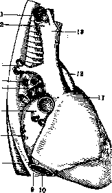
Figure 4. Pulmonary vessels and bronchi of the right lung. View from the right: 1-a. anonyma; 2-v. anonyma dext.; 3-v. azygos; 4-bronchus; 5-a. pulmonalis dext.; 6-superior pulmonary veins; 7-inferior pulmonary veins; 8-aorta descendens; 9-esophagus; 10-v. cava inf.; 11-a. pulmonalis communis; 12-aorta ascendens; 13-v. anonyma sin. (After Sauerbruch.)
Congenital diseases of the pulmonary artery are rare and concern the non-closure of the ductus arteriosus (see Heart defects). Traumatic injuries are also not frequent and in the majority are accompanied by damage to other organs, which extremely complicates diagnosis. Gunshot wounds of the pulmonary artery are accompanied by profuse bleeding, and patients usually die quickly. The presence of a gunshot or stab wound opening corresponding to the location of the vessels with profuse venous bleeding can indicate a wound of the pulmonary artery. If a wound of the pulmonary vessels is suspected, an immediate operation is necessary, consisting of suturing the pulmonary artery and ligating the small branches of the pulmonary arteries and veins. In recent years, embolism of the pulmonary artery has been frequently noted in the literature. Up to 1902, Lotheissen collected 55 cases of death from pulmonary embolism. Adolph-Hoppmann believes that death in therapeutic diseases occurs in 3.4% of cases from pulmonary embolism, and Martin and Opitz give a figure of 5.7%. According to Bodon's statistics, out of 12,861 autopsies (in persons older than 15 years), death from pulmonary embolism occurred in 115 cases (i.e., 0.89%), of which 58 cases were after therapeutic diseases, 54 cases after operations, and 3 after childbirth. The percentage of postoperative embolisms of the pulmonary artery varies from 0.035% (Kirschner) to 1% (Zweifel). Embolism is observed more often
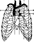
Figure 5. Projection of the heart and pulmonary vessels onto the anterior chest wall: 1- v. cava sup.; 2- a. pulmonalis.
exit from the heart (4 and 5); the unshaded triangle corresponds to the left auricle; 3 - ascending aorta; 6 and 7 - right ventricle; 4 and 7 - border of the atrioventricular orifices; 5 and 8 - site of the exit of vessels from the heart. (According to Sauerbruch.) occurs at the age of 40 to 70 years. Some authors note pulmonary artery embolism more often in women, others find no difference between the sexes. Among postoperative embolisms, the highest percentage falls to abdominal operations compared to operations in other areas. According to Capelle, this is expressed by a ratio of 21:11. Some authors note an increase in pulmonary artery embolism in the post-war period in connection with an increase in cardiovascular diseases. -- Etiology of pulmonary artery embolism (see Embolism, Thrombus). Factors contributing to the occurrence of pulmonary artery embolism can be divided into the following groups: 1) changes in blood circulation in the sense of slowing down the blood flow; 2) changes in the vessel wall; 3) changes in the composition of the blood. These factors are observed in heart defects, myodegeneration due to various diseases, endocarditis, diseases and injuries of the vascular wall, changes in blood composition due to chronic intoxications, leading in turn to cardiovascular weakness, as well as in various infections causing diseases of the cardiovascular system. Thus, Kuhn established cardiovascular disease in 78% of pulmonary artery embolism cases, Martin-Opitz in 65%. For the occurrence of postoperative pulmonary artery embolism, great importance is attached (Lister, Payer) to the weakness of the abdominal musculature and diaphragm, especially during abdominal operations, as a result of which a deterioration of blood circulation occurs with stasis in the veins of the lower extremities and the small pelvis, leading to thrombosis of these veins with subsequent embolism of the pulmonary artery. No small role is played by general anesthesia, which weakens cardiac activity, and subsequent prolonged lying in bed, which promotes stasis, as well as damage to the vessel walls during the operation and infection. Bodon notes, out of 54 cases of pulmonary artery embolism, thrombosis of the saphenous vein in 31 cases, hypogastric vein in 6 cases, prostatic, vesical, and iliac veins in 4 cases, without thrombosis in 13 cases; local infection in 5 cases, general infection in 6 cases. In the origin of pulmonary artery embolism, constitutional factors are also noted. Thus, according to some authors, embolism is observed more often in obese subjects. Blockage of the pulmonary artery, caused by a thrombus that arose in the pulmonary artery itself, or by an embolus brought from another place, can occur in different parts of the pulmonary artery: in the main trunk, in the right or left pulmonary artery, in the small branches. Depending on the location of the embolus and its size in terms of obstructing blood circulation, the clinical picture will be different. According to Bodon's statistics, out of 58 therapeutic cases, the main trunk was affected in 12 cases, both pulmonary arteries in 32, small vessels in 10, and small emboli in the lungs were observed in 4. Out of 54 postoperative cases: in 24 cases - the main trunk, in 25 - both pulmonary arteries, and in 5, small emboli were noted in the lungs. If the embolus is located parietally or in a small branch, then insufficient blood flow to the lung occurs, which leads, on the one hand, to venous blood stasis in the right ventricle and venae cavae, and on the other, to insufficient supply of oxygenated blood to the periphery. Depending on this, symptoms develop: pallor, and then cyanosis at the periphery, with cerebral phenomena due to a lack of O2. These same phenomena develop with complete blockage of the pulmonary artery or both of its branches, but only significantly faster and proceed more menacingly. With a parietal location of the embolus, there can be two outcomes: 1) the embolus will not increase and will resolve, and during this time the organism will adapt thanks to compensatory dilation of the pulmonary artery and heart, and the patient will recover, or 2) the embolus will increase and gradually cause complete blockage with subsequent death of the patient. In the second case, complete blockage of the pulmonary artery will suddenly cause an acute lack of arterial blood at the periphery with sharp stasis in the venous system, which sometimes leads to death within a few minutes. At times, during this period, there is improvement and then deterioration depending on the passage of blood or the displacement of the embolus. Clinical picture. Upon the onset of embolism, there is a sudden appearance of sharp facial pallor with cyanosis at the periphery due to insufficient supply of arterial blood because of the obstruction to the passage of blood in the pulmonary artery. The patient develops a feeling of pre-death anguish, a restless state, which is replaced by complete apathy and even an unconscious state, which is the result of insufficient blood supply to the brain. The accumulation of carbon dioxide and decay products due to blood stasis leads to a disorder of the respiratory center and the regulatory centers of cardiac activity. The patient develops sharp rapid breathing, and all the neck muscles take part in the respiratory movements, cardiac activity is disturbed, the pulse quickens, at times it appears and then disappears. The activity of the heart gradually weakens also because the heart muscle suffers due to the strain necessary to push blood through the obstruction, as well as from insufficient nutrition and poisoning by decay products. Gradually, dilation occurs. Figure 6. Instruments necessary for the operation of removing a pulmonary artery embolus: a - rubber tourniquet with a screw-on end for the curved forceps b; c - forceps for the embolus; d - vascular clamp. Figure 6. Instruments necessary for the operation of removing a pulmonary artery embolus. The initially appearing clapping 2nd tone on the pulmonary artery subsequently disappears, which indicates insufficiency. If the patency of the pulmonary artery is not restored, at least temporarily, then death from respiratory paralysis quickly ensues. When patency is restored, the phenomena of pallor are quickly replaced by a rush of blood, only to disappear again when blockage occurs. Such a state can be repeated several times.
The diagnosis of pulmonary artery embolism, despite the vivid clinical picture, is not always easy to make. Kitzman, out of 35 cases of pulmonary embolism established at autopsy, had a correct clinical diagnosis in only 6 cases. In Korte's 22 cases, 12 resulted in instantaneous death, 10 - gradually from 10 minutes to 3 hours, and in all cases a diagnosis of pulmonary embolism was made, while the autopsy established it in only 6 cases. A clinical picture similar to embolism can be caused by myodegeneration of the heart, sclerosis of the coronary vessels, and heart defects, acute insufficiency of cardiac activity. Characteristic distinguishing signs of embolism are initial pallor with gradually occurring cyanosis, a clapping 2nd tone of the pulmonary artery, and dilation of the heart to the right. Treatment of pulmonary embolism, according to Kirschner, should consist of prophylaxis. Since the disease is more common in cardiovascular patients, it is necessary to carry out appropriate treatment for them in advance. To prevent pulmonary artery embolism in the postoperative period in the corresponding categories of patients, preparatory treatment of the heart should be carried out even before the operation. The operation itself should
Figure 7. Incision of soft tissues, resection of the costal cartilages. Figure 8. Displacement of the pleura. Dissection of the pericardium.
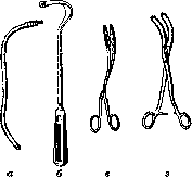
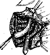
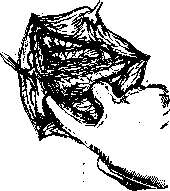
should, if possible, be performed under local anesthesia. To eliminate the causes contributing to venous stasis, an elevated position of the lower extremities, early movement, gymnastics in bed, massage of the extremities, deep breathing, and early rising are recommended in the postoperative period, but the latter should not be abused, as after major operations and especially with the ligation of many vessels, early rising may lead to the occurrence of an embolism from the unstrengthened thrombi of the ligated vessels. In case of cardiac weakness, it is necessary to use cardiac agents. Saline enemas of 1-2 liters per day have an extremely beneficial effect on weakened patients. In the event of thrombosis in the veins of the lower extremities, especially if it is progressing, ligation or resection of the affected section of the vein is recommended. In the event of a pulmonary artery embolism, it is necessary to energetically use agents that stimulate cardiac activity, large doses of morphine for sedation, and oxygen inhalation. The latter must be given until complete relief with the disappearance of the symptoms of embolism; upon the onset of life-threatening symptoms, one must immediately proceed to surgery. To avoid errors in diagnosis and to provide timely assistance, it is necessary for such patients to be under the observation of a surgeon with prepared instruments so that within 10-15 minutes, upon the onset of threatening symptoms, one can proceed to the operation. Surgical treatment, proposed by Trendelenburg in 1908, consists of removing the embolus from the pulmonary artery. The operation is usually performed without anesthesia, as the patients are in a semi-unconscious state. Special instruments have been proposed by Trendelenburg for the operation (Fig. 6). The incision is made along the left edge of the sternum from the I to the V rib, supplemented by a second incision along the III rib from the sternum. The skin with muscles is separated and the flaps are turned back. The III rib is resected for a length of 10 cm (Fig. 7). If necessary, the II rib can also be resected (Meyer). The internal mammary artery and vein are ligated, which at the beginning of the operation do not bleed even without ligation, but later may cause bleeding (one patient
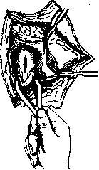
Figure 9. Insertion of a rubber tourniquet into the pericardial sinus behind the pulmonary artery and incision of the artery wall.
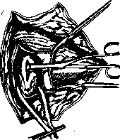
Figure 10. Extraction of an embolus from the branches of the pulmonary artery with the tourniquet tightened.
of Trendelenburg died from bleeding from the internal mammary artery). After ligation of the vessels, the pleura is opened or pushed aside, the pericardial sac is found and also incised (Fig. 8). A curved Trendelenburg probe is carefully inserted into the pericardial sinus and brought out on the right side of the aorta. After threading the drain, the probe with the latter is brought back out. The drain, encompassing the aorta and the pulmonary artery, is handed to an assistant (Fig. 9). When the rubber tube is tightened, the bases of the aorta and the pulmonary artery are compressed. The pulmonary artery is incised longitudinally above the exit from the heart, a sponge forceps is quickly inserted into it, and the embolus is extracted first from the right and then from the left pulmonary artery (Figure 10). If the embolus cannot be extracted entirely, it is recommended to remove it in parts, entering with the sponge forceps several times. According to Trendelenburg, clamping of the aorta and pulmonary artery can be performed for 45 seconds; with a longer duration, cardiac arrest may occur. Therefore, after the expiration of this period, if the embolus has not yet been completely removed, it is recommended to clamp the opening in the pulmonary artery with two fingers, loosen the drain, and allow a stream of blood to pass (Fig. 11). By this, on the one hand, the blood-engorged heart is relieved, on the other hand, the thrombus is expelled from the right ventricle if it descends from the pulmonary artery into the right ventricle, and, thirdly, the blood supply to the lungs is restored. Upon restoration of cardiac activity, the clamping of the vessels can be repeated and the embolus removed from both branches. After removal of the embolus, a delicate vascular clamp is applied to the wall of the pulmonary artery (Fig. 12), without completely disrupting patency, and the edges of the artery are sutured with interrupted silk sutures (Fig. 13), previously paraffined. The pericardium and pleura are sutured. A drain is inserted under the skin and the skin is sutured. The operation was first successfully performed in 1924 by Kirschner, who resected the II and III ribs for wide access and operated with an apparatus for increased pressure. Up to 1932, 7 cases with a good re
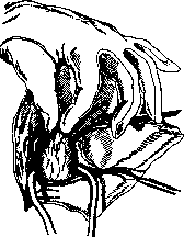
Figure 11. Clamping the opening of the pulmonary artery with fingers while the tourniquet is loosened. sult. Meyer, who improved both the operation and the instrumentation, has the largest material. Meyer proposed resecting the II and III ribs and approaching the pericardium without opening the pleura, which significantly facilitates the operation. Meyer's instrumentation
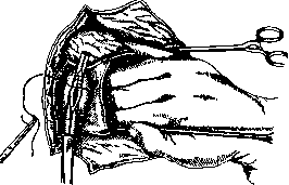
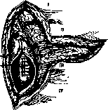
Figure 13. Suture of the pericardium.
Figure 12. Parietal application of a clamp and suturing of the opening. [It] consists of 15 items and is kept in a sterile state in a special metal box. The operation presents great difficulties if only because it requires perfect technique—one must approach the pulmonary artery in a few minutes and remove the embolus in a few seconds. A threatening moment for the patient's life is the compression of the aorta and pulmonary artery, which forces the already dilated heart to become even more engorged with blood, which threatens cardiac arrest. In case of severe engorgement, it is recommended to perform punctures with aspiration of blood. The second negative factor is the exclusion of the left heart from circulation, which leads to even greater damage to the brain due to its depletion of blood. Therefore, to eliminate these factors, Bond proposes, following Rehn, to clamp only the venae cavae, bypassing the heart with a drain from behind or with the fingers of the hand from below and behind, simultaneously compressing the atrium. The advantage of this method is that: 1) compression can be performed for several minutes without damage to the heart, 2) blood pressure does not drop as sharply as when clamping the aorta and pulmonary artery, and 3) the brain is less damaged. The Trendelenburg operation is complex and not every surgeon can perform it. The resolution of the issue of treating pulmonary artery embolism lies not in the operation, but in the discovery of agents that prevent or delay blood clotting.
Related articles
Mentioned in
Cite this page
“Pulmonary Artery.” Soviet Medical Encyclopedia. English translation of Bolshaya Meditsinskaya Entsiklopediya, 1st ed. (Moscow, 1928–1936), ed. N. A. Semashko. https://sovietmedicalencyclopedia.pages.dev/article/pulmonary-artery/