Pulmonary Tuberculosis
Historical document, translated for reference. It reflects medical knowledge of the 1920s–30s and is not medical advice.
Summary
This article from the 1928–1936 Soviet medical encyclopedia provides a detailed overview of the pathological anatomy of pulmonary tuberculosis. It covers the development of primary and secondary tuberculous complexes, the role of the Ghon focus, and the mechanisms of bronchogenic dissemination.
Encyclopedia article (1928–1936)
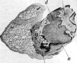
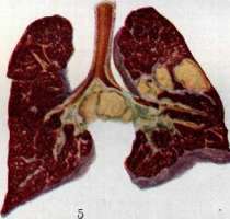
PULMONARY TUBERCULOSIS. Contents: I. Pathological anatomy...........110 II. Classification of pulmonary tuberculosis .... 124 III. Clinical picture.....................128 IV. Diagnostics ..................160 V. Prognosis..................... 190 VI. Treatment.....................196 VII. Pulmonary tuberculosis in children..........215 I. Pathological anatomy. The lung is the organ in humans most frequently affected by tuberculosis. This depends not only on the fact that the lungs are in the vast majority of cases the entry portal for tuberculous infection, but also on the fact that the lung tissue is particularly predisposed to the development of a tuberculous process within it; it has long been established (Baumgarten and many others) that, by whatever paths the tuberculous virus penetrates the organism and wherever the initial focus of changes may be, very often, during the subsequent hematogenous spread of the infection, the tuberculous process develops predominantly in the lungs. As is known, the pathologico-anatomical changes in tuberculosis generally appear different depending on whether it is a matter of the first contact of the organism with the tubercle bacillus (primary tuberculosis) or if there is a tuberculous process in an individual who has already undergone primary tuberculosis and is already relatively immune to tuberculosis; in these cases, one speaks of secondary tuberculosis, or reinfection. Therefore, it is natural to consider first the pathological anatomy of primary pulmonary tuberculosis, and then the pathological anatomy of secondary pulmonary tuberculosis in adults. Primary pulmonary tuberculosis, observed as a rule in childhood, is expressed in the development of the so-called primary complex [see separate table (p. 119-120), fig. 5]; the latter consists of a focus of primary affection, or the Ghon focus, located under the pleura of one or the other lung, and a tuberculous lesion of the regional lymph node of the lung hilum corresponding to this focus. The localization of the primary affection focus in the lungs varies. All authors (Ghon, Lange, Derman, and others) agree that it is more often found in the right lung (55%). Of the pulmonary lobes, the upper lobe of the right lung is in first place (26%), in second place is the upper lobe of the left lung (25%), followed by the lower lobes of the right (21%) and left (20%) lungs; in last place is the middle lobe of the right lung (6.7%). In all lobes, the focus of primary affection is significantly more often situated in the lower third of the lobe; in the region of the lung apices, the primary affection develops extremely rarely. Usually, there is only one focus of primary affection, sometimes (7-10%) there are two foci, rarely 3-4. By the nature of the changes, a fresh primary tuberculous affection is a focus of pneumonia with serous-fibrinous exudate containing an admixture of leukocytes and alveolar epithelial cells (tuberculous 'alveolitis'); the development of tubercles in such a fresh period is not observed. Around the area of such pneumonia, there is often a zone of serous perifocal inflammation, which subsequently disappears. Such a pneumonic focus occupies part of a lobule, or captures an entire lobule, or 2-3 lobules, more rarely more. Very soon, both the exudate and the alveolar septa in the area of the focus undergo caseation, and the focus turns into a cheesy focus, in which, only when stained for elastic tissue, the preserved alveolar structure is visible. Simultaneously with the caseation of the focus, non-specific granulation tissue rich in vessels develops around it, passing at the periphery into specific epithelioid granulation tissue, among which typical epithelioid tubercles with giant cells are interspersed; tubercles are also visible in the adjacent lung tissue and under the pleura. On the pleura in the fresh period, a small fibrinous coating may be observed. The zone of non-specific granulation tissue is gradually replaced by specific tissue, which subsequently undergoes fibrous transformation and scarring; the primary affection becomes encapsulated. A fresh Ghon focus has an irregular angular, sometimes triangular shape, whereas an encapsulated affection acquires a round shape, disturbed only by the fact that at the point of contact with the vascular-bronchial bundle, with which the affection is usually connected, a connective tissue strand extends from it, connecting it with the aforementioned bundle. Following encapsulation comes petrification of the cheesy mass and very often the development of bone tissue on the inner surface of the capsule. In a healed primary focus, the capsule is usually sharply demarcated from the adjacent slightly altered lung tissue; this circumstance, together with the development of bone tissue in the capsule, distinguishes the Ghon focus of a healed primary affection from the Aschoff-Puhl focus of a healed reinfection (see below). The lesion of the regional lymph node, which is part of the primary tuberculous complex, is expressed in the development of tubercles in it with significant perifocal inflammation of the entire tissue of the node, and the ensuing caseous degeneration captures the entire node completely. Often, small lymph nodules in the vascular-bronchial bundle leading from the lung hilum to the primary focus are also affected, as well as lymph nodules under the pleura; sometimes tubercles arise along the course of the lymph vessels in the direction from the focus to the regional node. In all these lymphogenous foci, the tuberculous process undergoes the same evolution as in the primary focus. Fresh caseous primary complexes with the beginning of encapsulation are encountered in childhood; in adults, however, we find only old petrified, i.e., healed, foci of primary complexes. Bacteriological examination (inoculation of guinea pigs) in 30% of such 'healed' foci reveals virulent tubercle bacilli. The aforementioned outcome of the primary complex into encapsulation, petrification, and ossification is the most frequent. However, in a number of cases, the process does not show a tendency toward stopping and delimitation, but progresses and spreads; this spread proceeds partly locally in the lung, and partly takes on a general character, expressed in the picture of general generalization. This course of the process corresponds to the second stage of Ranke with allergy II, and according to modern views, is an expression of a hyperergic reaction, which has as its basis a specific or non-specific increase in the sensitivity of the organism to the tuberculous virus. Sometimes a newly formed primary affection does not stop in its development, but steadily grows; around it, perifocal inflammation persists the whole time, which, as the focus grows, turns into tuberculous pneumonia and undergoes caseation. In the end, in the area of the primary focus, there is a more or less large bronchus, the wall of which undergoes destruction; the cheesy masses containing tubercle bacilli, having entered the lumen of the bronchus, move during expiration and inspiration and infect new territories of the given lung, and can also penetrate into the other lung. Bronchogenic (aspiration) dissemination arises, giving exudative foci of tuberculous pneumonia of various volumes: small acinous foci [see separate table (p. 119-120), fig. 2], corresponding to the acinus (see Acinus) and having the appearance of yellowish-gray nodules with scalloped edges, larger lobular foci, corresponding to a lobule, also yellowish-gray, of irregular shape with an uneven, scalloped edge, sometimes with disintegration in the center, and finally lobar pneumonias, capturing an entire lobe of the lung. Such lobar pneumonias usually capture the lower lobes in children; appearing at first in the form of serous-fibrinous inflammation with noticeable participation of the alveolar epithelium, they soon completely undergo caseous degeneration; the cut surface appears homogeneous, yellowish-gray, matte; if lobar tuberculous pneumonia exists for some time, a number of foci of softening and disintegration appear in it. On the pleura in lobar pneumonia, there is often a light fibrinous coating. The spread of the process from the lymph node belonging to the primary complex proceeds partly by contact to the adjacent tissue, partly along the lymph vessels to adjacent and distant nodes. Very often, in the capsule of the node and in the surrounding tissue, perifocal inflammation develops, easily passing to the wall of the adjacent bronchus and causing phenomena of inflammation of its mucosa (so-called bronchoadenitis); after the subsidence of the acute perifocal inflammation, sclerosis of the tissue surrounding the bronchus and the wall of the bronchus may remain, for a long time disturbing the state of its mucosa. A more severe picture arises during the transition of the tuberculous process from the node to the bronchus; in these cases, destruction of the bronchial wall occurs and the introduction of cheesy masses into its lumen, which has the same consequences as the destruction of the wall of an intrapulmonary bronchus (see above). Abrikosov (1904) established that besides gross destruction of the bronchial wall, sometimes so-called microperforation occurs, invisible to the naked eye; only a microscope reveals the presence of the transition of the tuberculous process from the node to the wall of the bronchus up to the mucous membrane and the possibility of the transition of tubercle bacilli into the lumen of the bronchus with very slight damage to the mucous membrane. From the lymph node of the lung hilum, the tuberculous process can directly spread to the lung tissue. It is important to note that all these changes can arise independently of the behavior of the primary affection in the lung.
It happens that the primary focus, being very small, undergoes encapsulation and becomes quite inconspicuous, whereas the lymph nodes of the lung hilum turn out to be sharply altered, large, caseous, with an active tuberculous process that causes the spread mentioned above. Furthermore, from the lymph nodes of the primary complex, the process spreads lymphogenously to neighboring mediastinal nodes, the hilum of the other lung, cervical, retroperitoneal nodes, etc., which already pertains to lymphogenous generalization. Primary tuberculosis can also be accompanied by
b g
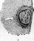
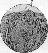
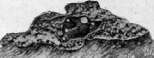
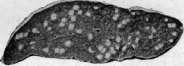
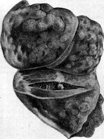
Fig. 1. Miliary tuberculosis of the lung; significant edema of the lung tissue. Fig. 2. Healed primary affect (under a magnifying glass): a—bony trabeculae with areas of bone marrow (c) between them; d—cheesy masses; b—fibrous capsule delimiting the affect. Fig. 3. Healed primary affect (under a magnifying glass). Fig. 4. Tuberculous tubercles (a) in the mucous membrane of the larynx. Fig. 5. Koch's bacilli on the wall of a cavity and in the lung. Fig. 6. Solitary tuberculosis of the spleen. Fig. 7. Healed primary affect of the lower lobe of the right lung in an elderly emphysematous patient (vesicular inflation of the anterior and upper parts of the lung is visible); the affect has the appearance of an ossified formation and lies immediately beneath the thickened pleura.
hematogenous generalization as a result of the penetration of tuberculous bacilli into the bloodstream directly or via the lymph (see Tuberculosis). In the lungs, hematogenous involvement manifests in various ways. In general miliary tuberculosis, an eruption of a mass of miliary tubercles is observed in both lungs [see separate table (pp. 119-120), fig. 3]; in these cases, the lung tissue appears hyperemic, and against this background, tubercles are visible, which, depending on their age and type, are either small, gray, translucent, or yellow, with irregular contours, and often larger. Often, according to the indication of Ribbert (1902), it is possible to notice that in adults, the tubercles in the upper regions of the lungs are somewhat larger than in the lower ones, which is explained by the more energetic growth of tubercles specifically in the upper sections of the lungs. If the penetration of tuberculous bacilli occurred into a branch of the pulmonary artery, then the miliarization involves only the corresponding part of the pulmonary territory. Microscopic examination during miliarization in the lungs can reveal different types of tubercles: sometimes these are typical productive epithelioid tubercles, having alveolar septa as their source of development; the tubercles grow, compressing and destroying the alveoli, and remaining productive tubercles for a long time; in other cases, exudation into the alveoli is quickly added to a tubercle that has begun to develop in the alveolar septum; finally, it happens that a tubercle seems to arise immediately in the form of miliary pneumonia, involving 2-4 adjacent alveoli (miliary alveolitis), and thus reveals an exudative character. Huebschmann and Schlessing put forward the point of view that every tubercle in the lungs begins in the form of miliary pneumonia and only later is surrounded by a productive zone. This point of view provoked categorical objections from major pathologists (Herxheimer, Aschoff, Sternberg, and others); furthermore, Grethmann (1928) established that hematogenous miliary tubercles in the lungs, as a rule, develop as productive tubercles from the walls of small vessels; exudation into the alveoli is a sequential phenomenon, frequent, but not mandatory. Hematogenous metastasis to the lungs can also occur in the form of the appearance in the lungs of one or several tuberculous foci; in its essence, such a form is a particular manifestation of organ tuberculosis arising in the order of late generalization of primary tuberculosis (see Tuberculosis). The fate and expression of such individual hematogenous foci in the lungs vary (see below). Simon's attempt to explain all apical scars encountered in the lungs of adults by hematogenous metastasis occurring in childhood did not meet with success among pathologists; such hematogenous metastasis to the apex of the lung in childhood is rarely encountered, and in pediatric cadavers, we do not encounter anything similar to the beginning of the formation of these foci; apical scars of the lungs are observed almost only in adults, and their frequency increases with age. Primary tuberculosis leads to the death of children either in connection with significant spread of the tuberculous process in the lungs or in connection with generalization, especially in the form of general miliary tuberculosis and tuberculous meningitis. In adults, primary tuberculosis of the lungs is very rare; most often this concerns those nationalities which in childhood do not come into contact with the tuberculous virus (see Tuberculosis). Secondary tuberculosis of the lungs represents what is accepted to be distinguished as pulmonary consumption of adults; in essence, this is an outbreak of the tuberculous process, or a reinfection in the lung in an individual who has already undergone a primary infection and is relatively immune to the tuberculous virus. The main feature of secondary tuberculosis, or reinfection, is that the process proceeds more or less in isolation in the lung, not showing a tendency toward lymphogenous and hematogenous metastases. If the pathological anatomy of pulmonary consumption of adults has long been developed sufficiently well and appears more or less clear, this cannot be said regarding the pathogenesis of the initial changes. In former times, when no distinction was yet made between tuberculosis of childhood and tuberculosis of adults, when nothing was yet known regarding primary and secondary tuberculosis, allergy, etc., two circumstances seemed completely obvious: 1) pulmonary consumption of adults is a consequence of aerogenic infection of the lung by the tubercle bacillus; 2) pulmonary consumption of adults always begins in the region of the apex of the lung. The study of the structure of initial foci of tuberculosis discovered in the apices showed (Birch-Hirschfeld, 1899; Schmorl, 1901) that in them, it is a matter of a tuberculous lesion of the wall of the apical bronchus and the development in such a place of bronchitis with peribronchitis. Abrikosov (1904), during his studies of initial tuberculous foci of the apices, came to a somewhat different conclusion; he found that the lesion usually begins lower, specifically in the region of the 2nd dorsal subapical bronchus of the upper lobe, and that the focus has a bronchopneumonic character. Abrikosov argued that the process begins from the wall of the intralobular bronchiole and quickly assumes a bronchopneumonic character. These studies provided, as it were, a factual foundation for the theory of the aerogenic origin of pulmonary consumption; partly in connection with this, the views of Aufrecht (1900) and Ribbert (1901) regarding the hematogenous origin of pulmonary tuberculosis of adults did not have any success. After the proposition regarding the pathogenetic difference between tuberculosis of childhood and tuberculosis of adults was put forward, and the essence of this difference was clarified in the doctrine of Ranke (see Tuberculosis), it became obvious that one cannot look at the genesis of pulmonary consumption of adults as simply as was done before. Once it turned out that pulmonary consumption of adults is tuberculosis of an organism that has already undergone a tuberculous infection, it naturally began to be said that at the basis of tuberculosis of adults lies a second infection, or reinfection with the tuberculous virus. The mechanism of this reinfection was understood differently. According to the doctrine of Ranke, reinfection represents nothing other than an exacerbation of changes relating to the primary tuberculosis of childhood (endogenous reinfection); thus, tuberculosis of adults is a continuation of tuberculosis proceeding from childhood, but exacerbating in an adult who is in a state of relative immunity to the tuberculous virus. In contrast to this, Aschoff and others began to insist that pulmonary consumption of adults in the vast majority of cases is a consequence of a new aerogenic infection of the lung, i.e., exogenous reinfection or superinfection of an organism that has already undergone a primary infection. In recent years, very many phthisiologists have inclined to the opinion that pulmonary consumption of adults is a consequence of endogenous reinfection via the hematogenous route. The source of such hematogenous metastasis to the lung is either foci of the primary complex in the lung and in the lymph node of the mediastinum, or a tuberculous focus in some organ that has developed in the order of organ tuberculosis. Another controversial point that has emerged in recent years concerns the place of the beginning of pulmonary consumption of adults. Until 1925, the doctrine that pulmonary consumption begins from the very apex of the lung and that scars encountered in the apices with or without calcifications are precisely the expression of such a beginning that did not receive further development (healed foci of reinfection) seemed completely unshakable. In 1925, Assmann, in 1926, Redeker, and in the near future, Ulrici, Romberg, Lydtin, and many others, on the basis of clinical and radiological data, began to insist that the above-mentioned apical foci have no relation to the beginning of pulmonary consumption, that pulmonary consumption begins not in the apex, but in that region of the upper lobe which projects directly below the clavicle; it is precisely here that the process begins in the form of a focus of pneumonia, which was designated as an infraclavicular infiltrate or early tuberculous infiltrate (German: Frühinfiltrat). Pathological anatomists (Aschoff, Graff, Huebschmann, Loeschke, and others) could not agree with such a point of view; they asserted that the infraclavicular infiltrate is not the anatomical beginning of pulmonary consumption, but only a clinical-radiological manifestation of a process that began in the apex and proceeded until then without clear clinical symptoms. At the special tuberculosis congress in Wildbad (1928) and at the congress of pathologists in Vienna (1929), this contradiction had not yet been resolved. However, in the very recent past, we see a significant softening of the indicated dispute, connected with the circumstance that, on the one hand, pathological anatomists (Aschoff, Sternberg, Pagel, Schürmann, and others) have inclined to the admission of the possibility of the beginning of the tuberculous process in adults from the infraclavicular region, and on the other hand, clinicians (Lydtin, Assmann, Starlinger, Rubinstein, and others) have agreed that the infraclavicular infiltrate is by no means necessarily an independent initial focus of tuberculosis; it can also develop sequentially from a pre-existing latent focus. At the same time, it began to be definitely said that the reinfection, which is the beginning of pulmonary consumption of adults, can have both an endogenous and an exogenous origin. Thus, at the present time, different modes of origin of pulmonary consumption of adults and different types of its onset are admitted.
Its origin can be the result of exogenous (aerogenic) and endogenous reinfection; endogenous reinfection develops hematogenously or directly from an old focus in the lung (by contact or bronchogenically). In terms of its onset, the process can originate from apical foci or begin in the infraclavicular region. To this must be added the possibility of the development of pulmonary phthisis in adults from tuberculous lymph nodes of the lung hilum. The latter method of formation of a reinfection in the lung is, however, comparatively rare; therefore, two methods of the onset of pulmonary phthisis have practical significance: from the apex of the lung and from its infraclavicular region. At the present time, the majority of pathologists and clinicians agree with this (Aschoff, Pagel, Schürmann, Assmann, Lidtin, Rubinstein, Ugryumov, Stefko, and others). However, there is still no complete agreement on which method of onset is more frequent; clinicians have a tendency to consider infraclavicular onset more common and point out that apical onset is observed only in 7-10% of cases of tuberculosis (Romberg). On the other hand, pathologists, on the basis of autopsy material, establish that cases with apical onset relate to cases with infraclavicular onset as 4:1 (Pagel, Ugryumov), i.e., that onset from the apex of the lung is 4 times more frequent than onset from the infraclavicular region. The question of how often aerogenic (exogenous) and how often hematogenous (as well as other types of endogenous) development of reinfection occurs is considered completely open. The majority indicate that the infraclavicular focus has a hematogenous origin, whereas apical foci can have an aerogenic and hematogenous origin. In contrast to this, Neumann and Starlinger believe that it is precisely the early infraclavicular infiltrate that is the result of aerogenic (bronchogenic) superinfection, whereas apical foci have, in the main, a hematogenous origin. Leaving aside these still unclear and controversial points of the genesis of pulmonary phthisis, let us touch upon the pathological anatomy: 1) of apical foci and 2) of infraclavicular infiltrates. 1. The concept of 'lung apex' is not the same for all authors; some understand by 'apex' only the very dome of the apex (German Kuppe), others—the upper third of the upper lobe. Different understanding is one of the reasons for the discordant interpretation by authors of the localization of apical foci. It must be noted that in adults, the entire upper third of the upper lobe of the lung, in which apical foci of reinfection are encountered, is predisposed to tuberculosis. The predisposition of this upper part of the lung to tuberculosis, according to modern views (Orsós, Loeschke), is connected with the fact that the influence of the traction of the diaphragm is most strongly felt on it, creating here anemia and an atelectatic state of the lung tissue due to the compression of vessels and bronchi; this manifests itself especially strongly in individuals with an asthenic chest. Regarding the number and localization of apical reinfections, the most precise data are presented by Schürmann: in 40.8% of all reinfections, 1 focus was discovered, in 26.4%—2-4 foci, in 23.2%—5-10 foci, in 9.6%—more than 10 foci. Multiple reinfections can have the same age, which speaks for their simultaneous formation, or they represent different degrees of development, which testifies to a different time of their origin and, possibly, dependence on each other. In 37.4% of cases, reinfections were present only in the right lung, in 30.1%—only in the left, in 32.5%—in both lungs. In 40% of cases, the foci lay in the very dome of the apex, in 60%—two fingers below the dome (Abrikosov also found that initial foci usually lie not in the dome itself, but in the region of the subapical bronchus). According to Strukov, 60.4% of foci are encountered in the right lung, 23.2%—in the left, 16.4%—in both lungs. The external appearance of apical foci can be different. Their dimensions fluctuate from 2 to 15 mm in diameter. Sometimes the focus represents simply a limited thickening of the pleura, on the cross-section of which a layer of dense cartilage-like connective tissue is discovered without any caseous nests and petrificates; in other cases, this is an anthracotic focus, also located pleurally; further come retracted scars connected with the pleura and accompanied by the formation of synechiae between the parietal and visceral pleura; they can be without petrificates or with them; finally, caseous and petrified foci are often encountered, located at a distance from the surface and being in connection with the pleura or lying freely in the lung tissue without connection with the pleura. There is a tendency (Stefko) to subdivide these foci into: 1) intrapulmonary and 2) pleuropulmonary (extrapulmonary, pleural) with a subdivision of intrapulmonary foci (Strukov) into: a) classical apical reinfections (aerogenic) and b) Puhl's foci (hematogenous). Others, excluding pleural foci as non-specific, consider Simon's and Puhl's foci separately, etc. The descriptions of these individual types of apical foci and their qualification by no means coincide among different authors, which forces one to consider the above-mentioned classifications, just as the definition of some foci as hematogenous and others as aerogenic, to be highly subjective and shaky. If pleural thickenings and anthracotic nodules without caseosis and petrificates are not considered as relating to tuberculosis, then it turns out that reinfections are encountered in 50-60% of all cadavers of adults; if, however, one qualifies as tuberculosis also pleural thickenings, small scars, and anthracotic nodules (as Aschoff, Focke, Anders, and others do), then it turns out that foci of reinfection are encountered on cadavers of adults no less frequently than the primary affect, i.e., in 95-98%. According to modern views, every reinfection in the very beginning of its development appears in the form of a focus of 'early infiltrate,' i.e., a pneumonic focus with very weak proliferative phenomena; this, of course, does not exclude the fact that even before the outbreak of the pneumonic process, there was a small tuberculous focus in the wall of the bronchiole, which is the beginning of the whole process. The focus of pneumonia can undergo reverse development, resorption; more often, however, it undergoes caseous degeneration and encapsulation. Encapsulated foci of reinfection, Puhl's or Aschoff-Puhl's foci, as they are called (in contrast to Ghon's foci of the primary affect), are very polymorphic in their appearance; they are sometimes round, sometimes oval, sometimes consist of a group of individual nodules, often oriented along the ramifications of a small bronchus; a peculiarity of them is the not sharply expressed and comparatively late petrification, the absence of the formation of bone tissue in the capsule, a thick non-specific capsule with coal and with the spread of connective tissue proliferation with lymphoid accumulations to the neighboring lung tissue; often the formation of connective tissue cords connecting the capsule with the pleura, with vessels, and bronchi. Sometimes around the focus, the lung tissue over a large extent is atelectatic, sclerosed, anthracotic, with bronchiectasis. As bacteriological examination of encapsulated and petrified foci of reinfection shows, they contain virulent tuberculous bacilli for a long time. Furthermore, microscopic examination often reveals fresh tubercles around such seemingly completely healed foci. There is no involvement of regional lymph nodes in reinfections; sometimes only individual tubercles are observed in the nodes. As already indicated, at the present time, the view that apical reinfections can be the source of pulmonary phthisis, i.e., progressive tuberculosis, is generally accepted. The development of the tuberculous process from apical foci occurs in 4 ways: 1) sometimes the formed reinfection directly increases and forms a large pneumonic focus (apical infiltrate), which subsequently progresses; 2) in other cases, next to the seemingly healed reinfection, fresh tubercles and foci of the pneumonic type appear by contact; 3) a focus of reinfection located at the wall of a bronchus gives a breakthrough into the bronchus and the development of bronchogenic metastases both near and at a distance from the focus; 4) from the reinfection, the process spreads lymphogenously along the course of the bronchus, giving tuberculous peribronchitis, and subsequently also tuberculous bronchitis with bronchogenic dissemination. Loeschke considers these last two paths to be the most frequent types of the onset of pulmonary phthisis in adults and even qualifies the infraclavicular infiltrate as a bronchogenic metastasis from an apical reinfection. 2. The infraclavicular early infiltrate from the pathological-anatomical side has been described by a small number of authors (Assmann, Zadek, Schürmann, Pagel, Stefko, Rubinstein and Pozharisky, Ugryumov); this depends on the fact that patients do not die in the period of the early infiltrate, as a result of which the early infiltrate in the majority of cases can be caught on the autopsy table only as an accidental finding in patients who died from other diseases.
Anatomically, an infraclavicular infiltrate represents a focus of acute serous-fibrinous pneumonia with collateral edema; such a focus may subsequently resolve, leaving behind only a small sclerosis of the lung tissue, or it may undergo caseous degeneration, presenting in this case as a homogeneous caseous field 2-3 cm in diameter; the caseous focus may become encapsulated and calcified; however, softening of the caseous mass and discharge of the decay through a bronchus often occur, which leads to the formation of a cavity with caseous, eroded walls, called an early cavity. On the periphery of the main infiltrate focus, a number of smaller foci of the same structure and with the same course are often observed. The decay of the focus and its communication with a bronchus are a prerequisite for the progressive spread of tuberculosis, i.e., the development of pulmonary consumption. The origin of infraclavicular infiltrates is apparently not always the same. There is no doubt that they can arise as an independent reinfection, probably of hematogenous (according to Starlinger—aerogenous) origin; on the other hand, one cannot deny the possibility of their development from older reinfections, which may be located near the infraclavicular region (Abrikosov, Schürmann, Aschoff), or even from apical reinfections in the spirit of the view of Leschke. The possibility of the formation of infraclavicular infiltrates via retrograde lymph flow from bronchial lymph nodes affected by tuberculosis is also indicated. If an infraclavicular infiltrate can have a different origin, then the thought naturally arises that the subclavicular region of the lung appears to be particularly predisposed to an outbreak of tuberculous pneumonia here, regardless of the pathways of penetration of the tuberculosis virus. The following should be considered rarer types of the onset of pulmonary consumption. 3. Many also distinguish a hematogenous form of onset as a special type of initial changes in pulmonary consumption. As signs of this form, in addition to the presence of a focus somewhere in the body capable of producing hematogenous metastases, the following are indicated: multiple small foci evenly distributed in the apex of the lung or multiple small homogeneous cavities amidst indurated or emphysematous tissue; these foci possess a comparatively small tendency to spread, especially bronchogenically. 4. Reinfection from tuberculous lymph nodes of the lung hilum occurs in those cases where, after the liquidation of the primary affect in the lung, the process in the regional lymph node or group of nodes continues to exist in a latent state; in the lung, in such cases, a process may flare up by contact directly in the adjacent lung tissue in the form of a hilar infiltrate, or retrograde spread into the lung occurs along lymphatic pathways, or finally, the process spreads from the lymph node to the wall of the bronchus and produces bronchogenic metastases, as happens in the period of primary tuberculosis.
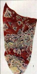
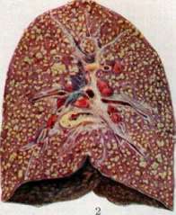
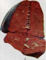
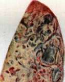
The process, having begun in one of the four ways indicated above (more often by method 1 or 2), spreads partly by contact, partly along the air passages of the lung, i.e., intracanalicularly, bronchogenically. As a rule, in pulmonary consumption of adults, the spread of the tuberculous process that began in the upper part of the lung proceeds in a direction from top to bottom; therefore, there are older changes in the upper parts, and the lower one goes, the fresher the changes are. The character of all these changes corresponds to what was indicated above regarding the pathological anatomy of tuberculosis in general (see Tuberculosis). Spread from the site of the onset of the process by contact is expressed either in the fact that the zone of pneumonic (exudative) changes rapidly expands, undergoes caseation, and decay [see separate table (cols. 119-120), fig. 4, and separate table (cols. 111-112), fig. 5] with the formation of one or several cavities (see), or, if the initial focus was surrounded by a zone of specific granulation tissue and tubercles, a predominantly productive, comparatively slow proliferation of this granulation tissue occurs, along with the development of new tubercles with compression and destruction of alveoli and weak pneumonic (exudative) manifestations. Subsequently, the indicated granulation growths [see separate table (cols. 35-36), fig. 1] may undergo caseation, decay, or show a tendency toward fibrous transformation, often accompanied by the development of non-specific connective tissue around them. Bronchogenic spread in pulmonary consumption of adults has dominant significance. Upon disruption of the integrity of the bronchus in the area of the initial focus or in connection with an already existing communication with the lumen of the bronchus, in view of the bronchopneumonic structure of the focus, caseous masses containing tuberculosis bacilli are moved during respiratory movements in the lumen of the bronchus and are carried by aspiration into new branches of the bronchial tree. As a result of the transport of infectious material into the small bronchi-bronchioles, predominantly exudative foci of tuberculous pneumonia flare up, corresponding to that part of the lobule which is called the acinus; they are called acinous foci; they have the appearance of yellowish-gray, angular foci about 2 mm in diameter ("small grain" of radiologists). An acinous focus, having a pneumonic character at first, may subsequently retain it and increase, capturing neighboring acini and eventually producing a larger lobular pneumonic focus. A lobular focus can also form immediately as such upon the distribution of infectious material at once to all branches of the lobular bronchus. A lobular focus has the appearance of a caseous focus with a scalloped edge; its size is about 1.5 cm in diameter ("large grain" of radiologists). On the other hand, an acinous focus may at the very beginning acquire a granulation zone, limiting its spread and giving it a predominantly productive character. A group of such acinous foci located nearby with a productive zone gives the so-called acinous-nodose focus [see separate table (cols. 119-120), fig. 1, apex of the lung—see the lower part of the figure]; usually, the focus has a central part consisting of atelectatic lung tissue rich in carbon pigment, and a periphery of a series of productive acinous foci; the size of the acinous-nodose focus is about 1.5 cm in diameter ("large grain" of radiologists). Both lobular pneumonic and acinous-nodose foci can merge with each other, as a result of which fields of various sizes of an exudative or predominantly caseous-pneumonic character are formed, or predominantly productive ones, consisting of nodules of various sizes, caseous in the center, and with granulation tissue on the periphery, and often even with fibrosis. Sometimes, in pulmonary consumption, an outbreak of a pneumonic process occurs that captures an entire lobe of the lung at once (lobar tuberculous pneumonia). In such cases, the entire lobe (usually the lower one) appears enlarged, dense, and gray from the side of the pleura; on a cross-section at the beginning of the process, the tissue of the lower lobe has a uniformly gray appearance; under a microscope, the picture is one of catarrhal-fibrinous pneumonia with a large participation of proliferating and desquamating alveolar epithelial cells (in the opinion of some researchers, some of these cells belong not to the epithelium, but to macrophages emerging from the connective tissue of the alveolar septa). With visible participation of the alveolar epithelium in the exudate, one speaks of desquamative, Buhl's tuberculous pneumonia. There is data suggesting that tuberculous pneumonias, even lobar ones, do not undergo...
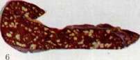
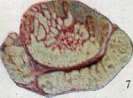
Plate 1. Lines of lung tuberculosis, infraclavicular focus of the lung with a cavity in the apex of the lung, caseous masses. Fig. 2. Acinous focus of the lung. Fig. 3. Lobular focus of the lung. Figure 4. Exudative focus of the lower lobe of the lung; caseous pneumonia of the lower lobe. Fig. 5. Primary tuberculous complex: pulmonary caseous focus and regional lymphadenitis; miliary tuberculosis of both lungs.
Tubercles undergoing caseation may undergo regression, i.e., resorption. They may also carnify with organization of the exudate by specific and nonspecific connective tissue. Most often, however, cheesy degeneration occurs in both the exudate and the alveolar septa (only the elastic tissue of the latter is spared); caseation begins in separate regions of the pneumonic area, which thereby acquire a yellowish, dry, opaque appearance, and then engulfs the entire pneumonic area completely. If the patient does not die in this period of lobar tuberculous pneumonia, multiple foci of softening and disintegration appear in its area, with the formation of sinuous cavities filled with pus-like mass.
Tuberculous pneumonias are sometimes also expressed as so-called gelatinous infiltration; by this is meant lobar or smaller extent tuberculous pneumonia, in which the exudate does not undergo caseation, but undergoes a special type of swelling, due to which it acquires a resemblance to gelatin. On the cut surface, the entire area of pneumonia has a yellowish, translucent, gelatin-like appearance. In such a state, pneumonia can remain for a very long time, without being accompanied by any cellular or enzymatic reactive manifestations. Only later does a sluggishly developing cheesy degeneration occur. With a widespread tuberculous process in one lung, the other lung rarely remains spared; most often, bronchogenic foci of acinous and lobar type arise in it. During pulmonary tuberculosis in adults, perifocal infiltrates may also flare up in the form of collateral serous pneumonia surrounding the foci of the tuberculous process. Epituberculous infiltrates are also not uncommon, by which are meant (Eliasberg, Pagel, Schürmann) regions of atelectasis of the lung tissue, which are a consequence of compression or obstruction of the corresponding bronchus; as observations by radiologists show, epituberculous infiltrates (atelectases) can be liquidated upon removal of the obstruction in the bronchus; in opposite cases, they undergo splenization and induration. Complication of pulmonary consumption by infection with various extraneous bacteria adds to tuberculosis elements of various banal pneumonias, suppurations, ichorous and gangrenous processes. All the above-listed components of the pathological anatomy of pulmonary consumption in adults can be combined in the most varied ways, which gives extremely diverse and variegated pathological-anatomical pictures of pulmonary tuberculosis in adults.
Upon autopsy of a patient who died from pulmonary consumption, one usually finds in the upper part of the upper lobe older changes in the form of indurated, coal-rich lung tissue with bronchiectasis and with dry cheesy, often calcified foci; usually, right there, there is a cavity with smooth fibrous walls or with a caseous inner surface (for more details on cavities, see Cavities). Below, one can see caseous foci of different sizes, either disintegrating with the formation of cavities and not sharply demarcated from the adjacent tissue, or dry, surrounded by dense connective tissue, passing without sharp boundaries into the indurated lung tissue. Still lower are confluent, and in the lower sections—isolated acinous-nodose foci; finally, in the very lowest parts of the lungs—only scattered acinous nodules. In other cases, along with the just-mentioned changes in the apices, dissemination in the underlying parts has a predominantly pneumonic, exudative character; in the upper sections, confluent lobular foci of cheesy pneumonia with a tendency to disintegration and formation of cavities are visible, below—lobular and acinous pneumonic foci; sometimes there is also lobar pneumonia. It often happens that older changes have a productive character (fibrous tuberculosis with cavities in the upper sections, acinous-nodose dissemination), whereas the fresh process, relating to the last time of the patient's life, bears all the signs of exudativity (lobular, acinous pneumonic foci, sometimes lobar pneumonias).
For a long time, a need has arisen to create a classification of the various forms of pulmonary tuberculosis. At different times, quite a few purely clinical classifications have been proposed; some also put forward clinico-anatomical classifications (Ribbert, Nicol, Aschoff, Tendeloo, Beitzke, and others), which had as a basic premise the anatomical characteristic of the process, important for clinicians for assessing the course of the process, prognosis, etc. As this basic characteristic, the productivity or exudativity of the tuberculous process was indicated. These classifications did not receive wide distribution for the following reason: each tuberculous focus individually can almost always be easily evaluated: whether it is predominantly productive or predominantly exudative (see Tuberculosis), but having before one the unfolded picture of pulmonary tuberculosis as a whole, only in a part of cases can one speak of predominantly exudative, or predominantly productive, or fibrous forms of pulmonary consumption; in the other part of cases, the changes turn out to be so mixed that it is difficult to indicate the predominance of either productive or exudative changes. It is even less easy for a clinician to do this, for whom the criteria for assessing an individual case serve not only a number of signs revealed by different methods of lung examination, but also a number of symptoms relating to the general condition of the patient. As a consequence of this, clinicians prefer to use classifications that include an assessment of both the process in terms of its volume, localization, and the state (degree of compensation) of the organism.
Abrikosov proposes the following designations for the pathological-anatomical pictures of pulmonary tuberculosis: I. Primary tuberculosis of the lung. 1. Pure primary complex. 2. Primary complex with spread of the process (perifocal inflammation, bronchogenic foci, etc.). II. Secondary tuberculosis of the lungs in adults (reinfection). A. Predominantly productive form. 1. Acinous tuberculosis, 2. Acinous-nodose tuberculosis, 3. Cirrhotic tuberculosis. B. Predominantly exudative form. 1. Acinous pneumonia, 2. Lobular pneumonia, 3. Lobar pneumonia.
For acute miliary pulmonary tuberculosis, the following designations are given: 1-productive form and 2-exudative form. It must be emphasized that the above-mentioned designations are proposed by Abrikosov not so much to designate the forms of pulmonary tuberculosis as a whole, but for the protocol registration of changes in pulmonary tuberculosis, with or without cavities, with encapsulation and with softening. Therefore, when evaluating the entire picture as a whole, it may happen that, for example, lesions in the upper lobe of the lung will be registered as a "predominantly productive form in the form of fibrous tuberculosis with a cavity," while simultaneous changes in the lower lobe are registered as a "predominantly exudative form in the form of foci of lobular and acinous pneumonia." Such registration undoubtedly has important significance for an understanding of the dynamics of the tuberculous process in individual cases. If we know (see Tuberculosis) that the predominant productivity and predominant exudativity of the tuberculous process usually have at their base a different reactive state of the organism, then a corresponding evaluation of all changes observed in the lungs of a deceased consumptive makes it possible to judge the shifts in his reactive state that occurred during the course of his disease. Thus, if in the upper sections of the lungs there is a fibrous, predominantly productive process, this speaks for the fact that the disease proceeded in a state of the organism that was relatively immune with respect to tuberculosis, an anergic state, as is characteristic of the "normal" course of pulmonary consumption in adults; if, however, there are simultaneously later, predominantly exudative changes with breakdown, etc., this indicates a decrease in immunity that has occurred and an increase in sensitivity to the tuberculous virus due to one or another changed condition. On the basis of the pathological-anatomical picture, one can also grasp the reverse, for example, that a process that proceeded as predominantly exudative later changed and acquired a predominantly productive, even fibrous character (sometimes due to changed living conditions of the patient, therapeutic measures, etc.). Among the complications in pulmonary consumption, pleurisies are encountered, developing as exudative inflammation of a perifocal type in the presence of tuberculous foci or even individual tubercles in the pleura. An outbreak of exudative pleurisy during a tuberculous process in the lung that has manifested itself little is an allergic manifestation. Upon the rupture of a cavity into the pleural cavity, pneumothorax or pyopneumothorax arises. Pulmonary tuberculosis is frequently complicated by hemorrhage (hemoptysis); on the autopsy table in these cases, one finds either the destruction of a vessel by a fresh exudative process with breakdown or the rupture of an arterial aneurysm that has formed in the wall of a cavity, often already fibrous and without signs of an active tuberculous process; less frequently, hemorrhage from varicously dilated veins of the cavity wall may take place. Miliary tuberculosis is observed in pulmonary consumption of adults much less frequently than in primary tuberculosis of children; its pathological-anatomical expression in the lungs and the structure of the tubercles with its variants does not differ in anything special from that which takes place in primary tuberculosis (see above). As for extrapulmonary changes in pulmonary tuberculosis, tuberculous changes in pulmonary tuberculosis are most often discovered in the intestine (60% of cases) and in the larynx (35-40% of autopsy cases); much less frequently in tuberculosis of adults are tuberculous metastases encountered in the spleen, kidneys, liver, and testicle [see separate table (cols. 119-120), fig. 7]. In fibrous forms of pulmonary tuberculosis, hypertrophy of the right ventricle of the heart is sometimes observed, but usually, the heart is atrophied. Asthenic type, emaciation, anemia, marantic edema, sometimes degenerative changes of organs, amyloidosis, and in exudative forms, hyperplasia of the spleen—these are the main changes discovered during the autopsy of corpses of persons who died from pulmonary tuberculosis. For the features of pubertal, juvenile tuberculosis, and senile tuberculosis, see Tuberculosis. A. Abrikosov. II. Classification of pulmonary tuberculosis. The creator of the first classification of pulmonary tuberculosis is Laennec, who distinguished the following three stages: 1) the stage of tubercle accumulation with bronchophony and dullness, 2) the stage of softening with coarse rales and bronchial breathing; 3) the cavernous stage. Subsequently, throughout the 19th century, a whole series of classifications appeared, based on some single clinical sign (acute-chronic, active-inactive, stationary-progressive, etc.). Koch's discovery of the pathogen did not reflect on the further development of the question of the classification of pulmonary tuberculosis. Until the end of the 19th century, not a single one of the attempts at classification received general recognition. Only the division into 3 stages proposed by Turban together with Gerhardt at the beginning of the present century was accepted for international use. This scheme is built on a single principle, very simple, clear, and easily understandable to everyone, namely: on the principle of the extent of the process. I stage. Mild disease, limited to a small area of one lobe, if it occupies both apices and does not cross the clavicle, and in a unilateral process does not descend below the II rib in front. II stage. Mild disease, more widespread than stage I, occupying a space of no more than one lobe, or a severe form involving half of one lobe. III stage. All diseases with an extent greater than II, as well as all cases with cavities. This classification does not require complex auxiliary examinations and great special training; by percussion and auscultation, one can more or less accurately determine the degree of local changes and thereby indicate the place for the given case in the general scheme. The simplicity and accessibility of Turban's classification ensured its wide distribution. But Turban's scheme has shortcomings that make it unacceptable at the present time. It gives only a primitive characterization of the process, without revealing the character of the pathological-anatomical substrate; it also says nothing about the dynamics of the process. The first attempt to create a classification of pulmonary tuberculosis on a pathomorphological basis belongs to A. Fraenkel. It is built on three principles: 1. Qualitative-anatomical: a) cirrhotic, b) nodular, c) caseous-pneumonic processes with cavities and without them. 2. Spread of the process. 3. Complications. A further stage on this path is the Nicol-Aschoff scheme, based on the old, dualistic teaching of Virchow and Orth. This teaching of Virchow says that in tuberculosis there exist two different anatomical processes—productive and exudative, i.e., the organism reacts to the introduction of the bacillus one time by the formation of specific granulation tissue, another time by the formation of specific exudate, and both these processes are histogenetically completely different. On the basis of these views, Nicol and Aschoff put forward the following scheme of pulmonary tuberculosis: A. Hematogenous forms: a) interstitial miliary tuberculosis, b) acinous miliary tuberculosis. B. Bronchogenous forms: a) productive forms—acinous-nodular and cirrhotic form, b) exudative forms—caseous-lobular and caseous-lobar form. The classification of A. I. Abrikosov (see above), adopted in 1923 at the All-Russian Congress of Pathologists—a modified Beitzke classification—is also based on the Aschoff scheme. A number of modifications of the Nicol-Aschoff scheme (Paweletz, Kraus, Gerhardt, Bacmeister, and others) were built on the account of the close interrelation between the clinical and pathomorphological picture in various forms of pulmonary tuberculosis, differing from each other only in the character and variety of the introduced individual clinical signs of a qualitative character, for the most part episodic, but not connected with each other into a single whole. The so-called qualitative diagnostics was created, which from the 33rd Congress of German Internists in 1918 to the last years became leading in the clinic of pulmonary tuberculosis and an integral part of our working platform. This striving to give a firm anatomical base to clinical concepts gave an impetus to a more deepened study and understanding of the various forms of pulmonary tuberculosis. In comparison with Turban's classification, the proposal of Nicol and Aschoff meant a step forward and opened horizons for the development of a truly valuable nomenclature of tuberculosis on pathomorphological foundations. However, these hopes were far from justified. Comparative pathological-anatomical study of clinical-roentgenological data showed the impossibility of distinguishing with certainty between exudative and productive forms not only at the patient's bedside but even on the autopsy table. There are no purely productive forms, just as there are no purely exudative ones, and a focus diagnosed macroscopically as productive turns out microscopically to be exudative, and vice versa. Furthermore, in the chronic forms of pulmonary tuberculosis usually observed by us, both forms—productive and exudative—are always encountered together, and therefore it is impossible to speak of the predominance of one form over the other (Ziegler). And X-ray examination often does not provide the possibility of determining with certainty the pathomorphological character of a given focus. Even greater difficulties are encountered by the attempt to create a concept of the clinical forms corresponding to them with a definite character of course and prognosis on the basis of X-ray-anatomical data. On the one hand, the exudative and productive reactions of the tissue are not independent, detached pathomorphological changes, but are closely dynamically connected with each other.
On the other hand, the prognostic indicator embedded in this nomenclature, unfortunately, has far from justified itself, and the resulting antithesis: exudative-malignant, productive-benign, turns out to be unviable and does not correspond to reality. A large number of exudative forms proceed quite favorably and, conversely, productive ones often proceed significantly more severely than exudative ones. The classification of Pulmonary Tuberculosis, not containing within itself solid prognostic elements, naturally could not satisfy the broad medical masses. But if the characterization of the process (exudative, productive, fibrous) cannot serve as a basis for classification, it still does not lose its significance for clinical observation. When in the clinic there is talk of an exudative, productive, or fibrous form, everyone invests in this concept the same qualitative prognostic and clinical content. We consider processes that are severe and stormy, leading rapidly to death with high temperature and severe symptoms of intoxication, to be exudative, and chronic, slowly progressing ones without significant intoxication symptoms—mainly productive-fibrous. Despite the difficulties noted above in the morphological differentiation of tissue reactions, in the majority of cases of Pulmonary Tuberculosis, it is possible to establish clinically and radiologically whether they are, at a given moment in their reactive tendency, more exudative, more productive, or more cirrhotic. And one cannot help but agree with Redeker, who, while rejecting anatomical qualitative diagnostics as the basis for classification, finds it impossible in the clinic to strike the concepts "exudative, productive, fibrous" from the qualitative diagnosis of Pulmonary Tuberculosis, since they would have to be replaced by new words which, in a clinical sense, would have an equivalent meaning. Qualitative diagnostics has become an integral part of our anatomical-clinical thinking, but it gives only a general clinical idea of the main tendency of the process at the moment of observing the patient. It cannot give more. Least of all can it become a method for the classification of pulmonary Tuberculosis. In the USSR, since 1922, the classification of the II Congress (Sternberg) has been applied, which, based on the Turban scheme, attempts to take into account the dynamics of the process by introducing the characteristic: compensation (A), subcompensation (B), decompensation (C). This scheme, in comparison with the Turban scheme, was a step forward, but with the expansion of our knowledge about pathogenesis and clinical practice and the growth of the staff of phthisiologists, the Sternberg classification also ceased to satisfy the demands of the clinic. At the IV Congress (Tiflis, 1928), an attempt was made to adapt the old classification to the newest views and requirements of the clinic, whereby individual, most important signs were taken from various existing classifications and grouped together as a single scheme. (Qualitative definitions of the process were introduced, definitions of Ranke phases and concepts of infiltrative outbreaks were introduced.) This variant, not presenting theoretical advantages, being more cumbersome and complex than the classification of the II Congress, signifies an undoubted step backward from the previous classification. What was valuable in the old classification—brevity, clarity, and generality of approach—disappears here among a multitude of unrelated trifles. Instead of a classification, what resulted, as A. Ya. Sternberg expressed regarding a similar issue, was a scheme for an epicrisis or a shortened individual diagnosis, having a descriptive character and representing a collection of information about the patient, and moreover, information for the most part not connected with each other. The attempt of the IV Congress did not take root in the practice of tuberculosis institutions. As can be seen, to this day there is no classification of pulmonary Tuberculosis that would satisfy our modern concepts. It exists nowhere—neither here nor abroad. It apparently cannot exist yet. The impossibility of constructing a classification of Pulmonary Tuberculosis that satisfies the requirements of practice forces us to go by other paths. We have the opportunity to isolate, in the process of observing tuberculosis patients, large groups uniting a whole series of individual syndromes that have common pathogenetic roots and are similar in the manifestations of the disease. With all the methods available to us—physical, radiological, and biological—we must attempt to outline these individual clinical pictures of Pulmonary Tuberculosis and base the establishment of prognosis and therapy on the results of our thorough clinical observations, which is the primary and ultimate goal of any clinical differentiation at the patient's bedside. With such an approach to this issue, the clinical scheme of Pulmonary Tuberculosis should consist of two parts: I. basic clinical types, representing individual phases of the development of the tuberculosis process, encompassing and uniting specific clinical syndromes; II. necessary elements of qualitative diagnostics, providing a characterization of the given syndrome—clinical and anatomical. 1) Clinical characteristic. The course of Pulmonary Tuberculosis is wave-like: we observe either rises in the form of outbreaks or a subsiding of the process, an interval (Redeker). An outbreak corresponds to a state of decompensation and subcompensation, an interval—to a state of compensation. 2) Anatomical characteristic determines the phase of anatomical development at the moment of examination. It includes the exudative phase, the phase of disintegration and dissemination, the phase of resorption and induration. Furthermore, it is necessary to note: 3) the extent of the process—by fields and ribs—since it undoubtedly has great prognostic significance, and finally 4) the excretion of bacilli, i.e., constant (BK+), intermittent (BK±), or absence of excretion (BK-). On these principles, the following scheme of clinical forms of pulmonary Tuberculosis is built: A. Basic clinical types. 1. Primary complex. 2. Tuberculosis of the bronchial glands. 3. Focal-disseminated forms: a) forms of lympho-hematogenous origin—1) acute miliary Tuberculosis, 2) subacute and chronic miliary Tuberculosis; b) focal forms of unclear origin. 4. Apical Tuberculosis. 5. Pulmonary infiltrate. 6. Pulmonary cavernous consumption. 7. Exudative-caseous pneumonia. 8. Cirrhoses. 9. Pleurisies. B. Elements of qualitative characterization. 1. Clinical-anatomical dynamics: a) infiltrative-exudative phase (outbreak—B, C); b) phase of disintegration and dissemination (outbreak or subsiding); c) phase of resorption and induration (subsiding—A, B). 2. Extent by fields and ribs (I, II, III). 3. Bacillarity: BK+, BK±, BK-. Example: right-sided infiltrate the size of a plum between the II and III ribs with disintegration and bronchogenic dissemination at the apex of the heart, with BK in the sputum, in the phase of subcompensation. Preliminary testing of the proposed scheme in a number of tuberculosis institutes and tuberculosis dispensaries allows us to expect that it will satisfy our clinical demands, ensure the further development of problems of pathogenesis, and will not complicate the statistical processing of materials of tuberculosis institutions in the slightest. On the basis of the above-mentioned groupings, the clinical pictures of each of these syndromes are given below.
G. Rubinstein. III. Clinic. The doctrine of the evolution of pulmonary tuberculosis. The last decade has been for the clinic of pulmonary tuberculosis a period of significant breaking of previous concepts. The old clinic, not having at its disposal X-rays, had to be guided partly by the subjective complaints of the patient, and partly by data from physical examination methods, which have limited significance for diagnosis. Pathological anatomy naturally allows one to reveal only retrospectively the general major stages of pathomorphological changes that occurred in the lung during the course of the disease, but it cannot provide the full dynamics of a tuberculous disease in each individual case. Modern clinics, thanks to X-rays, have gained the possibility, even during the patient's lifetime, to create for themselves a correct idea of the continuous course of development of anatomical changes in each individual case, to link them with their clinical observations, and to form a picture of the complex dynamics of the tuberculous process. The most important tenets of the modern clinic, which have received a different resolution than before, concern mainly the following three fundamental questions. The first is the question of the anatomical character of newly forming fresh tuberculous foci. X-rays and simultaneously pathological anatomy show that any fresh tuberculous formation almost always has an infiltrative-exudative character and that foci of a primarily productive character are either completely absent on the plate or on the autopsy table, or they are rarely encountered. The tubercle, according to Hübschmann, is not an independent new growth, but only a productive stage of the same development in which the exudative component has significantly receded; likewise, caseous inflammation is not an independent disease, but only a particularly strongly expressed exudative phase. In this, it is completely indifferent whether this newly formed focus appears for the first time in a completely intact lung, whether it forms in a healthy part of the lung in the presence of old foci in other places of the lung, or whether it is a relapse at the site of a process that was there before but has now resolved. On this cardinal question, representatives of most modern schools converge—both German and French, starting from Redeker and Rist and ending with Aschoff and Hübschmann, as well as A. I. Abrikosov. Modern clinics have fully adopted these pathological-anatomical concepts not because they gleaned these views at the autopsy table, but because their observations on X-rays led them to these concepts. The second question, which has undergone significant evolution in recent years, concerns the pathogenesis of pulmonary tuberculosis. The doctrine of the primary localization of the early tuberculous focus in the apex was previously considered an irrefutable axiom, which for its part led to certain practical and therapeutic conclusions and gave a certain direction to our sanatorium measures. At the same time, it was thought that the entire process of the development of consumption originates precisely from this focus that first germinated in the apex, that the focus itself gradually grows and increases, and that it becomes overgrown with new tubercles developing around it. Subsequently, these new foci merge and disintegrate in the center, and this is followed by further bronchogenic advancement of the process, which proceeds continuously from one section to another, from lobule to lobule, i.e., in an apico-caudal path, until it captures the entire side and passes to the other lung, where it also gradually captures the entire lung from top to bottom. With the development of X-ray diagnostics, it became clear that our old ideas about the beginning of pulmonary consumption in the apex and the close connection of consumption precisely with this apical focus by no means reflect reality, at least on the exclusive scale that it seemed before. In recent years, the focus of attention has been the infiltrate, partly as a prototype of the initial fresh process in a hitherto intact lung, but for the most part as a perifocal inflammation around old foci, as a link connecting closed forms with open ones and transferring them through disintegration into chronic bronchogenic consumption. The study of these infiltrates (Schürmann, Pagel, Rubinstein, and Pozharisky, et al.), especially serial pathological-anatomical sections of apices in cases with fresh infiltrates that ended up on the autopsy table, showed that the apices in such cases often contain no focal or cicatricial changes. These anatomical studies and clinical observations on the course of apical forms showed that there is no reason to recognize an obligatory connection of fresher foci, especially those of a larger infiltrative character, with preformed older scars or foci in the apex. The clinic still does not deny the possibility of such a connection in some cases, but it believes that this connection is not mandatory for all cases, that new foci can develop in any part of the lung by both exogenous and endogenous-hematogenous routes without any participation of older foci, and that the development of consumption through an infiltrate proceeds significantly more often without the participation of old apical foci than with their assistance. Finally, the third question concerns the latest views of the clinic on the anatomical and clinical dynamics of the process in pulmonary tuberculosis. The old clinic of pulmonary tuberculosis did not admit the acute onset of a more or less significant anatomical tuberculous process and considered such forms of development (and the clinic certainly could not fail to see them) atypical, because it found no explanation for them. For the old clinic, the subjective complaints of the patient with pulmonary tuberculosis constituted an integral part of the clinical syndrome from the very beginning of the onset of the disease, and accordingly, to the anatomical ideas about the gradual continuous cranio-caudal spread of the process, the clinical course of consumption itself was also thought of as slow, chronic, the severity of which increases just as slowly and gradually. The best expression of these views on the gradualness in the course of pulmonary tuberculosis is the well-known Turban scheme with its three stages, of which each subsequent one is a direct continuation of the preceding one. However, observing the dynamics of the spread of the process on serial X-ray images, one can be convinced that it proceeds by no means in a contact-continuous way, but, on the contrary, in a jump-like manner, jumping not only over individual small sections but sometimes over entire completely healthy lobes, often throwing itself into the other lung. This new idea about the spread of the pulmonary process in the lung is now finding confirmation among pathologists as well. Thus, Leschke says: 'Pulmonary tuberculosis in all its stages spreads only to a comparatively small extent continuously from acinus to acinus; on the contrary, it infects one acinus after another in a jump-like manner along the path through the bronchi, and these jumps, with an increase in the caliber of the bronchi, also gradually become smaller.' Careful study of the course of tuberculosis has shown that initial pulmonary tuberculosis usually does not give definite typical complaints, and if it does, they are either too short-lived, or so insignificant that the patient does not pay attention to them, or these complaints are masked by other diseases, usually of a cold, more often influenzal nature. Only when an infiltrate appears, only then do complaints appear. At the same time, the process in more than half of the cases begins clinically acutely, without prodromal phenomena, like a real acute infection. Patients usually claim that they felt well until now, had never been ill with their lungs before, and had no painful sensations, and that they have been ill for only a few weeks or months, etc. This acute phase passes after some time, and then, in accordance with the jump-like anatomical advancement of the process, after some time, deterioration occurs again. These jumps, i.e., any formation of new foci, any more or less significant changes in them, if they manifest themselves clinically in any way, we call an outbreak (Schub in German, poussée in French). The entire clinical essence of the tuberculous process, the entire complex of pathomorphological changes and clinical phenomena of the tuberculous disease are determined by the character of these outbreaks, the course and outcome of which depend partly on nonspecific endogenous-constitutional and exogenous-conditional factors, and partly on specific moments, depending on the degree of virulence of the focus and the immunobiological characteristics of the given subject. An outbreak is a purely clinical concept and corresponds to the clinical idea of an exacerbation of the process. When the acute phase passes, then, as with other diseases, improvement, a lull, occurs, followed after a certain time by a new exacerbation. The period between two outbreaks we call an interval. In the interplay of these two phases—the outbreak and the interval (this nomenclature is only to a certain extent covered by the concept of 'compensation and decompensation' introduced by A. Ya. Sternberg into the clinic of pulmonary tuberculosis, which is so popular among phthisiologists)—the entire kaleidoscope of the tuberculous process passes. Naturally, the duration of the interval is determined by the frequency of outbreaks, and likewise, the equilibrium achieved during the interval depends on the activity and fate of the focus that caused the outbreak. And the fate of the focus can be different—from complete resolution to caseous degeneration and disintegration. B. M. E. Vol. XXXIII. 182
The clinical presentation corresponds to this. In some cases, flare-ups follow one another so rapidly (galloping consumption) that no interval is visible at all; we have a type of permanent flare-up, if one may put it that way—a fire phase. In other cases, we see short intervals between two flare-ups (pubertal phthisis); in still others, the intervals become long (ordinary tertiary bronchogenic consumption); sometimes the process progresses extremely slowly and imperceptibly, the flare-ups are so sluggish and insignificant that it seems they are not there at all, as if a permanent interval is present (sluggish apico-caudal forms with a tendency toward cirrhosis). Between these two extremes—between a permanent flare-up and a benign permanent interval—and in the interweaving of these diverse phases with one another, lies the entire clinic of pulmonary tuberculosis, so rich in the variety of its forms. But in this huge, boundless mass of variants, individual syndromes emerge, individual, frequently recurring clinical-radiological types of waves, often observed at the patient's bedside. The first flare-up in the patient's life is the primary, so-called Ghon focus, a typical alveolitis, i.e., a tuberculous exudative-pneumonic focus, which always draws the nearest regional glands into the process. The first manifestation of tuberculosis infection in children clinically, as a flare-up, usually passes unnoticed, and only the next stage, and primarily the infiltration around the primary complex, the so-called primary infiltration, makes itself known both clinically and radiologically. Both the primary focus and even the next flare-up—infiltration—are usually resolved; only a solid primary complex or an induration field remains; the ensuing “interval” can last a lifetime. Only in rare cases does true primary pulmonary consumption develop from this primary focus. The next group of flare-ups is fed by infectious material lodged in the bronchial glands during the period of development of the primary complex. The range of action of these flare-ups, the so-called secondary allergic period, is significantly wider than in the previous period, which was limited to the area from the primary focus in the lung to the venous angle. Here, all paths are already open to infection: broncho-, lympho-, and hematogenous, and there is not a single organ that would be insured against the seeding and development of a tuberculous process in it during this period. We see here flare-ups from the mildest to the most severe—from innocent tuberculids and phlyctenules up to fatal meningitis. And in the lungs, we see in this period flare-ups of varying intensity, starting from insignificant perifocal inflammation in the region of the hilus and individual hematogenously seeded foci, up to infiltration encompassing the entire lung and continuous miliary dissemination. Depending on the extent and character of the process, flare-ups are sometimes shorter and milder, sometimes longer and more severe with short intervals. In the course of the various forms of this period, the center of activity of which lies predominantly in the bronchial glands, we see, in connection with the stages of the process's progression, frequent rises and falls of the curve with usually unstable intervals, which clinically often simulate the patient's complete well-being and stand in sharp contradiction to the picture of the pathological-anatomical changes present in the patient. And while the entire large group of perihilar infiltrations, closely connected with tuberculosis of the bronchial glands, gives a good prognosis in the final result, despite the tendency toward frequent relapses inherent in it, flare-ups among hematogenously disseminated forms lead in a significant number of cases to severe further proliferation of the process with an unfavorable outcome. These phases of waves—the alternation of flare-up and interval—run like a red thread through the clinical course of the so-called early subclavicular infiltrate. Flare-ups reach maximum intensity here at the first manifestation of the infiltrate itself, during the development of decay, and during subsequent aspiration bronchogenic seeding from the cavity. The severity and duration of the first flare-up are extremely diverse. Sometimes subjective sensations are insignificant, sometimes the flare-up takes the form of a severe infection with all the subjective sensations characteristic of these states. And the duration of the flare-up is different. Beginning in most cases suddenly, without prodromal symptoms, it lasts only a few days, sometimes weeks, then everything seems to subside, an interval occurs, and the patient feels healthy or almost healthy. This interval can be long, sometimes for many years, if the infiltrate resolves or indurates, but in most cases, the course is different. The infiltrate softens, a cavity is formed. And this flare-up sometimes proceeds, although rarely, without a special reaction on the part of the organism, but most often the subjective complaints of patients in this phase are quite significant. In other cases, the flare-up of the decay phase merges with the previous flare-up of the infiltration development phase; the cavity develops quickly, so that there is no interval between the two flare-ups. And after the development of the cavity, an interval usually occurs soon, when the body's forces stabilize and when the patient's subjective complaints and objective changes characteristic of the flare-up phase subside. And this interval can often last a long time, sometimes for years, allowing the patient to be fully capable of working, but every interval has an end, and especially this one. From the cavity, sputum is carried to other parts of the same lung or to the other lung. New foci develop at the site of the seeding, which merge, forming so-called daughter infiltrates. These daughter infiltrates, like the maternal infiltrate, go through the same cycle of development; they show the same phases of waves in their course, i.e., flare-up and interval. Each new focus becomes a new source of decay and further seeding, new infiltrates are formed, and each new stage gives a corresponding clinical flare-up, and between two flare-ups in the subsidence phase, an interval occurs. In this period, when we already have a clearly expressed chronic consumption, both the flare-up and the intervals can vary significantly in both quality and duration depending on exo- and endogenous conditions, and it is quite natural that there can be an infinite multitude of variants for each individual case. Thus, in general terms, the course of the tuberculous process proceeds in a constant change of flare-ups and intervals, and thus the gradual progression of the tuberculous process in the lung occurs. With each flare-up, a new section of lung tissue is involved, so that further spread proceeds from many sources, which seed the remaining healthy parts of the lung. It is quite natural that in the last phases of the disease, when the sources for the formation of new foci are infinitely numerous, reinfects appear simultaneously in different parts of the lung, and clinically they correspond to flare-ups that overlap each other, sum up together, leaving no time for an interval. And just as with some acute exudative-infiltrative forms, a rapid spread of the process occurs from the very beginning, so in this terminal stage, when the body's forces are broken, we see a continuous clinical flare-up, anatomically a severe exudative-caseous phase, which lasts until death itself. The tuberculous process begins with an exudative flare-up; it usually ends with an exudative phase. But in life, at the patient's bedside, we cannot be satisfied with identifying only one evolutionary phase of the disease. We are interested to no less a degree in the entire complex of the disease, the clinical appearance of the patient, i.e., the entire sum of semiotic, pathophysiological, and radiological signs, which give the given case the picture of a specific clinical syndrome. Clinical forms of pulmonary tuberculosis, symptoms, and course. 1. Tuberculosis of the bronchial glands. The first syndrome—the “primary focus and primary complex”—is extremely rare in our conditions in adults and is observed mainly in children (see below—Pulmonary tuberculosis in children). As for tuberculosis of the bronchial glands, although this syndrome is also encountered relatively rarely in adults, until recent years it occupied, and still continues to occupy, a fairly significant place in the clinic of so-called latent tuberculosis. This group usually includes so-called bronchoadenitis, glandular-pleural forms, juvenile tuberculosis, etc. For the diagnosis and therapy of cases with a real anatomical basis of tuberculosis of the bronchial glands in adults, the same indicators as those set forth for tuberculosis of the bronchial glands in children are valid; but if the difficulties of diagnosing tuberculosis of the bronchial glands increase in children in parallel with the child's age, and if it is already difficult for schoolchildren to reveal objective changes in the glands with our research methods, then in adults these difficulties become particularly serious. Since infection with tuberculosis usually occurs at an early age, the involvement of the glands, especially the development of large caseous forms, so-called “potato” glands, which provide palpable diagnostic indicators, falls precisely on the immediate period after infection, i.e., the early childhood period. With age, the tumor-like glands begin to recede into the background; only non-tumor-like forms remain.
Although caseous foci, which are a source of intoxication and even metastasis of the process, can persist in the tissue of the gland, it is not always possible to detect these foci with certainty either by physical methods or by X-ray. In adults, this is possible only in one case: if the infection occurred recently. Then, as in young children, there is an enlargement of the glands, which can be diagnosed. True, symptoms associated with the compression of the trachea, vessels, and nerves by enlarged glands are already absent in adults due to the large size of the chest, but X-ray sometimes reveals the paratracheal group, especially on the right, and the bronchopulmonary glands on both sides and the bifurcation glands. Often, in cases of late infection, swelling of other lymph glands is found in adults, especially cervical and axillary, and typical infiltration of the root zone—these so frequent and faithful companions of tuberculosis of the bronchial glands in children. Various manifestations of the exudative phase, so characteristic of this period of the evolution of the tuberculous process, are also found, and just as in children, the entire clinical picture of tuberculosis of the bronchial glands unfolds with its typical features, and at the same time with an extraordinary variety of individual episodes (see below—Pulmonary tuberculosis in children).
The question of the diagnosis of bronchoadenitis in adults, in whom the primary infection occurred long ago, in early childhood, is different. Into this group, most doctors are still inclined to classify patients with a certain clinical syndrome, but without objective changes. The main clinical sign of this syndrome is an abundance of complaints, which stands in glaring contradiction to the almost complete absence of objective changes, especially in the lungs. The complaints are extremely diverse and can be interpreted as signs of neurasthenia, neuropathy, rheumatic, toxic, and others. Most often, dizziness, headache, emaciation, insomnia, sweats, weakness, rapid fatigue, lack of appetite, increased irritability and excitability, phenomena from the gastrointestinal tract, menstrual complaints, pain in the sacrum, and often a dry cough, pain in the chest and between the shoulder blades, etc., are noted. One can distinguish a triad that is very characteristic of this syndrome. First of all, an asthenic constitution is often encountered among this group of patients. Further, the main complaint is weakness. Patients tire quickly, are little capable of work, and in the morning feel as if they had not slept at all. Next come prolonged, monotonous subfebrile temperatures—around 37.5°C, and in some women, temperatures that fall to normal only before and during menstruation.
The question of other etiological possibilities for this symptom complex was almost not debated until recently, and only in the last years has a certain turn and a more critical approach to the clinical interpretation of these forms been noticed. On the one hand, the same picture can be produced by a whole range of diseases that have nothing in common with tuberculosis, such as chronic sepsis, diseases of the nose, tonsils, chronic pyelitis, appendicitis, adnexitis, Basedow's disease, pituitary disease, constitutional features, etc. On the other hand, long-term observation of such patients does not reveal any objective pathological changes in them and does not show any changes over a long period, and most importantly, the frequency of the development of active, progressive forms of tuberculosis among this group is no higher than the figure for tuberculosis disease among the entire population that is usual for the corresponding age and social groups.
One can consider oneself justified in diagnosing tuberculous bronchoadenitis in adults only under the following conditions: a) if we are dealing with people who, due to their living conditions, were exposed to a late infection and in whom bronchoadenitis can be detected, just as in children, by complex diagnostic methods; b) when there are any pulmonary or extrapulmonary manifestations of a tuberculous process; and c) when we are dealing with adolescents in the transitional age. In these age groups, tuberculosis of the bronchial glands undoubtedly still plays a greater role, and it is precisely among them that we see various forms of tuberculous diseases that did not arise on the basis of old changes in the lungs, but are connected with flare-ups of foci in the bronchial glands. It is completely impossible to deny this fact, and it is precisely in these patients that one can often be convinced of the existing continuity between childhood tuberculosis and tuberculosis of later ages. Before our eyes, patients constantly pass whom we knew earlier—in early or school age—in whom there were then completely indisputable tuberculous changes in the lungs or glands. These changes gradually resolved, leaving only small foci and fibrous strands, light indurated fields, as if already devoid of any activity. The patients felt impeccable, considered themselves completely healthy, their living conditions in no way predisposed them to the disease, there was no visible source of superinfection either, and yet, in these patients, suddenly, quite unexpectedly, a fresh infiltrative process appears, sometimes exceptionally severe. Where did it come from? It is impossible in these cases not to take into account the possibility of endogenous reinfection from foci in the glands that developed in childhood many years ago. It should not be forgotten that the tubercle bacillus, as recently completed experiments (Rubinstein and Trius) have shown, can lie in foci for decades without causing any reactive manifestations from the surrounding lung tissue, but while retaining full virulence. Transferred to new soil, it is capable of causing generalized tuberculosis, and under certain favorable conditions, it can also cause local flare-ups, as we observe daily. There is a series of observations made by Braeuning, Langer, and other authors showing that in the transitional age, it is predominantly those who were subjected to severe superinfection in childhood who fall ill. All this suggests that in the transitional age, the bronchial glands undoubtedly still are a factor playing a role in the pathogenesis of pulmonary consumption and that one should by no means ignore their significance in these age groups. In adults, however, the role of the bronchial glands in the clinical picture of tuberculosis is apparently extremely insignificant. The prognosis in terms of the possibility of the process progressing in adults with tuberculosis of the bronchial glands is in most cases satisfactory, in any case better than in children. Manifestations of generalization are usually not intense either in character or in extent and rarely lead to fatal forms of dissemination, especially meningitis. But it is always necessary, however, to remember that a patient with tuberculous bronchoadenitis is a tuberculous patient over whom hangs the sword of Damocles of sometimes catastrophic complications. Therapy for actual bronchoadenitis is reduced primarily to the prevention of any irritations capable of causing a flare-up of tuberculosis (hyperinsolation, pregnancy, abortion, excessive physical exertion, the use of strong irritants), the regulation of labor, and the creation of favorable external conditions for the patient. In some cases, tuberculin is indicated, but only in skillful hands. The remaining "bronchoadenitis" cases without a clearly proven tuberculous etiology need first of all the establishment of the cause that gave rise to the given symptom complex, and only depending on this can appropriate therapy be prescribed. In many cases, psychotherapy yields much better results than special treatment in a sanatorium. In any case, these patients do not need to be sent to tuberculosis sanatoriums. 2. Hematogenously disseminated forms. Hematogenously disseminated forms are understood to be those forms of pulmonary tuberculosis in which a more or less uniform eruption of small exudative-caseous, or purely productive, or transitional from exudative to productive, approximately equal-sized foci is observed in all or several organs where Koch's bacilli can only get through the blood. The first and most important source of the dissemination of tubercle bacilli are the lymphatic tuberculous glands, and primarily the bronchial ones, infected from the primary focus. From the glands, through large lymphatic vessels, the bacilli enter the blood. The strongest and most frequent lesions in miliary tuberculosis are found in the lungs, which is quite understandable, as they are the nearest organ that the bacilli encounter on their path after they have entered the blood. The second path is the rupture of a caseous-disintegrating focus into an adjacent vessel. This focus can be in the lung, in a caseous bronchial, mesenteric, or other lymphatic gland; it can also reside in the greater circulation, most often in the bone marrow, prostate gland, testicle, ovary, appendage, adrenal gland, etc. Upon rupture, when the possibility of a massive influx of softened virulent product into the blood is given, the conditions of infection are different and more severe than with the periodic, rather slow intake of bacilli from the lymphatic glands. Clinically, the following forms of miliary tuberculosis are distinguished: Acute miliary tuberculosis. It proceeds with the picture of a severe infectious disease. Depending on the prevalence of general and organ phenomena, 3 forms are distinguished: 1) Typhoid. The prodromal period is short. The disease begins acutely, with severe general phenomena, and only in rare cases are there complaints of weakness, headaches, and malaise even before the disease. Clouding of consciousness sets in very quickly. At night, and sometimes during the day, delirium appears. Regarding internal organs, no special deviations from the norm are noted. The spleen, although enlarged, is difficult to palpate due to its softness. Very often, there is leukopenia and eosinopenia. The picture does not differ in any way from typhoid fever, especially if roseolae also appear, which is observed not infrequently. The temperature is not constant, as in typhoid, it stays within 39-40°, but gives significant fluctuations not characteristic of typhoid fever; the pulse is small, rapid, and the patients lose weight quickly. After a week, phenomena associated with the eruption of tubercles in the lungs and meninges already appear. Breathing becomes accelerated—up to 40-60 per minute; cyanosis appears on the pale face and extremities. Regarding the lungs in these forms, no special deviations from the norm are noted upon auscultation, and only an X-ray, and even then most often only a radiograph, reveals the true nature of the disease. The X-ray picture shows a significant eruption of tubercles, sometimes sharply defined, sometimes with blurred edges in the presence of an exudative zone around them. Death usually occurs after 2-3 weeks. These patients very often, even in large hospitals, if an X-ray is not taken, go until the end of the disease under the diagnosis of typhoid fever, and only at the end do meningeal phenomena occur, or the true nature of the disease is revealed only at autopsy. 2) Pulmonary form. This is the most frequently encountered form. Here, from the very beginning of the disease, phenomena from the lungs dominate. The disease begins acutely with a strong, dry cough, sometimes in the form of attacks. The temperature is high, between 39-40°, with fluctuations as in the typhoid form, in some cases hectic with profuse sweating; as a striking symptom, severe dyspnea appears here. Breathing is superficial, strongly accelerated—up to 50-70 per minute. The face is pale, with clear cyanosis, especially of the lips and cheeks. Regarding the lungs, at the beginning of the disease, only diffuse bronchitis is noted, but already from the second week, small, moist, rather resonant rales are very often heard, similar to those found in pulmonary edema. Tubercle bacilli are often absent in the sputum. The boundaries of the lungs are often expanded in connection with emphysema developing as a result of catarrh of the small bronchi. In connection with this, the percussion sound is loud, deep. Death usually occurs 4-7 weeks after the onset of the disease. In rare cases, there are remissions with a temporary improvement in the general condition, but a subsequent flare-up still, even if after a few months, leads the patient to the grave. Recoveries from true acute miliary tuberculosis are unusually rare. 3) Meningeal form. Symptoms of tuberculous meningitis (see) dominate. 4) Extremely rarely, a type of acute hematogenous tuberculosis is encountered in which, due to complete anergy of the organism, the process does not reach the development of tubercles at all. Necrosis of individual small sections of the lung occurs. The lung is permeated as if with small abscesses, ranging in size from a pinhead to a pea and larger, filled with liquid pus. Under a microscope, the purulent necrotic areas consist of a pure culture of tubercle bacilli. The process usually originates from tuberculous mesenteric glands with bovine-type bacilli. The prognosis of these cases, which are known in the literature under the name sepsis tuberculosa acutissima, or typho-bacillose Landouzy, is absolutely poor.
If at one pole we have the fatal miliary pulmonary tuberculosis, then at the other we see the so-called abortive pulmonary forms, devoid of any activity, and between these two extreme groups stand the subacute and chronic forms. Subacute forms can be both large-focal and small-focal. The former represent a type of focal, lobular, exudative-caseous pneumonia scattered throughout one or both lungs. These forms usually begin acutely, sometimes gradually, but more often proceed with frequent flare-ups and short intervals and lead to death in a relatively short time, although stabilization and transition into chronic consumption are sometimes observed. These forms are classified by many authors as belonging to the hematogenous group, but in some cases it is impossible to shake the impression that they arise along the usual path of other, non-specific lobular pneumonia. Subacute small-focal forms represent a type of hematogenous seeding with soft foci during a clearly expressed general and tissue exudative phase. Like the large-focal group, they are also sometimes accompanied by the fusion of foci and the formation of infiltrates, which soon disintegrate, forming multiple cavities. They also cause severe complications of an exudative-inflammatory nature from the larynx and relatively quickly lead the patient to the grave. In other cases, the process stabilizes in the productive phase and passes into a chronic hematogenous form. Forms of chronic dissemination represent a large group. Subjective symptoms from the lungs are either absent or weak, but the anatomical changes in them are more or less significant. The significance and role of these forms were first evaluated and described in detail by Grau in 1918. In recent years, we have encountered them quite often, and the more closely we look at them, the more we find them. We are talking about cases of the development of small foci, sometimes single, sometimes scattered in a part of the lung, or in the form of a dense rash throughout both lungs. The development of the process is slow, chronic; very often patients do not know that they are ill, and the disease is discovered in them accidentally during an X-ray examination. Frequently, not only are the subjective complaints of the patients insignificant, but the data from physical examination do not at all indicate the presence of special anatomical changes in the lungs. And yet, the X-ray plate sometimes shows a continuous eruption of tubercles, by which the entire lung is literally permeated. Since the tubercles lie in unchanged lung tissue, the percussion and auscultatory data are not very characteristic. Where there is already a fusion of foci, there is dullness and a change in respiratory sounds, but where there is no fusion, one can sometimes ascertain only a slight tympanitis and a shortening of the percussion tone with rather harsh breathing. Coarse bronchial sounds are sometimes observed but are inconstant. It is necessary to point out one diagnostic sign that often helps to orient oneself even without an X-ray—these are fine moist rales, rather dry, scattered throughout the lung, especially in the paravertebral region. The number of these rales is not very large; sometimes there are so few of them that it is only possible to provoke them after coughing at the end of a deep exhalation or during an immediate deep inhalation. This bronchiolitis is extremely characteristic, and we have learned to value it as a rarely deceiving clinical symptom of disseminated pulmonary tuberculosis. The diagnosis is decided by an X-ray image, on which small tubercles of more or less the same size are visible, scattered across several fields—from the tip of a pin to a pinhead, sometimes even larger. The apices in fresher forms are usually free, but in later phases they also already contain tubercles. The root is almost always enlarged, juicy. Grau attaches special importance to this sign. Subjective complaints are not characteristic, are very different depending on the spread of the process, and are of an intoxication nature: fatigue, headaches, weakness, various functional disorders of internal organs. Complaints from the lungs are almost absent; patients do not feel like pulmonary patients. Tubercle bacilli in the sputum are found no more often than in 25% of cases, sometimes they are excreted only periodically. The temperature is sometimes normal, more often subfebrile, within 37.5 degrees. Grau draws attention to the connection of these disseminations with primary initial exudative pleurisies. Out of 18 cases with pleurisy, he found tubercles scattered throughout the lungs in 15 and asserts that initial exudative pleurisy is the result, and therefore a sign, of a hematogenous eruption that has occurred in the lungs. Redeker and Braeuning also emphasize the connection of hematogenous forms with pleurisies, but at the same time point out that the latter are most often at the height of the eruption period. Often, extra-pulmonary tuberculous changes are found in these patients, or they are noted in the anamnesis as having been suffered in childhood or even at a later age. As for the further evolution of these chronic forms, all the same laws that are observed during the evolution of any other tuberculous process apply to it. On the one hand, we see further spread, which is expressed either in repeated eruptions—protrahierte Durchseuchungsperiode (Schürmann's period of protracted generalization)—or in the formation of infiltrates with subsequent disintegration. Infiltrates are of two origins: either the fusion of a group of foci, forming shadows on the X-ray in the form of roundish or rhomboid balls (Simon's Ballungen), or the actual development of perifocal inflammation around a group of foci. Infiltrates are in most cases multiple, located in both lungs without any particular regularity. And as in pulmonary tuberculosis in general, as long as infiltrates have not formed, nothing special threatens the patient. He is capable of working, feels well, and only the infiltrate, only the cavity following it (sometimes rapidly growing, as if stamped out in the lung tissue, often localizing in the root zone), which forms against its background, puts the seal of doom on this case. We then see two processes going side by side—hematogenous and bronchogenic. The prognosis upon the appearance of a cavity becomes unfavorable, and even an effective pneumothorax does not always give the results here that are obtained in infiltrative-bronchogenic forms. One of the most frequent complications is a lesion of the larynx, which is often generally one of the first symptoms and signals the disease when the patient has no other subjective complaints that could force him to see a doctor. Changes in the larynx are localized mainly in the outer ring of the larynx, which can often be used for differential diagnosis between bronchogenic and hematogenous forms (Dashevskaya, Boadgard, Binshtok, Dobromyslov). In other cases, it does not come to the formation of infiltrates and the development of phthisis. There is an increased development of fibrous tissue along the course of blood and lymphatic vessels; the foci are replaced by fibrous tissue. On an X-ray image, striped shadows are visible, sometimes in the form of cords, sometimes in the form of dashes or stars and other figures of irregular shape; a picture is obtained known as Schürmann's reticular lymphangitis (lymphangoitis reticularis fibrosa). Subsequently, this fibrosis can gain more extensive spread; from these hematogenously disseminated forms, real cirrhoses develop with all the clinical signs and course inherent in this syndrome: chronic bronchitis, emphysema, general emaciation, and associated phenomena from the heart. In other cases, only single or grouped petrificates scattered throughout both lungs remain from the eruption, especially if it was not abundant, often the only witnesses to a past generalization that played out in early childhood. In a number of cases, even with continuous seeding of both lungs, complete resorption of the process is observed with the leaving of small fibrous-productive changes, especially in the apices. It is necessary to point out that it is precisely these hematogenously disseminated forms that possess a huge tendency toward resorption and the development of fibrosis and that this tendency is also inherent in the cavities of these forms to a much greater degree than in the cavities of other groups. The so-called abortive forms also belong here, i.e., small-focal eruptions limited to a small area, which stopped in their development at the very beginning and are preserved either in the form of productive-fibrous foci, especially in the apices, or in the form of petrificates or remnants of induration fields. These forms can proceed without any painful sensations on the part of the patient. Often, however, they are found in subjects with a significant number of complaints that in no way correspond to the presence of objective changes in the lungs. The tendency of these abortive forms to transition into consumption is small, although here too, as in old induration fields in general, flare-ups with infiltrative formations can appear. The so-called masks of disseminated processes also belong here, i.e., certain clinical syndromes without a clear pathological-anatomical basis of tuberculosis, to which some authors, such as Bahr, Neumann, Starlinger, Gerhardt, and others, attribute a tuberculous etiology.
This includes extremely diverse diseases with various syndromes, of which the most important are rheumatoid, serous (polyserositis), and vegetative-endocrine; also included here are all kinds of diseases falling under the diagnosis of bronchoadenitis, tuberculoid, and glandular-pleural forms. All these are patients with a significant number of the most diverse complaints, but in the absence of a clear localization of tuberculous foci and other certain or probable indications of a tuberculous etiology (Mikhailov's reaction, tuberculin reactions—see diagnostics), it is very difficult to distinguish them from syndromes of a different etiology, and sometimes, even with prolonged and comprehensive clinical observation, it is almost impossible. It is difficult to say what role hematogenously disseminated forms play in the general framework of pulmonary tuberculosis. Graul believes that hematogenous forms account for half of all cases of pulmonary tuberculosis. The latest data from Schürmann, Pagel, and others, obtained at the autopsy table, indicate 30–40%. Pagel provides the following anatomical signs of hematogenous processes: 1) uniform eruption, 2) uniform emphysema, 3) perifocal infiltration around small groups of coalesced foci, 4) stamped cavities, 5) sclerosis limited to the edge of a lobe or apex, which developed from miliary tubercles. It is very difficult to determine clinically the share of participation of hematogenous forms in the development of consumption, since this diagnosis is possible only in more or less early forms or where there are series of radiographs. In more advanced cases, where there are already cavities with aspiration seeding, differential diagnosis is impossible, and this undoubtedly explains the discrepancy often found between the clinic and the pathologist. The prognosis of hematogenously disseminated forms in the general mass, although better than that of infiltrative-bronchogenic ones, is far from favorable and depends on the extent of the process, the density of the eruption, and undoubtedly also on the reactive properties of the organism, constitutional, and all kinds of nonspecific conditions. All acute forms have a hopeless prognosis; subacute forms have an equally bad one—the only difference is in the duration of the course. The largest group of chronic forms, although their course is long, sometimes 10–15 years, gives an unfavorable final prognosis in the majority of cases: these patients, in the end, also do not escape infiltration, or they become incapacitated due to developing fibrosis with emphysema and myocarditis. Only abortive forms with an insignificant extent of the process have a favorable prognosis. As for the therapy of disseminated forms, naturally, everything depends on the degree of extent and the phase of development of the process. Abortive forms or cases with great extent, but in the reparative phase, without the presence of foci of coalescence or infiltrative phenomena, require only careful observation, and sometimes, especially in labile cases, placement in a sanatorium. These cases deserve significantly more serious attention when limited coalescence of foci or infiltrative formations begin to manifest. These patients should already be considered as severe patients in need of the entire arsenal of anti-tuberculosis measures. Previously, all phthisiatricians were very conservative and, even when a cavity had already developed, refrained from artificial pneumothorax. This was a kind of tradition, which stated that in hematogenously disseminated forms, artificial pneumothorax brings nothing good. Now they are beginning to gradually move away from this template, and the near future will likely show how correct the new guidelines adopted on this issue are. 3. Tuberculosis of the apices. Tuberculosis of the apices is not a homogeneous pathogenetic group. It is quite natural that the clinical picture of apical tuberculosis cannot be homogeneous either. The greatest role here is played by subjective complaints and the general condition of the patient; physical examination data are of less importance. Slight dullness of sound at the apex is of no essential significance. It can be a consequence of scoliosis, increased development of musculature, especially on the right side, as well as atelectasis of the apices due to previously suffered diseases of the lungs and upper respiratory tract. Auscultatory changes are quite diverse depending on the nature of the anatomical changes. Breathing is always altered, most often it is harsh, with more significant fibrosis it is quite high-pitched, with a bronchial shade. With atelectasis, breathing is weakened. Often, fine moist or crackling rales are heard. The decisive word for diagnosis belongs to the X-ray. On the image, we see, depending on the nature of the anatomical process, either large or small, sometimes the size of a pinhead, hard productive foci, sharply standing out with their dense, saturated shadow. Often observed atelectasis is displayed in the form of a dense veil. Pleural deposits on the apex are outlined as real caps with either round or zigzag edges, with protrusions that give the apical shadow the appearance of a Venetian window. The correspondence between the auscultatory and radiological picture is far from always complete, especially in cases that are not extensive in scope, which is natural if one takes into account the different physical basis of both research methods. Physical changes are always much more pronounced in extent than one would expect from changes on an X-ray. It is not possible to determine the duration of the disease from an X-ray, even approximately, and with seemingly completely identical proliferative foci, a reliable inquiry yields different anamnestic data about the onset of the process, which in the overwhelming majority of cases has a multi-year duration. It is also impossible to claim that there is a definite connection between the nature of the anatomical process and the complaints and general condition of the patient. On the one hand, among these patients, there are people with a large number of complaints, poor general well-being, poor nutrition, loss of strength, and reduced ability to work; in other cases, complaints are minimal or completely absent. Subfebrile temperature is a common phenomenon among these patients; hemoptysis is often observed. The discharge of bacilli occurs in barely 1–2% of all cases (Redeker, Lidtin, Romberg). In some cases, there are accompanying phenomena, such as chronic purulent processes, neuroses, deviations in the endocrine system, etc., the elimination of which leads to significant improvement in both the general condition and the underlying tuberculous process. It is necessary to especially emphasize that many of these patients with apical tuberculosis undoubtedly belong to a specific selective group of vegetatively stigmatized individuals with significantly reduced nervous resistance and that therapy directed in this direction often yields much better results than special treatment in a tuberculosis institution, which the overwhelming majority of these patients (with the exception of a small group of fresh apical tuberculous processes, flare-ups, and exacerbations against the background of induration fields after previous infiltrates localized in the apices, the so-called late Redeker forms) do not need at all. The question of the role of apical processes in the development of chronic pulmonary consumption in adults is of great practical importance. The dominant role of apical processes in the old clinic of pulmonary tuberculosis is sufficiently well known; all our previous ideas about the development of pulmonary tuberculosis were closely connected with the apices, and only the last decade, thanks to new modern guidelines in the pathogenesis of pulmonary consumption, has introduced a significant correction to the firmly rooted old views. First of all, it is necessary to point out that there are true and false apical processes. By the latter, they understand the syndrome called by Braeuning and Neisser "tuberculoid," which, however, is rich in other various diagnostic synonyms, of which the most frequent is apical catarrh. This syndrome is devoid of an anatomical and radiological basis and is built exclusively on the subjective complaints of the patient, in whom corresponding objective changes in the lungs are not found. These patients with invariably harsh breathing and prolonged exhalation at the apex, with the famous veil of the apex and an enlarged root of the lung, without visible tuberculous changes in the apices, were registered everywhere as true initial forms of chronic pulmonary consumption. The entire apparatus for fighting tuberculosis was directed precisely at this huge group of patients; they were primarily sent to sanatoriums for long periods and repeatedly, and this preferential referral of precisely these forms to sanatoriums was motivated as a preventive measure against the further development of a still insignificant apical process.
Recent studies (Redeker, Braeuning, Rubinstein) have shown that this entire abacillary, X-ray-negative group has a mortality rate from tuberculosis of all organs, and in particular of the lungs, no higher than the general population group that is not considered selectively tuberculous, and that even incidental findings of a tuberculous nature, and in particular changes in the apices, are encountered on the autopsy table among these forms no more frequently than among other groups of the population, and that, therefore, this entire huge group with a specific clinical syndrome should be excluded from the tuberculous contingent of our tuberculosis institutions. There remains another group of apicites, a group of true apical lesions with anatomical changes clearly visible on X-ray and on the autopsy table. Here, the so-called new doctrine has also introduced a significant correction, and, as always with the appearance of new ideas that radically change old, habitual views and attitudes, it has not been without excesses in one direction or another. The main reason for the discrepancies in the question of the significance of apical tuberculosis in the pathogenesis of pulmonary phthisis is that apical tuberculosis, as already mentioned above, is neither pathogenetically, nor pathologically-anatomically, nor clinically a single, clearly defined concept; unfortunately, it is also delimited differently by various authors according to the extent of the process. Many, apparently, do not take into account at all the generally accepted convention, according to which, clinically, apical tuberculosis should be understood only as such tuberculosis in which the changes are projected above the horizontally lying clavicle when the tube focus is set at the IV thoracic (spinous process) during the dorso-ventral path of the rays. These authors include here cases in which the changes lie also below the clavicle, i.e., various parainfiltrative indurative processes, very often indurative fields of recent development, possessing a significantly greater capacity for flare-ups than processes limited to the area of the apex of older and, moreover, likely hematogenous origin. A further reason for the discrepancy is that apical tuberculosis is considered as a single whole group or, which is even much worse, individual, particularly severe forms are singled out, and the results of observations on them are generalized and attributed to the entire group. For a correct understanding of the problem of apical tuberculosis, it is necessary to differentiate the various types of apical tuberculosis in connection with the developmental possibilities inherent in each of these types, to consider these processes not in their statics, but exclusively in their movement, in the dynamics of their development, i.e., to identify the evolutive phase in which the given process is located. Under these conditions, a correct assessment of the place of apical tuberculosis in the clinic of pulmonary tuberculosis, as well as its prognosis and therapy, is possible. Among apical processes, we see 4 main, most frequently encountered groups, the clinical essence and significance of which turn out to be far from identical. The 1st group is characterized by the presence of fresh productive tubercles in the apices without sharply expressed reparative phenomena of fibrosis (1-2% of all tuberculous autopsies). Inoculation of the tissue of the foci into guinea pigs yields up to 68% generalized tuberculosis in the experimental animals. The 2nd group is with fresh exudative-caseous changes. These cases have a completely clear pneumonic exudative-caseous character and must be anatomically classified as fresh reinfections, and clinically as fresh infiltrative forms (about 1/2% of all tuberculous autopsies). Both of these groups represent types of fresh apical tuberculosis with all the qualities inherent in a fresh tuberculous process. At the opposite pole from these 2 groups stands the 3rd group, which encompasses cases with fibrous changes in the apices, in which there are neither caseous nor calcified foci, and even less so fresh tubercles. This group includes the oldest forms of apicites, scar remnants of all kinds of processes lying far in the past, about which today it is no longer possible to say whether they are of specific origin or not. This includes scars leading to shrinkage with atelectasis at the periphery, further pleural caps, representing a fibrous thickening of the dome of the apex, remnants of scarred single or group productive Simon foci, remnants of caseous-pneumonic processes in which the foci are entirely permeated with connective tissue, and finally small scars, detectable only with special technique, which pathologists find in the majority of people (90-95%). None of them, neither upon culture according to Ghon nor by inoculation into a guinea pig, yield a single positive result. The next, 4th, group consists of late stages, in which the period of the onset of the anatomical process lies far in the past, often tens of years ago, which during the time of their existence over a number of years have undergone changes. These are healed reinfections, in the vast majority having been exudative-pneumonic processes. We see here caseous foci, most often impregnated with lime and surrounded by a thick fibrous capsule, the connective tissue of which often permeates the focus itself. Among the forms of this group, caseous or fibro-caseous bronchitis is often detected. Around these old apical foci or against their background, fresh flare-ups can occur with the appearance of fresh eruptions. These old caseous, lime-impregnated foci in 40% of cases still harbor live Koch's bacilli and, when inoculated into a guinea pig, cause generalized tuberculosis. Pathogenetically, this 4th group is heterogeneous. It encompasses, on the one hand, hematogenously disseminated forms with an outcome of fibrosis and calcification of the tubercles. Radiologically, if the foci are small, we qualify them as single or group Simon metastases. On the other hand, represented here, and in the predominant number, are old "reinfections" that began as infiltrative forms but stopped in their further evolution. These reinfections are either single-focal, or most often multi-focal, ranging in size from a millet seed to a hazelnut. Among them are both old primary Ghon foci (see Ghon focus), and reinfections (Puhl), and late apical forms (Redeker), post-infiltrative forms not very old in duration, occupying that evolutive series which came closest to chronic phthisis, but stopped in its further development. It must be assumed that the cases of intracanalicular development of an infiltrate from the apices, described by Leschke, also fall precisely into this fourth category of apical tuberculosis. Thus, at one pole among this group stand small abortive foci of the Simon type, at the other the so-called late apical forms of Redeker, subclavicular infiltrates pulled upward during the shrinkage of the lung. Both forms can be of the same origin — hematogenous or exogenous — and between them both there is a series of all possible transitions. The closer the case is to the initial period, the more potentially active it is — such are the Redeker late apical forms, and, conversely, the further behind the initial phase lies, the fewer the chances for reactivation. Such are the Simon foci and the overwhelming majority of the remaining apicites. The forms of this group can proliferate further in various ways. Here are possible: 1) the endobronchogenic path of aspiration metastasis (Leschke), 2) the development of fresh tubercles as a result of either a new hematogenous eruption or the passage of bacilli through the fibrous capsule of an old focus (lymphogenous path), and finally 3) the reactivation of individual sections or the entire indurative field, radiologically manifesting in the form of infiltration. All this is not only theoretically possible, but is also encountered in reality, as we constantly see in the clinic. The whole question is only how often this is encountered. From the premise that almost half of these forms contain virulent bacilli in their foci, one cannot yet draw the conclusion that half of these forms progress into chronic pulmonary phthisis. An answer to this question can be given only by the clinic with the help of using X-rays and observing these patients over a long period of time. On the basis of the enormous material collected in recent years in world literature, it can be said that the percentage of development of phthisis from apical tuberculous processes, taken together as a single group, is small. The following table gives these approximate figures: Author, Observation period, Transition into acute form (in %), 3 years, 2-8 years, 8 years, 1-17 years, 3-7 years, 2-5 years, 3.6, 7.0, 2.98, 7.6, 6.0, 3.3, Kaijser-Petersen. These figures refer to the apices, taken together as a single group. Somewhat different figures are obtained if these processes are differentiated. Rubinstein, on his material, obtained the following figures for the transition into phthisis: fibrous apicites — 0%, focal apicites — 5.7%, indurative fields below the clavicle — 33.3%, indurated apical infiltrates — 61.0%. As can be seen, different groups of apical processes, depending on their anatomical evolutive phase, the character of the process itself, as well as localization, possess a different potential possibility for the transition into pulmonary phthisis.
Regarding induration fields and "late" apical forms, all authors have the same high figures for progression to phthisis (Redeker, Rubinstein, Khmelnitsky), but these are not apical processes. Clinical apicitis, as we said earlier, is not a strictly anatomical concept, but merely a rough roentgen-clinical one, and therefore only processes that do not descend below the clavicle should be included here. The entire mass of apical processes must be regarded only as old forms of late stages, the clinical significance of which as sources for chronic phthisis is small. Absolutely all clinicians and pathologists agree with the position on the extreme benignity of apical forms. Roentgenologically established focal forms of apical processes must be registered at the dispensary; they require observation, in moments of exacerbation even treatment, some of them in the flare-up phase are also subject to sanatorium treatment, and in rare cases, active intervention is possible. 1. Pulmonary infiltrate and chronic pulmonary phthisis. The doctrine of pulmonary infiltrate is a new stage in the development of our knowledge about the pathogenesis and clinical picture of pulmonary tuberculosis. According to modern concepts, phthisis in an adult begins with the development of a fresh focus or the flare-up of an old one, around which perifocal inflammation-infiltrate soon develops. Assmann characterized the infiltrate as an isolated tuberculous pulmonary focus at the clinical onset of the disease. Redeker calls perifocal-inflammatory reactions during the development of fresh foci infiltrates. For Redeker, both the focus itself and the perifocal inflammation surrounding it are equally important. Romberg and his school, however, bring perifocal inflammation to the fore and combine all forms of infiltrates—early, late—and various types of infiltration into one general infiltrative group, leaving the question of the nature of the focus open. An infiltrate is by no means an anatomical, but exclusively a roentgen-clinical concept, in which the focus and perifocal inflammation are closely intertwined in their interrelations and represent something integral. In the appearance of an infiltrate, the clinic sees a manifestation of the activity of the process and the beginning of probable severe complications for the patient in connection with the potential possibilities of rapid disintegration inherent in the infiltrate, and along with this, further bronchogenic spread of the process in the lungs. For the clinic, the infiltrate is the first tangible manifestation of the critical moment that has arrived in the development of the process (Leschke); it also signals an impending danger and the need for urgent measures. The factors contributing to the development of an infiltrate are apparently diverse. First and foremost, all authors emphasize the importance of superinfection, especially repeated superinfection. Redeker views superinfection as autotuberculinization, i.e., the massive exogenous entry of bacilli into the lungs causes not so much the development of new foci as the sensitization of tissue, which, like an increased tuberculin dose, paves the way for bacilli circulating in the blood to an endogenous metastasis or a flare-up with a perifocal tissue reaction. Next follow intercurrent infectious diseases, large doses of tuberculin, hyperinsolation, menstruation, abortion, pregnancy, unfavorable external living conditions, etc. At one time, the question was debated whether an infiltrate is a primary focus or one of the metastases of the primary complex phase. But the presence of primary Ghon foci with calcified regional glands in people who have fallen ill with an infiltrate forced a rejection of this notion. The emergence of an infiltrate is possible, as confirmed by numerous observations and special studies (Leschke, Rubinstein), via exogenous, endobronchogenic, and hematogenous routes. Regarding the question of the anatomical structure of the infiltrate, there are currently disagreements
seems to be absent, and previous conceptions of it as an exudative-pneumonic process with greater or lesser caseous changes do not evoke objections from anyone. The most important clinical characteristic of an infiltrate is its X-ray picture, which can be detected in a period when all other clinical phenomena are either completely absent or are expressed extremely weakly. The X-ray picture of an infiltrate is somewhat diverse, and in this diversity we have learned to recognize the various tendencies inherent in individual forms toward further evolution, in particular toward disintegration. We distinguish the following clinical-radiological types of infiltrate: 1. Isolated round or oval shadows of uniform density, the edges of which are distinctly but not sharply separated from the surrounding lung tissue, ranging in size from a hazelnut to a large plum. This form refers to the classic Assmann-type infiltrate. It results in disintegration in half of the cases. Forms with sharply defined edges at the periphery and with indurative changes appearing amidst the homogeneous shadow in the form of separate strands rarely lead to disintegration and are subsequently documented as Puhl foci. 2. The infiltrate does not have sharp outlines; its shadow is soft, homogeneous, and gradually fades at the periphery without a clear boundary. The tendency toward disintegration is in 70-80% of cases. 3. Large cloud-like homogeneous infiltrates with a dense shadow, which have lost their rounded shape, involving a large part or the entire lobe (lobitis). Forms 2 and 3 are usually multifocal. Almost all of them result in disintegration. 4. The infiltrate is not homogeneous but consists of separate flocculent soft foci, united by a background of not very dense perifocal inflammation. The extent of this type of infiltrate is quite significant. This is one of the most malignant forms of infiltrate. The onset of the disease is acute in approximately half of the cases. Patients usually claim that they became ill suddenly, while in perfect health, that until then they felt well, and had not been ill before. In these cases, the clinical picture of the disease has the clear character of an acute infection, which is usually qualified by both the patients themselves and the physician as influenza. General phenomena and subjective symptoms characteristic of consumption are still little or not at all expressed here. Patients complain of weakness, sometimes of pain in the chest and back, probably in connection with local limited pleurisy. Cough is absent in some cases, while in other cases it appears from the very beginning of the disease. The amount of sputum is sometimes negligible, but often Koch's bacilli are found in large quantities even in a small mucous lump. Sometimes a small, completely unexpected hemoptysis, while in a generally satisfactory condition, brings the patient to the physician. Sometimes the outbreak phase of an infiltrate does not produce any subjective complaints and passes unnoticed by the patient himself. Physical examination methods, especially percussion, do not always provide sufficient data for diagnosis. Upon auscultation, one sometimes hears, posteriorly in the paravertebral region, at the level of the scapular spine, harsh breathing with rare moist rales. Barely audible noises of a special character, developing due to swelling of the bronchial mucosa and creating the impression of an air current passing along rough walls, were described by G. R. Rubinstein as nascent rales, and in cases where they are detected, they can be used diagnostically as an indication of an infiltrate. Assmann points to lag in respiration on the affected side in the lateral subclavian areas of the chest as a constant early symptom of an infiltrate. Rubinstein cannot confirm this. Among objective signs in the first 5-10 days of the disease, elevated temperature and an altered hemogram with a shift to the left and accelerated ESR are observed. The temperature stays within 38.0°. As for the hemogram, it is, as Redecker particularly points out, valuable as an indicator of the activity of the tuberculous process not by its positive, but by its negative results, i.e., a smaller shift and less accelerated ESR, which in this phase is significantly less pronounced than in other non-specific pulmonary diseases. This tetrad—temperature, leukocytosis, shift to the left, and ESR—usually returns to normal after some time, although the process itself not only does not stop in its development but even progresses. The infiltrate is most often located in the subclavian zone, more laterally, but is also encountered in any other parts of the lung. Statistics provide the following average figures for the localization of the infiltrate: in the apex—2.1%, under the clavicle—4.8%, below the clavicle—53.1%, in the middle field—28%, and in the lower field—12.2%. Higher-located infiltrates lie more dorsally, lower-located ones more ventrally. Differential diagnosis between specific and non-specific infiltrative-pneumonic forms in the absence of Koch's bacilli in the sputum or in the absence of sputum is not always easy. Clearly limited, laterally located Assmann-type infiltrates hardly give cause to confuse them with any other formation. In an extreme case, doubt may arise here only regarding a tumor, but it can easily be dispelled by taking into account the entire complex of the given case. It is much more difficult to differentiate between cloud-like tuberculous infiltrates and non-tuberculous pneumonias. The following points can be used for correct orientation. 1. In tuberculous infiltrates, even in large cloud-like forms, auscultatory changes may be almost completely absent, whereas in non-specific pneumonia, altered breathing with a bronchial tinge or rales are almost always heard, depending on the phase of the disease. In induration fields of a tuberculous nature with clear changes on X-ray, auscultatory phenomena are also not sharply expressed, while in non-specific sclerosis, rales of various calibers are heard with great consistency, and radiological changes may be very insignificant. The difference in auscultatory phenomena in both cases is explained by the fact that in tuberculous processes, patients, not only with limited infiltrates or eruptions, but sometimes even in severe forms with a large extent of the process, produce very little or no sputum at all. A lot of sputum is found in patients with non-specific processes, with sharply expressed catarrhal phenomena despite insignificant changes on X-ray. One can say: in tuberculous processes, much is seen but little is heard, while in non-specific ones, it is the opposite—little is seen but much is heard. 2. Foci that are harder, defined, or softer are always suspicious for tuberculosis. Soft foci appear very early around infiltrates, especially cloud-like ones, apparently as a result of lymphogenous dissemination. The presence of such foci speaks for the tuberculous nature of the process. 3. The draining track. In all fresh infiltrates of a tuberculous nature, there is almost always a draining track running from the infiltrate to the root of the lungs, which should be considered an integral part of an active infiltrate: it is rarely encountered or, in any case, is significantly less pronounced in non-specific pneumonic processes. 4. The structure of induration fields is also different in specific and non-specific processes. In the first case, there is a definite strandiness, i.e., a network of usually parallel strands, owing their origin apparently to fibrous perivasculitis and perilymphangitis, among which more or less hard productive, and sometimes petrified, foci are interspersed. There is also a hard draining track to the pulmonary root. In non-specific indurative processes, these long, parallel-running strands are not observed, and a draining track is also not noted. Here, fibrous threads and strands of varying length and thickness are seen, scattered irregularly and as if unsystematically. Furthermore, in non-specific sclerosis, a picture is often observed that is not characteristic of tuberculous changes, namely: honeycombing, consisting of either round or irregular honeycomb-like formations of various sizes—from a pea to a hazelnut and larger, usually with thin walls—representing most often bronchiectasis, less often independent fibrous ring shadows. 5. Finally, the tetrad, which has already been mentioned, consisting of elevated temperature, leukocytosis, accelerated ESR, and a shift to the left, is significantly less pronounced in tuberculous infiltrates than in non-specific pneumonias. In addition, in the latter, the changes in the hemogram do not disappear until the end of the resolution of the process, whereas in tuberculous infiltrates, these deviations disappear early, while the further evolution of the infiltrate proceeds in its usual way. That in all doubtful cases the detection of Koch's bacilli in the sputum ultimately resolves the question does not need to be emphasized, but one should be warned, upon finding single acid-fast bacilli, against interpreting these cases as necessarily tuberculous: one should not forget that in abscess and bronchiectasis, acid-fast bacilli are often found, which can simulate tuberculosis.
With the help of the indicated signs, while taking into account the entire clinical picture, it is usually possible to establish a differential diagnosis between specific and non-specific processes, although there remain individual cases in which only further observation makes it possible to correctly identify the nature of the disease. The further course of an infiltrate is the same as that of any other tuberculous focus: 1) the infiltrate may resolve, sometimes completely, sometimes leaving insignificant strands or individual hard foci. The resolution of tuberculous infiltrates, especially small ones, is undoubtedly much more frequent than was previously assumed. The resolution of the shadow usually occurs gradually, typically from the periphery, although areas of rarefaction appear in limited sections in the center of the infiltrate as well. Resolution is rarely complete; usually, even if insignificant, traces remain as remnants of the process that previously existed in this place. 2) The infiltrate may become indurated. In some cases, especially with infiltrates of the first group, the entire infiltrate becomes indurated, i.e., it is entirely permeated by connective tissue and subsequently shrinks, decreases in volume, more or less retaining its configuration. It is often impregnated with lime. As a result, large foci are obtained, apparently identical to the Puhl-Aschoff reinfects. In other cases, especially with cloud-like infiltrates, the area of perifocal inflammation is also drawn into the process of fibrosis. Connective tissue here develops predominantly around blood and lymphatic vessels, as well as around the bronchi, and often in the walls of the alveoli. In place of the homogeneous infiltrative tissue, strands remain, usually running parallel to the hilum, between which individual productive and petrified foci are interspersed. This picture is called an induration field. Auscultatorily, harsh breathing is heard here, and often catarrhal changes as well, though not very intense. It is necessary to point out immediately that these induration fields, which represent relatively healed forms of tuberculosis, are by no means distinguished by particular stability and often produce local flare-ups, which, although they subside in the majority of cases, nevertheless result in disintegration with the formation of a cavity in approximately 25% of cases. These two outcomes of an infiltrate—resolution and induration—representing a favorable path of evolution for an infiltrate, unfortunately, are not encountered so often. In the overwhelming majority of cases, an unfavorable development is observed, namely, the transition of the infiltrate into caseation with subsequent softening and disintegration. A cavity is formed, a process which shifts the patient into a state of pulmonary consumption. This episode also proceeds in the form of a flare-up with clinical signs characteristic of the exudative phase. And the development of a cavity, like any flare-up, proceeds by no means identically in every individual case: sometimes it is very weakly expressed, and patients appearing before a doctor assure that they have not noticed any particular changes in themselves. Sometimes, however, the subjective complaints of patients in this phase are quite significant; patients especially complain of weakness, and that they cannot recover after having had "the flu." In other cases, the flare-up of the disintegration phase merges with the previous flare-up of the infiltrate development phase, with the cavity developing quite rapidly, sometimes within 5-6 days, so that there is no interval between the two flare-ups. Disintegration usually begins where the focus of the infiltrate sits, i.e., in the center, where its shadow has the greatest density. But this is far from always the case, and cases are not rare where the development of a cavity is noted in the peripheral parts of the infiltrate, where the shadow is transparent and, as far as one can judge by its intensity, is caused not by a focus, but exclusively by perifocal inflammation. These observations suggest that not only the nucleus of the infiltrate, its focus, but the perifocal inflammation itself around the focus can turn into caseation and subsequently disintegrate. On an X-ray, disintegration initially manifests as a small, as yet unclear rarefaction with uneven, zigzag edges, in the form of an irregular circle, square, or oval. A true rounded cavity with clearly delineated edges is formed only after the softening of the focus during the evacuation of the caseous mass through the bronchus. This phase usually proceeds with the picture of a rather severe flare-up with all the clinical and biological features characteristic of it. In this period, there is almost always already a cough, often caused not only by the formation of the cavity but also by irritation of the pleura at the site of the latter's development. In the sputum, there are tubercle bacilli and elastic fibers. And this flare-up of the cavity development phase, unfortunately, often still goes under the diagnosis of "the flu" if the patient has not been subjected to an X-ray examination. It, like previous flare-ups, subsides and gives way to an interval when the patient again feels well, when his "compensation" returns to him. The patient starts working, he is often quite capable of working, and even the attending physician does not yet suspect that the fate of his patient is already decided, because this stage in the life of a pulmonary tuberculosis patient—the stage of cavity development—is decisive; here help is still possible, here the application of an artificial pneumothorax is still possible, capable of saving the patient's life before adhesions have formed, which would make further attempts in this direction illusory. The cavity comes onto the scene and begins to dominate, which itself, regardless of the nature of the process, predetermines the course of the subsequent period and the severe complications associated with it (see Cavities). The cavity is dangerous mainly because, in addition to the various dangers that lie within it (hemorrhage, spontaneous pneumothorax, etc.), it becomes a center with a huge supply of infectious material, from where the latter is scattered by aspiration throughout both lungs, forming a significant number of new foci, which also disintegrate and, for their part, become a source of further dissemination. As a result of this permanent aspiration dissemination, already proceeding from various centers, one section of the lung is gradually affected after another, until the entire lung is seeded. The process of bronchogenic dissemination of the lung does not, however, proceed continuously from one section to the adjacent one, but, on the contrary, jumps from one lobe of the lung to another or even from one lung to the other. The development of aspiration metastases does not proceed unsystematically and without pattern. Dissemination is localized in certain selected areas; a certain regularity is apparently caused by the influence of cumulative forces that are inherent in the anatomical structure and branching of the bronchi, in coughing impulses, as well as in various pathological complications, such as the presence of adhesions and the loss of elasticity of the lung tissue and bronchi. Metastases are distributed by frequency as follows. On the right: a) the region of the dorsal branch of the eparterial bronchus, which supplies the posterior half of the lower third of the upper lobe, b) the region of the first ventral bronchus, going to the middle lobe, c) the horizontal branch of the apical bronchus, approximately between the middle and lower third of the upper lobe, d) the apical part of the lower lobe, e) the apical region of the upper lobe. On the left, the most frequently seeded areas are: a) the lower part of the upper lobe (the region of the anterior branch of the hyparterial bronchus) and b) the lower part of the upper field (the region of the posterior dorsal branch of the apical bronchus). The left lung first of all seeds itself and only afterwards the right side. The right lung, on the contrary, most often infects the left lung and only afterwards itself. While on the right the ratio of same-side dissemination to cross-dissemination is 1:4.5, on the left it is 1:2.5; this means that for one dissemination on the right, there are 4.5 disseminations into the left lung, and for one self-dissemination on the left side, only 2.5 transfers into the right lung, i.e., almost two times less. From this, it follows that right-sided cavernous processes, as they much more frequently spread to the other side, are more dangerous than left-sided ones. Familiarity with the laws of metastasis localization also has practical diagnostic significance. In doubtful cases, where a cavity is not visible on an X-ray, they often reveal the existence of a hidden cavity and confidently indicate the presence somewhere in the lungs of a cavity, which is the source of dissemination. A different character is borne by aspiration foci formed during hemorrhages. Here, cross-metastasis is very rare, and as a rule, blood is poured almost exclusively into the same lung from which the hemorrhage originates. And auscultatorily, during aspirations after hemorrhages, rales are heard in the lower part of the same lung, which often serves, in cases of bilateral involvement, as an indication of which side the hemorrhage is originating from. The entry of an aspiration transfer during hemorrhage mostly into the same-named lung is probably explained by the fact that liquid blood, by the law of gravity alone, flows straight down, without lingering on the way and without adhering to the walls, whereas for the transfer of viscous and sticky sputum, which adheres to the walls of the bronchus, the influence of various forces is necessary, which were mentioned earlier and the resultant of which gives them a definite direction.
It must be pointed out that in the first 24–28 hours after bleeding, an X-ray may not show any changes, while auscultatory phenomena in the form of fine moist rales are clearly audible. An X-ray usually becomes positive only when aspiration pneumonia begins to manifest. The greatest danger from the latter consists in its transition into cheesy pneumonia, which in many cases already becomes fatal for the patient. The aspiration bronchogenic dissemination itself, originating from a cavity, usually consists of foci ranging in size from a pinhead to a pea, which, like other tuberculous foci, can be resorbed and indurated, but in most cases merge and form new infiltrates, so-called daughter infiltrates, which in their turn disintegrate and become new sources for further dissemination and spread of the process. These two forms of flare-up—aspiration dissemination and daughter infiltrate—following sometimes immediately after the development of a cavity, and sometimes after many years of complete well-being, transfer the patient immediately into the phase of tertiary consumption. His fate is already decided. The newly formed aspiration foci, for their part, follow the same path as that traversed by the maternal infiltrate; they subsequently also either resorb, or indurate, or soften and form new cavities, which in their turn again give rise to dissemination and bring to life grandchild infiltrates, and so on. Along this path, as before, one observes either flare-ups, corresponding to the phase of the appearance of new foci, the phase of disintegration and dissemination, or—in the periods of the process subsiding between flare-ups—intervals. There is nothing new here. In these phases of the disease, we already have a fully developed chronic cavernous productive-caseous consumption. The anatomical and clinical picture of chronic consumption is extremely variegated, and for all the apparent template of these forms, there are hardly two cases that are completely similar to each other. Along with fresh exudative foci, older productive changes are discovered, with or without cheesy degeneration; along with cases with already huge destruction in the lungs, there are cases with only isolated small cavities; cirrhotic changes are sometimes barely noticeable, sometimes very significantly expressed. In one case, only one lung is affected, in another both, and differently on both sides. In a word, there is an infinite number of variants, and it is quite natural that in connection with this, both the general clinical and radiological picture, as well as the data of physical examination, will vary. Thus, for example, patients are often encountered who, despite severe changes and even destruction in the lungs, feel well for a long time, sometimes years, and are fully capable of working, and only in phases of severe flare-ups do they temporarily and usually for a short time drop out of action. And only in the penultimate and final phases of the disease, when flare-ups follow one after another, overlapping each other and summing up, no longer leaving room for subsidence, does the patient become decompensated and unable to work. Patients gradually lose weight, not only adipose tissue begins to disappear, but muscles also atrophy, the patient begins to waste away, emaciation reaches the final degrees, and the patient turns into a typical severe consumptive with his general appearance, with secondary anemia, with gradually increasing cachexia, often while retaining the euphoria characteristic of consumptives. The larynx and intestines are almost always affected and show pictures of extremely severe ulcerative processes. The duration of the disease varies depending on a whole range of causes and complications and fluctuates between 2 and 7 years. (For an assessment of the symptoms playing a role in the clinic of chronic pulmonary consumption, see Diagnostics.) 1) Temperature. One of the properties of temperature in tuberculous patients is its extreme lability, manifesting itself under the influence of various factors. Elevated temperature is not a characteristic ingredient sign of pulmonary tuberculosis. Patients are often encountered with multiple cavities and extensive changes in both lungs with a normal or slightly subfebrile temperature for months and even years. Elevated temperature is always found during flare-ups of tuberculous processes, in their acute exudative phase, but this elevation is small and not prolonged, disappearing after some time (1–2 weeks), although the process in the lungs continues to develop and no signs of improvement are noted. However, a temperature of 38.0°C and higher lasting for more than a few weeks in tuberculosis, if there are no other complications, speaks with great confidence for an exudative-caseous character of the process. With the course of the disease, especially already in the pre-terminal and terminal period, the temperature becomes either constant or gives remissions of 2–3°C with large rises towards evening and a drop towards morning, usually accompanied by profuse sweats. In some cases, rises in temperature are noted in the mornings; this so-called inverse type is considered characteristic of tuberculosis and prognostically unfavorable. 2) The weight of the patient is an important scale for the course of the disease. Emaciation is a constant phenomenon in tuberculosis. Weight loss speaks for deterioration, while weight gain is valuable as an indicator of the body's good resistance. True, the weight and nutrition of the patient do not in any way reflect the anatomical course of the process. Patients are often encountered, especially children, who are well-nourished and gaining weight, in whom the process of destruction in the lungs, although slowly, is still moving forward. 3) Sweats have been considered a sign of tuberculosis since ancient times. Sweats are one of the early signs of tuberculous disease, when other symptoms are not yet present. With the progression of the disease, the sweats increase and become especially profuse in the hectic stage, reaching an intensity that is not noted in any other disease except severe forms of sepsis. The height of the temperature and the intensity of sweating do not run parallel; sweats can be present at a low temperature and, conversely, absent at a high one. 4) Pain is not a constant symptom in tuberculosis. Many patients do not feel any pain throughout the entire disease, while others complain of colic in the chest, especially in the interscapular spaces. Pain is often noted with infiltrates and chronic cirrhosis and is with high probability a manifestation of pleuritic changes. 5) Hemoptysis (see) is considered one of the most frequent and serious complications of pulmonary tuberculosis, and in many cases, it is the first complaint that brings the patient to the doctor. 6) It cannot be said that cough (see) is the property of only certain forms of pulmonary tuberculosis; it is encountered in various clinical tuberculous syndromes, although most often in more advanced cases, already complicated by catarrhs or specific changes in the larynx and trachea. S. Exudative-caseous consumption. Exudative-caseous consumption represents pneumonia with the formation of cellular, rapidly caseating exudate in the alveoli and is encountered in the form of lobar and lobular forms. Clinically, the exudative form of consumption is less variegated than the productive one and differs significantly from the latter. The comparatively rare lobar caseous pneumonia proceeds violently with sharply expressed phenomena of intoxication, resembling the picture of severe sepsis. Patients fall ill acutely, by the type of croupous pneumonia, the sputum takes on a rust color, dullness appears, bronchial breathing with crepitant rales, the X-ray picture also does not differ in any way from the picture of ordinary pneumonia. In later phases, rales of great height and sonority are heard, called by Neumann caseous. Already after a short time, the picture begins to change: the sputum becomes purulent, greenish, the temperature that previously held within the limits of 39–40°C loses its constant character and begins to give remissions, the general condition of the patient becomes severe, his strength falls rapidly, profuse sweats appear, the face becomes pale, cyanotic. Bacilli are almost always absent at first, they are discovered only with the beginning of disintegration; in the sputum, elastic fibers in the alveolar structure are in significant quantity. On the part of the nervous system, clouding of consciousness, sometimes delirium, sometimes drowsiness, as in typhus. Patients often perish already after a few weeks, sometimes after a few months, but they die not from the destruction of the lungs or from cachexia, but from severe intoxication. This is death from a real, direct tuberculous infection. Other forms proceed less acutely, i.e., not in the form of one permanent flare-up, but in the form of a series of flare-ups with insignificant intervals. Among them are observed both more severe cases with an absolutely bad prognosis, so-called galloping consumption, and not so severe ones, such as the tuberculosis of the transitional age, the so-called Pubertatsphthise of Aschoff, which can stabilize in the phase of tertiary allergy and pass into ordinary chronic fibro-caseous cavernous consumption. Individual cases of lobitis, especially right-sided upper-lobe ones with cavities, often pass into cirrhosis with a comparatively good prognosis. The lobular form consists of individual small acinous more or less dense shadows with blurred edges at the periphery without a sharp boundary.
Subsequently, the fusion of foci often occurs, and the process then proceeds like lobar exudative-caseous pneumonia. This lobular form produces types of both galloping and transitional-age phthisis, but in general its course is not so stormy; between flare-ups there are, although not long, intervals, and in some cases, after a certain time, a tendency develops toward the type of ordinary productive-fibrous cavernous consumption. The prognosis, however, is poor here as well. Percussion-auscultation changes are the same as in nonspecific pneumonia and depend on the size and density of the foci, as well as the degree of their fusion with one another. Caseous-ulcerative bronchitis and bronchiolitis and tuberculosis of the mucous membrane of the trachea and larynx, usually without deeply penetrating ulcerative decay, are almost constant companions of exudative-caseous forms of pulmonary tuberculosis. Ulcerative tuberculosis of the intestine is rarely encountered here and clinically does not present special phenomena. Koch's bacilli in the sputum in large quantities, elastic fibers show a characteristic picture of alveolar structure. The nutrition of the patient in these subacute forms remains for a very long time, sometimes until the end of life, comparatively good, and the cachexia so characteristic of chronic productive-fibrous consumption is absent here. True, this satisfactory general condition is only apparent: a pasty, swollen, somewhat cyanotic face with a peculiar pallor and eyes with their typically dull "languid" luster, giving the face a look of some kind of helplessness, which are so characteristic that the mere sight of them, rarely deceiving, already makes it possible from a distance to diagnose with almost certainty "exudative tuberculosis"—all this already at first glance eloquently speaks of the serious condition of the patient. While acute exudative forms perish from intoxication, which drives the patient to the grave even before the lungs have had time to be destroyed, in subacute and subchronic cases, such enormous destruction is found in the lungs as is not encountered in other forms of pulmonary tuberculosis. One can only sometimes be amazed, as Ulrici rightly remarks, at how a person could exist with such insignificant remnants of lungs. Exudative pneumonias often develop against the background of old productive-fibrous processes, especially after influenza, measles, whooping cough, diabetes, and other diseases. A special form is exudative pneumonia, developing in tuberculous patients in the terminal phases of their life. Both hematogenous eruptions and exudative changes develop here as a result of a breakthrough of allergy and the onset of a phase of anergy, when the body's strength is broken and the infection has gained the upper hand. The tuberculous process begins with alveolitis, with an exudative flare-up, and it usually ends with an exudative phase. 6. Cirrhosis of the lungs. Cirrhosis represents forms that have developed against the background of, in the majority of cases, productive, usually bilateral tuberculosis, and sometimes also exudative processes, especially right-sided upper-lobe lobitis. Small-nodular, large-nodular, and diffuse fibrosis are distinguished. Productive foci in the phase of induration, together with foci that have developed on the basis of collapse-induration, constitute the base from which cirrhotic nodules are formed. But besides them, the independent development of perivascular and peribronchial connective tissue is simultaneously noted, creating a picture of diffuse cirrhosis. Fibrous nodules also contain caseous foci, often cavities. The tone of the entire anatomical, radiological, and clinical picture of pulmonary cirrhosis is set by the fundamental property inherent in every fibrous process, namely, the tendency toward shrinkage, which leads to a significant reduction of the organ. This shrinkage of the lung, in which an entire lobe can sometimes decrease in volume to the size of a medium-sized apple, naturally cannot fail to have an influence on the surrounding organs. It is also understandable that in unilateral cirrhosis the picture will be different than in bilateral. In unilateral cirrhosis, especially that which has emerged from lobitis, a displacement of the upper part of the mediastinum is observed with a constriction of the trachea and the aortic arch toward the diseased lung, as well as the root, which is pulled upward and outward. The descending aorta itself, firmly fixed to the spine, cannot be pulled upward, which is why a slight elevation of the heart occurs, which, admittedly, is not anatomically striking. While the vessels and bronchi of the cirrhotic upper lobe are compressed in the longitudinal direction, the vessels and bronchi of the lower lobe, on the contrary, are stretched, assuming a plumb, almost vertical direction. In the lower lobe of the cirrhotic lung, compensatory emphysema develops; although it, even with the displacement of the mediastinum to the diseased side, is not always able to cover the excess in the pleural cavity formed under the influence of the shrinkage and upward pulling of the upper lobe, the diaphragm and liver, under the action of the suctioning negative pressure in the pleural cavity, rise upward and thereby reduce the pleural cavity. It has been calculated from the colossal negative pressure present in some cases of cirrhosis in the pleural cavity (up to 26) that such pressure signifies an attractive force equal to 2.5 kg per 10 cm2, i.e., a force quite sufficient to lift and hold the liver. In bilateral cirrhosis, there is naturally no displacement of the mediastinum, but the upward pulling of both roots, the heart, and the diaphragm, and the development of emphysema in both lower fields are clearly expressed. Changes in the chest, so characteristic even by external appearance, are caused not so much by the process itself as by adhesive pleurisy, the bands of which, during shrinkage, pull the ribs toward each other and thereby reduce the volume of the chest. The X-ray picture fully corresponds to the anatomical one and is extremely instructive. In unilateral cirrhosis, the constriction of the trachea can be so strong that it lies next to the spine, often in the form of an arch slightly curved outward. When the heart is displaced to the left, either the entire spine or its right edge is completely exposed. The aortic arch in left-sided cirrhosis is pulled up almost to the clavicle, and in right-sided cirrhosis, the shadow can shift onto the spine to such an extent that it completely disappears. The picture of bilateral cirrhosis of the upper fields is very typical, which is not encountered in any other disease. The lower border of the upper lobe, due to shrinkage, sometimes rises by 2 intercostal spaces, forming a line convex upward; it is natural that both hila are also pulled upward in this process. The aortic arch, pulled upward, pulls the heart along with it, which assumes the typical shape of a drop heart: it stands narrowed, stretched straight, in the middle in front of the spine, and the edges of the pericardium, pulled upward, enclose the figure of the heart with two straight lines. The picture of the pulmonary pattern is extremely peculiar and characteristic: the vessels stand vertically, like lines drawn with a ruler, and resemble the drooping branches of a weeping willow. In the lower parts of the lungs is a typical picture of emphysema. The cirrhotic process originating from productive tuberculosis develops slowly, over years, and accordingly, the clinical symptoms are revealed only gradually. The chest is for the most part narrow and flat, the affected side is clearly narrowed. The supraclavicular fossae form deep depressions, the shoulders are lowered, the ribs stand obliquely, the intercostal spaces are narrowed. In the affected areas, usually in the upper fields, there is significant dullness with sharp bronchial breathing and a small number of fine, sometimes resonant rales. There is diffuse bronchitis throughout both lungs. Displacement of the mediastinum toward the diseased or most affected side is not always easy to prove by percussion. The following symptom, proposed by Rubinstein and named by him the "fork" symptom, makes it possible to easily establish this displacement. One enters with the index and middle fingers into the suprasternal notch between both sternocleidomastoid muscles. Normally, both fingers feel the edges of the trachea and penetrate to the same depth into the lateral tissue; however, when the trachea is displaced to one side, one finger encounters the trachea without feeling its outer edge, while the second falls into the fossa quite far inward, clearly feeling the edge of the trachea. This symptom makes it possible to determine cirrhosis with shrinkage unmistakably without an X-ray. Karpilovsky, for the same purpose, proposed using auscultation of the tones of the large vessels on symmetrical areas of the chest from the front. The tones of the vessels are heard much more distinctly on the side toward which the cardiovascular bundle is displaced. Secondary changes are always observed on the part of the heart. Forced to pump blood through a shrunken lung with partially obliterated vessels, the heart reacts to these increased demands placed upon it with hypertrophy, and clinically with dilation to the right and intensification of the second tone on the pulmonary artery. But the heart is not in a condition to satisfy the increasingly intensifying demands; it begins to gradually fail, dyspnea appears when climbing stairs or walking, cyanosis appears, which, together with the especially pale, grayish color of the face, gives cirrhotics an appearance that allows one to diagnose pulmonary cirrhosis by appearance alone. The patient gradually becomes incapacitated.
The course of pulmonary fibrosis is slow, lasting many years, sometimes even decades, with long intervals, without the violent flare-ups that are so characteristic of ordinary consumption, and without any particular complaints, especially without fever. The general condition of the patient, as well as their nutrition, is usually reduced. In the sputum, Koch's bacilli are rare or entirely absent. Cirrhoses that have developed from lobitis can also proceed for years without any symptoms, and among these patients, who always have cirrhotic cavities in the lungs, one often finds people engaged in heavy physical labor, such as stevedores, metalworkers, and even athletes. But sudden hemoptysis or unexpected aspiration seeding with the development of an infiltrate with decay can suddenly change the entire situation and provide an impetus for the rapid further development of the process. In purely cirrhotic consumption, the patient dies not from tuberculosis, from which they have effectively already recovered, but from secondary changes in the lungs (chronic bronchitis, emphysema) and secondary parenchymal changes in other organs, primarily the heart. 7. Pleurisy—see Pleurisy. The course of pulmonary tuberculosis is varied, and the causes leading a tuberculous patient to death are diverse. In general, there are 2 main causes (Ulrici): one is the tuberculous process itself, this is true death from tuberculosis; the other is complications in the lungs themselves or other organs that have developed in connection with the tuberculous process. These include: 1) Death from intoxication: acute caseous pneumonia, acute tuberculous sepsis, acute miliary tuberculosis. 2) Death from destruction of organs, especially in exudative forms, where decay and caseation lead to almost complete destruction of the lungs. 3) Death from exhaustion—especially with complications of intestinal or laryngeal tuberculosis. Life in these patients truly fades away, "just like a little lamp without oil." 4) Death from catastrophe: profuse hemoptysis, embolism, spontaneous pneumothorax. 5) Death from cardiac decompensation—in chronic fibrosis, when the patient dies not from the tuberculosis itself, from which they had almost recovered, but from secondary changes—emphysema, chronic bronchitis, and the closely related myocarditis. 6) Death from complications (meningitis, empyema, perforated peritonitis, amyloidosis). G. Rubinstein. IV. Diagnostics. The diagnosis of pulmonary tuberculosis rests on the assessment of general phenomena: elevated temperature, loss of appetite, asthenia, decreased ability to work, etc., and the establishment of local changes in the lungs and pleura. Focal changes in the lungs in some cases do not communicate with the bronchial passages, and then we are dealing with closed forms of tuberculosis; in others, the development of the process creates a connection between the focus and the bronchus. In these cases, they speak of open tuberculosis, since with careful examination of the sputum, tubercle bacilli are usually discovered. The detection of bacilli in the sputum is an indisputable confirmation of the diagnosis of tuberculosis. Repeated negative examinations for tubercle bacilli should raise doubts regarding the etiology of the condition. The diagnostics of tuberculosis also provide for: 1) determination of the degree of spread of the process, 2) characterization of the main clinical syndrome, the clinical type of the disease, and 3) identification of the phase of anatomical development, namely: resorption, indurative, infiltrative, cavernous. All these data are a condition for a reasonable prognostic judgment and timely, rationally constructed therapy. It is extremely important to skillfully synthesize and correctly evaluate anamnestic data and data obtained with the help of various research methods. Anamnesis and symptoms of the disease. When collecting an anamnesis, it is important to establish whether there were diseases of a tuberculous nature in the parents, to learn about the nature of diseases in the family (favorable forms, diseases with a fatal outcome). The development of the individual, childhood, school period, and the period of puberty are studied. Next, the duration of contact with relatives and loved ones who are sick with tuberculosis is clarified. It is determined whether the given individual remained in an infected room after the death of a bacillus carrier and for how long. Intramural contact with a patient with open tuberculosis or similar contact at work must also be noted. Social and living conditions (housing, nutrition, etc.) and habitual intoxications (smoking, alcohol) are characterized in detail. The influence of past infectious diseases is carefully evaluated, and especially all phenomena that may be evidence of the manifestation of tuberculous infection in the distant past, e.g., enlargement of lymph nodes, scrofulous skin changes, phlyctenules, etc. Special attention has long been paid to pregnancy and childbirth as factors weakening the body's resistance in certain cases, especially in the presence of tuberculous changes in the body. Conditions of professional labor are studied, e.g., work in dusty production conditions. In the period when local changes in the lungs are not yet pronounced, a tendency to catarrhal phenomena of the mucous membrane of the nose and throat often attracts attention. Indications of long periods with a cough and sputum production (recurrent bronchitis) are important. Indications of past dry or exudative pleurisy and hemoptysis that occurred in the past, which are often the first heralds of developing tuberculosis, are noted quite separately. Of the general symptoms, an increase in temperature is especially characteristic, and this symptom is often evaluated incorrectly. It is necessary to remember that in some individuals the normal temperature fluctuates around 36°C, in others around 37°C (Krehl). In perfectly healthy women, temperatures of 37.1–37.3°C in the evenings are not uncommon. According to Hayek, a temperature indicating irritation of a focus usually does not fall during evening rest. A progressive tuberculous process is accompanied by an increase in temperature during evening rest, which increases as a result of any irritation, e.g., after unusual physical exertion, in connection with mental trauma, etc. A stationary compensated process corresponds to normal temperatures, which rise only with stronger irritation. In practically healthy, stably compensated individuals, the above-described temperature fluctuations usually do not occur. A premenstrual rise in temperature and the persistence of elevated temperature during the menstrual cycle often accompany active tuberculous lesions of the lungs. It is known that chronic tuberculosis proceeds with an alternation of exacerbations and remissions. A subfebrile temperature in the presence of other clinical data accompanying the tuberculous process is an important diagnostic sign, but a monotonous subfebrile temperature, e.g., 36.9°C in the morning and 37.2°C in the evening, is not necessarily characteristic of a tuberculous process. It is important to pay attention to the amplitude of temperature fluctuations during the day and its more significant swings in tuberculosis. It is known that subfebrile temperatures often accompany chronic tonsillitis, endocarditis, chronic diseases of the paranasal sinuses, chronic inflammation of the biliary tract, etc. All this should be kept in mind during differential diagnosis. Careful measurement of temperature has great diagnostic significance, and in addition, thermometry data, along with consideration of weight fluctuations, are a significant aid in assessing the course of the disease. Temperature is measured in the armpit, in the mouth, and in the rectum. Here in the Union, the first method is the most popular, so it is necessary to take care of its correct application, especially in dispensary practice. Temperature should be measured at the same time of day and always on the same side, given some difference when measuring on both sides. The thermometer is placed in a dried armpit. To identify all the features of the daily temperature curve in a hospital, for the first 2–3 days, the temperature is measured every 2 hours, and then in the morning, at noon, after dinner, and before bedtime. Sometimes it is useful to determine the temperature reaction after physical exertion, e.g., after an hour's walk. In cases where the processes have not completely subsided, one can observe a more or less significant temperature jump in such cases, and if the temperature rise is associated with irritation of the focus, it does not smooth out during the hours of evening rest. Instability of the autonomic nervous system is often encountered. In some tuberculous patients, sensitivity to meteorological fluctuations is pronounced—sleep disturbances, depressive states, asthenia, etc., are associated with them. True, these phenomena in cases with a large, widespread process are also associated with manifestations of respiratory failure and in such cases depend on an anoxemic state. In many cases, a whole range of disorders of the endocrine system is observed, such as: of the thyroid gland, disturbances and even cessation of the menstrual cycle. In some cases, increased excitability of the genital sphere is noted. At the very beginning of the disease, such trophic disorders as hair loss and changes in skin turgor are sometimes observed, even when weight loss is still relatively insignificant. Often, early companions of tuberculous toxemia are functional disorders of cardiac activity, especially vasomotor disorders, tachycardia. The frequency of the hypotonic symptom complex is pointed out. Sometimes the true nature of the disease is masked by phenomena from the gastrointestinal tract (tendency to diarrhea, spastic colitis). Sokolovsky pointed out these symptoms in his time. Thus, chronic.
Pulmonary tuberculosis is initially accompanied by a number of phenomena that are also encountered in carriers of so-called irritable weakness—neurasthenia—and only in the period of irreversible exhaustion do phenomena of depression come to the fore. Although in individual cases euphoria is observed even in the very last days of life, this does not happen as often as is commonly said.
The main symptoms of pulmonary involvement are cough (see), sputum production (see), hemoptysis (see), and to a lesser extent, pain and respiratory disturbance. Pain is often localized in the chest area and is associated with pleural reaction; in other cases, these are Head's hyperesthetic zones. Often, "pleural phenomena" are what first compel a careful examination of the state of the lungs (a radiograph is mandatory!). The radiograph sometimes reveals the presence of dissemination, the consequence of which is a pleural reaction. With mediastinal changes, painful sensations are localized paravertebrally and are often accompanied by a number of phenomena from the nervous system (dizziness, increased excitability, tachycardia upon the slightest movement).
Evaluation of physical examination methods. Inspection and measurement. A careful inspection of the patient gives the physician much valuable information. The manner in which a patient with symptoms of tubercular intoxication undresses is already characteristic; in his entire appearance, one can see a haste accompanied by rapid fatigability. In rare cases, a hectic flush (vasomotor phenomena) is visible on pale cheeks, along with bright eyes, which in individual cases are associated with hyperfunction of the thyroid gland. More often, especially in initial lesions, this is absent, and sometimes the outwardly blooming appearance of patients does not even allow one to suspect the presence of serious changes. Emaciation is by no means an obligatory companion of the early period of the disease of tuberculosis, especially if the patient lives in good social and living conditions. On the other hand, in cases with pronounced progression, the classic habitus phthisicus is often encountered—a narrow, flat, and long chest with protruding shoulder blades due to exhaustion and a decrease in muscle tone.
The chest is carefully inspected, skeletal changes are established, such as: scoliosis, kyphosis, etc., and the constitutional type is characterized (conveniently according to Kretschmer—athletic, asthenic, pyknic). Asymmetry, depressions of areas of the chest during phenomena of shrinkage, and bulging during intrapleural effusion are recorded. The width of the intercostal spaces is noted (emphysema). An assessment of the manner of breathing is always instructive, for example, a sharp lag of one side during pleural pain, or the inability to inhale a more or less significant amount of air in connection with processes of shrinkage of the lung and pleura. Changes in the type of breathing are noted. Simultaneously with the inspection, it is convenient to palpate the neck area (glands along the sternocleidomastoid muscle) and the muscles of the shoulder girdle to establish tenderness and their increased tension in the presence of focal changes in the lungs, as pointed out by Pottenger. Intercostal spaces are not elastic in the presence of significant adhesions. In conclusion, it is useful to record the data of certain chest measurements. The difference in the circumference of the chest at the nipple level during inhalation and exhalation, according to Yanovsky, is 7 cm, and according to Besancon, 6–9 cm. It is useful to orient oneself regarding the degree of impairment of respiratory and circulatory function using simple tests, for example, with the help of a spirometer to determine vital capacity, tidal, supplemental, and reserve air, the breath-holding time according to Stange or Sabrazes, etc.
Percussion. Both comparative percussion of the lungs—and then staccato is better—and delimiting percussion to determine the boundaries of organs and focal changes in the lungs are used (see Percussion). At the beginning of the examination, comparative supraclavicular percussion during inhalation and exhalation is willingly used. Dullness that clears upon inhalation is associated with a significantly smaller change in the pulmonary parenchyma, if it is not a question of an apical cavity (Klemperer, Einis). Auscultation. The importance of auscultation for recognizing tubercular changes in the lungs in combination with radiological examination is very great. It is important to adhere to the following rules during auscultation: 1) initially, only breathing is listened to (respiratory sounds, vesicular breathing, bronchovesicular, bronchial, and amphoric); 2) then, adventitious sounds are listened to (dry and moist rales, fine-, medium-, and coarse-bubbling, resonant); 3) if rales are not heard during a routine examination, they may appear upon coughing, sometimes only upon a very careful one. Normal vesicular breathing is heard as a "soft velvety" double sound corresponding to inhalation and exhalation. Changes in breathing relate to its intensity (weakened, intensified). This may concern both phases or only inhalation or exhalation. The timbre of breathing may be changed—it becomes harsh. Finally, the duration of any of the phases of breathing—inhalation or exhalation—may be increased. The disruption of the continuity of vesicular respiratory sounds, described for the first time by Jackson in 1833 and observed during consolidation of lung tissue, is called saccadic breathing. Bernard's reminder is correct that only clearly expressed changes in breathing deserve attention. Minor changes in the strength, timbre, and length of breathing phases are sometimes observed as a physiological phenomenon over the right apex in cases of kyphoscoliosis, or with changes in the upper respiratory tract, e.g., deviation of the nasal septum, hypertrophy of the nasal turbinates, and adenoid growths. A reliable indication of anatomical changes (consolidation) in the pulmonary parenchyma is only a pronounced change in breathing, its timbre, and the replacement of vesicular breathing with bronchovesicular or bronchial, and sometimes amphoric (cavernous). Finally, an indisputable indication of parenchymal changes are adventitious sounds—mainly various rales (crepitant, fine-, medium-, and coarse-bubbling), and, according to Yanovsky, the latter are evidence of destructive processes in the lungs. The resonant, consonant character of the rales, together with other data, especially the quantity and quality of sputum, usually speaks for the presence of a cavity of significant size, and only in rare cases, e.g., with the displacement of large air-containing channels, the trachea, or a large bronchus, can the latter act as a resonating factor and also influence the character of the heard rales. In addition to ordinary moist rales, in the presence of destructive changes in the lungs, rales similar to the crackling of folding parchment, the characteristic squeak of a cavity, etc., are heard. Classic phenomena of a cavity, such as: various changes in Wintrich's sound, Gerhardt's (see Wintrich's sound change, Gerhardt's sound change), etc., are not encountered very often. Among 354 cases of a cavity, Einis discovered Wintrich's symptom in 49 cases, i.e., in 13.8%. But sometimes even significant cavities are not detected during auscultation; for example, Einis encountered "silent cavities" 18 times in 354 cases of cavernous pulmonary tuberculosis. As for dry bronchial rales (wheezing, buzzing, etc.), these phenomena are encountered more often in old cirrhotic processes and are only rarely an early sign of the lesion. In the presence of large cavities in the chest and the presence of freely mobile fluid in them, e.g., in seropneumothorax, the classic succussio Hippocratis is heard, the characteristic splashing of fluid upon movement. A completely special character of adventitious sounds, "close to the ear," is noted in dry pleurisy. In sharply expressed cases, this friction can even be palpated. In some indistinct cases, it is extremely difficult to differentiate it from crepitant rales in the lungs.
Graphic representation of physical examination data. To document the results of the physical examination, percussion, and auscultation, special designations according to the international scheme proposed by Guinard are used. These designations, given below, are entered onto a diagram of the chest according to the topography of the detected changes. Designations not included in the scheme should be written in the margins, providing them with appropriate references or arrows. Clinical evaluation of laboratory examination data. Examination of sputum (see). It is necessary for the attending physician to personally familiarize himself with the macroscopic appearance of the sputum, establishing features significant from the point of view of diagnosis, such as—186 International scheme for designating lung examination data. Normal breathing. s weakened absent ....... 1\ intensified Exhalation prolonged......... Inhalation and exhalation harsh....... 4- Bronchial breathing...... ch= ch- Rales and sounds accompanying this Tracheal sounds...... » pleural..... Amphoric breathing . . . I Dry buzzing rales . . » dry clicking . . » moist clicking crepitant and alveolar ......... "t-Cr J5 crepitation, », "subcrepitant fine »", "»", "medium »", "I)", "coarse »"
with an amphoric shade. Friction sounds............ Percussion data. Slight dullness .... Significant dullness. Dullness........... Intensification of the percussion sound. It is: lumpy character, purulent, layered with fibrinous casts, with shreds of tissue, cheesy particles, lenses, etc. Where sputum is not expectorated or is swallowed, a so-called laryngeal swab is used: the patient is asked to cough onto a cotton swab inserted at the level of the glottis; then a smear is made on a glass slide, subjected to standard staining. Armand-Delille recommends the examination of fasting gastric lavage fluid for Koch's bacillus. This method is used mainly in pediatric practice, as is the examination of stool for the same1 purposes. As for other examinations, blood examination is most often resorted to for characterizing the process. - B l o o d. The blood picture in Pulmonary Tuberculosis is little changed, and phenomena of secondary anemia are observed only in severe cases with cachexia or with concomitant extrapulmonary tuberculous lesions. Morphological changes in the blood in tuberculosis do not present any features peculiar only to tuberculosis. The study of the so-called Schilling hemogram is very popular. But in Schilling's opinion, 'in the clinic, in more severe cases, the lymphocytosis described in textbooks is a rarity, and from the clinical point of view, we should be interested more in the presence of a nuclear shift than in its magnitude, although in secondary infections a parallelism is observed in this regard. Purely tuberculous processes, such as, for example, tuberculous pneumonia, do not show a nuclear shift (to the left), therefore the absence of a shift is not necessarily a favorable factor, but persistent neutrophilia and nuclear shift are factors that worsen the prognosis even in afebrile cases.'
The most commonly used practice at the present time is the study of the erythrocyte sedimentation rate (see), the acceleration of which is associated with a change in the physical-chemical properties of the blood under the influence of the absorption of toxic substances and decay products. Along with the prognostic value, the reaction indicates the significance of the method in monitoring the influence of therapeutic measures. The progression of changes is accompanied by a more or less significant acceleration of erythrocyte sedimentation, while stabilization, on the contrary, is accompanied by a slowing down. True, in some, even very severe cases, more often accompanied by cachexia, due to a disturbance in the absorption process, no acceleration of sedimentation is observed. Likewise, acceleration of erythrocyte sedimentation may be absent with good encapsulation of the focus. All these are exceptions; the rule is a parallelism between the severity of the process and the speed of erythrocyte sedimentation. In cases with a delayed reaction, 24-hour observation of erythrocyte sedimentation helps. The sedimentation reactions of Daranyi, da Costa, Vernes, and others find limited application in practice. At the heart of these reactions lies an attempt to determine the fluctuations in the degree of dispersion of blood serum colloids. The Vernes test, in which resorcinol is the reagent, is used with success in France. The results are analogous to those of the ESR. Tuberculin diagnostics occupy a special place among research methods and are based on identifying the degree of sensitivity of an infected organism to tuberculin. For diagnostic purposes in the clinic, three methods of using tuberculin are accepted: 1) subcutaneous administration of tuberculin—the classic Koch test; 2) intradermal administration according to Mantoux; 3) application of tuberculin to a scarified area of skin according to Pirquet. As a result of the subcutaneous administration of tuberculin, in the presence of tuberculous changes in the body, the following occur: a) a general reaction (increase in temperature, malaise, fatigue, etc.); b) a focal reaction in the lungs, expressed primarily in coughing, an increase in the amount of sputum or its appearance if it was not present, sometimes in the appearance of bacillary sputum; catarrhal phenomena increase or appear upon auscultation, and local pain sensations arise; c) a local reaction at the site of the needle puncture, accompanied by induration and hyperemia. The technique of the classic Koch test is as follows: before the test, the temperature is measured every 2 hours for two days. Then, according to Klemperer, the first injection of 0.1 mg is given; in the absence of a reaction, not earlier than 48 hours after the first administration, a second injection of 1 mg is given; the third injection—after the same interval of time in an amount of 3-5 mg, and finally the last—10 mg. Romberg gives 0.1-0.5-1.0-10 mg respectively. A negative result of the reaction as a rule indicates the absence of a tuberculous lesion. But a negative result can also occur in the presence of tuberculosis, although only in extremely emaciated patients. A febrile reaction is an increase in temperature of not less than 0.5° 8-10 hours after the injection. Sometimes a late reaction is observed, even after 48 hours. A positive result, when taking into account other clinical data, can help in some cases with differential diagnostic difficulties to resolve the question of the presence of a latent tuberculous focus. But great caution must be exercised in evaluating the result of the reaction. The test should not be used in cases of pronounced neurasthenia, in patients with organic lesions of the heart or kidneys, or in cases of arteriosclerosis or epilepsy. The use of subcutaneous tuberculin tests is absolutely contraindicated in the presence of radioscopically detectable foci: infiltrates, small focal dissemination, and in general in any fresh processes. The application of the test should take place in a clinical setting. Among skin tuberculin tests, the Pirquet reaction (see) is the most popular. In the case of a negative Pirquet reaction, one resorts to the more accurate intradermal test according to Mantoux (see Mantoux reaction). Rubbing in tuberculin or tuberculin ointment (Tuberculini, Lanolini aa) or Merck ointment (Merck) does not find application in adults. In childhood, especially in early childhood, positive skin and intradermal tuberculin tests, in addition to indicating a past tuberculous infection, may accompany an active process, but usually, on the basis of positive tuberculous tests, one can only speak of an infection that has occurred, and not of the disease. A negative test in the presence of tuberculosis is observed after infectious diseases, such as: measles, whooping cough, influenza, etc. Of particular importance is a negative tuberculin test in emaciated tuberculous patients; it indicates negative anergy according to Hayek. The conjunctival test (ophthalmoreaction) of Wolff-Eisner-Calmette has lost its practical significance. In some cases, when monitoring tuberculin therapy and in differential diagnostic difficulties, the so-called titration with Koch's old tuberculin is recommended. According to Kutsch and Wolff-Eisner, with cutaneous and intradermal application of various dilutions of tuberculin, the dilution at which a reaction still appears represents, depending on the method of application, the cutaneous or intradermal titer. Model and Sidelnikova recommend determining the subcutaneous titer after preliminary establishment of the Pirquet (4 incisions, to which 1%, 5%, 25%, 100% ATK are applied). With a positive reaction to 1% tuberculin, it is recommended to start titration from the lowest dilution of ATK No. 8 or No. 7; with a reaction to 5%, one starts from the 5th or 6th dilution. Tuberculin injections are given every 72 hours and then proceed to the minimum dose capable of causing a clinically expressed reaction (injection reaction, general, focal, or one of these manifestations). The study of quantitative fluctuations of white blood cell elements in connection with the subcutaneous administration of tuberculin, such as the reactions of d'Amato, Kraske, and F. A. Mikhailov, is beginning to enjoy increasing popularity. The latter has found application in the practice of a number of Soviet tuberculosis institutions and promises certain assistance in characterizing the activity of the tuberculous process. The methodology of this reaction is as follows: according to the Dungern method, the absolute number of eosinophils in 1 mm³ of the patient's peripheral blood is counted in a chamber. Immediately after taking blood from a finger, 0.1 of Denys' tuberculin solution No. 00 or ATK 1:1,000,000 is injected under the patient's skin. After 30 minutes, a second count of eosinophils is performed. If their number decreases by more than 5% (with small amounts of eosinophils, by no less than 20 per 1 mm³), then the reaction is positive. If the number of eosinophils does not change or increases after the injection, then the reaction is negative. An intermediate position—the reaction is indeterminate. Standing apart is the reaction of Grafe and Reinwein, based on the study of the fluctuation of erythrocyte sedimentation under the influence of tuberculin. Serodiagnostic tests. As for serodiagnostic tests, they have not found practical application. More than others, the complement deviation reaction according to Besredka with Negre and Boquet antigen and Wassermann promised results. But precisely in the case of initial tuberculosis and in latent forms, where the reaction should have significance, it proves to be ineffective (Patrick et al.). The synthetic use of anamnestic data and the results of a comprehensive clinical examination of the patient allows for the timely recognition of not only open forms of tuberculosis but often clarifies the etiology of closed focal changes in the lungs. The latter task, the task of early recognition of tuberculous lesions, is one of the main ones, and its correct solution will make our preventive and therapeutic achievements more effective. The skillful, combined use of modern diagnostic techniques, classic clinical methods of examination, X-ray examination, and laboratory methods usually ensures a satisfactory resolution of this task. The second task is the characterization of the main clinical forms of tuberculosis, the clinical types of the disease (see below). The third, also essential task, is the characterization of the activity of the process, its tendency to progress—here, alongside the above-mentioned methods, one must learn to use biological methods of diagnosis more widely and look for new paths in this direction. And finally, the fourth task relates to the resolution of differential diagnostic difficulties. Braeuning quite rightly points out that the diagnosis of pulmonary tuberculosis is timely only when it comes to establishing forms of the lesion that are still favorable in prognostic terms, i.e., closed forms. How to look for these forms, we are now learning. We know what great importance a correctly constructed examination of the patient and especially X-ray examination acquires in this. But in order to timely detect closed forms, one must first of all keep in mind the most vulnerable groups of the population. These are, first of all, members of the family of a tuberculous patient (family contact), then those surrounding him (household contact), and those in contact with him at work. Among 1,331 persons who had contact with relatives suffering from open tuberculosis, in 71, i.e., in 5.8% of cases, a lung process requiring treatment was discovered, i.e., 1 case per 15-20 examinations.
The same Breuning recommends subjecting all family members remaining after the death of a tubercular patient to periodic X-ray control for 10 years, and all young people at least up to the age of 25. All those in contact with tubercular infection, especially children, must be subjected to medical supervision and necessarily to X-ray control at least 2 times a year; after the cessation of contact with the patient, 1 time a year. It remains to answer the question of what forms of pulmonary lesions in adults should be kept in mind from the point of view of early diagnosis. The following forms are considered significant from the indicated point of view: 1) various types of apical foci accompanied by clinical phenomena; 2) sclerosing lymphangitis and perilymphangitis, often in the interclavicular-root zone (Bernard), and perihilar pulmonary-pleural lesions; 3) chronic forms of hematogenous dissemination; 4) infiltrates, primarily small, limited ones; 5) a group of pleurisies (dry and exudative). The combined use of all research methods with careful fluoroscopy and necessarily a well-developed radiograph will ensure correct diagnosis.
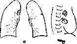
Differential diagnosis of pulmonary tuberculosis. As is known, a significant number of diagnostic errors are associated with the practice of recognizing tubercular lesions of the lungs. And this is understandable if one recalls how masked pulmonary tuberculosis proceeds in a number of cases. Very often it is difficult to distinguish general symptoms associated with tubercular toxemia from symptoms caused by other causes also of infectious origin. In the local pictures of processes, both during physical and radiological examination, there are also so many similar features that only the most careful synthesis of all clinical data can insure against gross diagnostic errors. For example, classic lobar pneumonia and the onset of caseous tubercular pneumonia have a number of common features—a stormy onset, stabbing pain in the side, high temperature, and only careful observation of the course of the disease, which in caseous pneumonia is distinguished by the absence of a crisis, and repeated careful examination of sputum for Koch's bacilli help to reveal the etiology of the disease. Very often in outpatient and even hospital practice, difficulties arise in differentiating grippal pneumonias and tubercular infiltrates, and here sometimes quite unexpectedly, data from X-ray examination and the presence of bacilli in the sputum contribute to a correct diagnosis. From the point of view of medical practice, three main groups of diagnostic difficulties and errors should be kept in mind: 1) errors associated with the interpretation of individual symptoms; 2) methodological errors, depending, on the one hand, on the technically incorrect application of one or another research method (e.g., a technically defective radiograph), and on the other, on insufficient or incorrect evaluation of the obtained research data; 3) the third group includes defects in research data and, in general, all materials collected about the patient. These are errors of diagnostic synthesis. Careful collection and study of anamnestic data, a pedantic attitude toward every research method used in the clinic, and a cautious, critical generalization of all data collected about the patient protect against gross medical errors. In the future, difficulties will be indicated that arise when interpreting both general phenomena accompanying various lung diseases and local changes in organs, with attention focused mainly on diseases more frequently encountered in the clinic. General symptoms. Such symptoms as general weakness, increased fatigue, loss of appetite, elevated temperature, especially of a subfebrile nature, sweats and sweating, etc., can be observed in a wide variety of diseases of both infectious and non-infectious origin. In the latter case, we are talking about disturbances of thermoregulation associated with deviations from the endocrine-vegetative system, either independent or depending on an infection suffered in the past. But a number of such diseases as malignant neoplasms of the lungs, echinococcus of the lungs at the beginning of the disease, can also cause general phenomena similar to initial tuberculosis. Therefore, a pedantic attitude toward collecting anamnesis and registering all clinical facts characterizing both the current state of the patient and the course of the pathological process in each individual case is extremely important. In the presence of the above-mentioned general symptoms, it is necessary to carefully examine all organs in order to thus either establish or reject the presence of a focus—the source of these phenomena of a toxic order. Here, of course, one should first of all remember the oral cavity, the upper respiratory tract, and in particular the accessory nasal sinuses (chronic inflammation in the maxillary sinus, frontal sinuses, etc.). Inflammatory processes in the roots of teeth, chronic tonsillitis, and pharyngitis can also cause a whole range of complaints and symptoms that are very similar to the picture of tubercular intoxication. In all these cases, fatigue and malaise are observed, often in the mornings; all three diseases can be accompanied by subfebrile temperature, and in all three diseases, a high erythrocyte sedimentation rate, etc., is observed. In diseases of the accessory nasal sinuses, a shadow corresponding to the anatomical localization of the process, purulent discharge from the corresponding sections during nasal examination, and leukocytosis are often detected on the radiograph. In tonsillitis—corresponding changes in the tonsils. As for pulmonary tuberculosis, it is necessary to keep in mind that often even very significant focal changes in the pulmonary parenchyma can be "silent," i.e., not audible, and they can often be discovered only by technically correct fluoroscopy together with a well-developed radiograph. Often in such cases, delicate pictures of limited dissemination or limited infiltrative foci are discovered on the radiograph, and sometimes even extensive tubercular lesions of the lungs previously unnoticed by the patient (phthisis inappercepta—unnoticed tuberculosis—of Breuning). In children and adolescents, and very rarely in adults, the cause of these general phenomena can be adenoid growths, causing difficulty in nasal breathing. The latter is established by a careful examination of the upper respiratory tract and the nasopharynx in particular. But in childhood, a frequent cause of general symptoms and especially unstable subfebrile temperature can be tubercular involvement of the bronchial glands. In this case, in addition to the above-described general phenomena, a lag in the development of the child or adolescent, pallor of the skin, and muscle lethargy, chest pains are often observed. With tumor-like glands: coughing in the form of attacks, sometimes sonorous, circulatory difficulties with the development of collateral pathways in the superficial skin vessels of the chest. Often the disease is accompanied by a tendency to bronchial catarrhs that worsen during transitional seasons of the year. The radiograph is convincing only with more or less pronounced tumor-like forms and especially with a shot in an oblique position, revealing the picture of the retrocardiac space (Fig. 1).
By the way, regarding subfebrile temperatures, one must first of all remember what was said in the diagnostics section on the question. Figure 1. Scheme: a - tuberculosis of the bronchial gland; b - the same.
X-ray in the oblique position, on the question of temperature fluctuations within the normal range. Monotonous subfebrile temperatures are not necessarily characteristic of tuberculosis. Where they are accompanied by marked emaciation, one should also keep in mind thyrotoxicosis, which can be established by a thorough clinical examination even in cases of mildly expressed forms of the disease (increase in basal metabolism!). According to some, vegetative neuroses, even without any provoking infectious onset, are also characterized by subfebrile temperature fluctuations, although in such cases it is recommended to search for a painful focus in all organs. Such a focus, the source of general phenomena, is often found in the biliary tract, in the endocardium, sometimes in the urogenital sphere, etc., but of course, in many cases, the cause of the general phenomena turns out to be tuberculosis indeed. Often, repeated sputum examinations help to establish the tuberculous origin of these general symptoms—the detection of tubercle bacilli in the latter, either by ordinary bacilloscopy, or by culture according to Ghon, or by introducing sputum into a guinea pig. The study of the formed elements of the sputum often provides the physician with indisputable data: for example, the presence of heart failure cells, eosinophils in bronchial asthma, pulmonary epithelial cells, etc. In some cases, Koch's tuberculin test supports a presumptive diagnosis. A positive reaction of F. A. Mikhailov (introduction of therapeutic doses of tuberculin with a count of eosinophils before and after introduction) usually indicates the presence of active tuberculosis in the organism. In the hemogram (Schilling), the presence of toxemia is confirmed by a nuclear shift. All this, together with the signs of a local process, upon careful synthesis of all clinical data, aids in diagnosis. Local signs of the disease. Along with the described general symptoms, the assessment of local manifestations of the disease often causes difficulties. Such symptoms as pain, cough, dyspnea, hemoptysis, and hemorrhage occur in various diseases of the lungs. Pain arises most often with concomitant lesions of the pleura. In some chronic forms, especially in fibrous processes, dry pleurisy is often encountered. Acute pain, "stabbing in the side," sharp cutting pain arise with pleurisy, acute caseous pneumonia, and with spontaneous pneumothorax, but persistent, steady pain, localized and radiating, is observed as a frequent sign in malignant tumors of the lungs. As for dyspnea, only a relatively small part of pulmonary tuberculosis patients (less than 1/3) suffers from marked dyspnea. When complaining of dyspnea, it is necessary to carefully examine, first of all, the circulatory apparatus and the kidneys. When examining the heart, one must remember about tuberculous lesions of the pericardium: exudative pericarditis is often the cause of dyspnea. Dyspnea in pulmonary tuberculosis more often manifests itself only during physical exertion. It may depend on the state of the nervous system, poisoned by specific toxins, but it can also be caused by a decrease in the respiratory surface, both as a result of consolidation of the pulmonary parenchyma and as a result of concomitant emphysematous changes. In some cases (e.g., after hemoptysis) and in tuberculosis, consolidation of the pulmonary parenchyma may be associated with atelectasis. In tuberculous cirrhosis of the lungs, dyspnea is associated mainly with disturbances of cardiac activity and congestive phenomena in the lesser circulation. The sudden appearance of dyspnea may be associated with spontaneous pneumothorax, observed most often in tuberculosis and very rarely in pulmonary emphysema. The accumulation of fluid in the pleural cavity can also cause severe dyspnea. By analogy with heart failure, the Brauer school describes pictures of respiratory failure associated with changes of central origin, with the exclusion of the respiratory surface, with a violation of the permeability of the alveolar lining in acute infections (grippal pneumonosis), etc. Cough is caused by the most diverse processes affecting the bronchial pathways, lungs, and pleura. A persistent cough is often a companion of more severe tuberculous processes in the lungs, but it is also encountered in the form of attacks ("hacking cough") in malignant neoplasms of the lungs, with irritation of the pleura, with tumor-like changes in the bronchial glands, and in Hodgkin's disease. Cough is often accompanied by the expectoration of sputum, sometimes even a mouthful. The latter is observed in bronchiectasis of various origins. Often, cough irritation arises in the mornings and is accompanied by the expectoration of a small amount of sputum. Often, sputum is expectorated after eating warm food. This is often observed in compensated fibrous processes of a tuberculous nature. In some cases, blood-tinged sputum and sometimes pure blood are expectorated during coughing. Most often this is observed in fibrous forms of tuberculosis, but not infrequently in fresh infiltrative processes of tuberculous origin. True, any damage to the pulmonary parenchyma, of whatever origin it may be, is capable of causing hemoptysis. In inflammatory processes in the lungs and bronchi, this occurs either as a result of active hyperemia or as a consequence of damage to blood vessels. But even with passive hyperemia, with congestive phenomena in the lesser circulation, hemoptysis is observed. Most often, hemoptysis of pulmonary origin is observed in pulmonary tuberculosis, then in malignant neoplasms, syphilis of the lungs, bronchiectasis, and finally in acute diseases: abscesses and gangrene of the lungs. Initial parenchymal hemoptysis in tuberculous lesions of the lungs is very similar to similar symptoms in echinococcosis. Profuse hemoptysis is more often of cavernous origin. Hemoptysis is not rare in grippal pneumonia, especially often observed in the 1919 epidemic. In such cases, hemoptysis is often associated with sharply expressed inflammation of the upper respiratory tract (tracheitis). Anamnesis, the presence of massive infection in the past, the entire clinical picture, the onset and course of the disease speak for hemoptysis of tuberculous origin (tubercle bacilli in the sputum are often not found during hemoptysis). Physical and even radiological data also often cannot help in the etiological characterization of the disease. Sometimes the nature of the disease is revealed only in the further development of the process after hemoptysis, e.g., during the formation of new inflammatory foci of an aspiration nature in the lungs. The course of the disease is characterized especially vividly by repeated X-ray examinations. In primary lung cancer and the hilar location of tumors, along with hemoptysis, phenomena of compression of neighboring organs are observed: bronchi (atelectasis), nerve trunks (paresis of the vocal cord, diaphragm), localized pain, etc. Along with such hemoptysis, hemoptysis is known in congestive phenomena in the lesser circulation, for example, in mitral stenosis, profuse and small hemoptysis with erosion of the bronchial wall by an aortic aneurysm (left side), etc. Finally, many speak of the possibility of hemoptysis in thyrotoxicosis, in phenomena of hysteria, although in the latter case it is more often a matter of sucking blood from the gums, and not of pulmonary hemoptysis. Usually, in the latter case, there are only squamous epithelial cells in the mucous sputum. Blood appears more often in the mornings, sometimes repeatedly and for a long time. Such hemoptysis is often associated with the phenomenon of hemorrhagic diathesis. Hemorrhages in sclerosis of the pulmonary artery and from varicose veins are a rarity. Differentiation of clinical pictures of the disease. For the purposes of differential diagnosis, it is practical to keep in mind the following anatomically easily characterizable main groups of changes in the lungs. 1. Consolidation of lung tissue. Here we are talking about the recognition of acute and chronic infiltrative and pneumonic processes occupying more or less significant areas of the lung, as well as small-focal consolidations of a disseminated type. 2. Processes accompanied by the formation of cavities, i.e., sequestering and suppurative, as well as cavities of a bronchiectatic nature. 3. To the third group should be attributed tumor-like formations in the lungs, benign and malignant tumors, and formations of parasitic origin (echinococcus). 4. Diffuse sclerotic processes. The early recognition of pulmonary tuberculosis has already been mentioned above. Consolidation of lung tissue. The classic acute onset of lobar pneumonia, accompanied by a sudden increase in temperature, stabbing in the side, etc., is well known to the physician. These phenomena are rarely observed during the onset of even very extensive tuberculous infiltrates. More often, the latter are accompanied by a grippal symptom complex with catarrh of the upper respiratory tract and sharp malaise, especially in the first days. In these cases, objectively, during physical and X-ray examinations, there is a picture of more or less sharp consolidation of lung tissue with dullness of the percussion sound, bronchial or harsh breathing, and various moist rales, sometimes sparse at first. On the radiograph, there is a more or less widespread darkening from a small limited infiltrate the size of a silver ruble to a shadow occupying an entire lobe (Fig. 2).
While in croupous pneumonia, along with the end of the classical cycle, the objective data also gradually disappear, with pulmonary tuberculosis infiltrates it is more often the other way around. The tempera-
ture
After an acute flare-up, sometimes accompanied by hemoptysis, the temperature either falls to normal or decreases to subfebrile levels. The general condition improves, but the data from physical and X-ray examinations remain unchanged for a fairly long time. Upon resolution and fibrous transformation, the local phenomena subside, but during disintegration, coarse bubbling rales appear, sometimes resonant and audible against the background of bronchial breathing. Only gradually, depending on the evolution of the process into fibrosis or disintegration, does a corresponding change in objective data occur. In childhood, rapidly passing consolidations of the perifocal inflammation type (epituberculosis—Eliasberg and Neuland) are often observed, frequently in the region of the pulmonary roots and often involving the interlobar pleura. The center of such inflammations is often tuberculous glands. One often has to observe these pictures in children with a whole range of signs which were described by old clinicians in the chapter on scrofulosis (blepharitis, phlyctenular conjunctivitis, etc.). A carefully collected anamnesis helps in diagnostic orientation and the establishment of the tuberculous etiology of the ailment, as well as indications of massive infection with tuberculosis, tuberculous changes in the past, and indications of provoking moments, for example, some preceding infectious disease—measles, typhoid infections, or sometimes (rarely) some vaccination. Decisive for the diagnosis is the course of the process and the presence of tubercle bacilli in the sputum; the presence of elastic fibers alone does not allow one to settle on one etiology of the process or another, since they are also encountered in suppurative processes in the lungs. It is well known how often, especially in outpatient practice, a tuberculous infiltrate passes under the flag of either influenza or some typhoid infection, and especially typhoid fever. With long-lasting catarrhal phenomena in the lungs after the influenza syndrome has smoothed out, one must always remember the possibility of a pseudo-influenzal picture, behind which the tuberculous nature of the process was hidden. Pneumonic infiltrations are observed in processes of various etiologies. Thus, in tertiary syphilis of the lungs, alongside the picture of chronic inflammatory processes, acute pneumonic flare-ups are observed, accompanied by hemoptysis, hectic temperature, and more or less sharp exhaustion. Here, of course, the anamnesis, a positive Wassermann reaction and sedimentation reactions, and the presence of specific syphilitic changes in other organs, for example, aortitis, gummatous skin changes, periostitis, etc., will provide much information. And in these cases, repeated negative searches for tubercle bacilli help to reject tuberculous changes, which are always possible even in a syphilitic. Interstitial-pneumonic changes in bronchiectasis are easily deciphered by the entire clinical picture, the general appearance of the patient, the characteristic abundant three-layer sputum, and sometimes a characteristic radiograph even without filling the bronchi with a contrast mass (Lipiodol), etc. Chronic infiltrating interstitial pneumonias are also observed in actinomycosis. The detection of fungal filaments and spores in such cases settles the question. Special mention should be made of the erroneous interpretation of hemorrhagic pulmonary infarction, especially in lesions of the lower lobes. Often, this disease is also treated for a long time as pulmonary tuberculosis. The onset of the disease in a carrier of a heart defect, after operations in the abdominal cavity, especially in the presence of inflammatory processes in the lungs in the past, an acute onset with hemoptysis, stabbing pain in the side and shortness of breath, a triangular shadow with the base toward the periphery on a radiograph, crepitant rales with pleural friction in a limited area, and the absence of bacilli in the sputum will help with orientation in the interests of timely rational therapy. Data obtained with the help of X-ray examination have taught the recognition not only of solid infiltrates occupying more or less significant areas of the lung but also of disseminated lesions of various types. In relation to tuberculosis in such cases, we are talking about various hematogenously disseminated forms, starting from acute miliary tuberculosis and ending with cold forms of hematogenous dissemination, to which the French gave the expressive name 'granulie froide'. The characteristics of all these forms are given above, and therefore one should dwell here only on some moments of the differential diagnosis of disseminated lesions in the lung. As for acute miliary tuberculosis, the meningitic form is usually not difficult to recognize. The typhoid and pulmonary forms present great difficulties. Difficulties often arise when differentiating in relation to typhoid fever. Here the situation is clarified only by observing the course of the disease. Often in acute miliary tuberculosis, a variable temperature curve of an indefinite type is encountered, often with increasing shortness of breath and cyanosis. Intestinal phenomena are rarer than in typhoid fever. The pulse is usually rapid (in typhoid fever, bradycardia), sometimes irregular slowing is encountered with meningitic phenomena. In the blood, there is relative lymphopenia. Throughout the entire disease, the diazo reaction is invariably positive. Timely blood culture and the Widal reaction will help with orientation. The presence of a picture of scattered foci in the lungs on a well-developed radiograph confirms the diagnosis. But there are cases where there is a syndrome very similar to acute miliary tuberculosis with a picture of scattered small-focal changes on a radiograph, which is considered as miliary tuberculosis. True, more often in these cases there is not such a high temperature as in miliary tuberculosis, and the diazo reaction is negative. These so-called chronic disseminated forms of tuberculous origin, especially cold disseminations, in the absence of bacilli in the sputum, can present significant difficulties in differential diagnosis. A picture similar to them is sometimes observed in pneumoconiosis, especially in chalicosis, but the occupational anamnesis and persistent abacillarity help to exclude tuberculosis. True, combined pictures of pneumoconiosis and pulmonary tuberculosis are often encountered. Processes accompanied by the formation of cavities. More often, caverns are of tuberculous origin and are the result of sequestering and suppurative processes associated with caseous necrosis of inflammatory foci in the pulmonary parenchyma. But the formation of cavities often accompanies chronic pneumonic processes of non-tuberculous etiology or arises as a consequence of an abscess or gangrene of the lungs. An acute onset, chills, high remittent fever, leukocytosis, sudden emptying of the focus with abundant discharge of pus and sometimes initially with blood, blowing bronchial breathing, a cavity with a horizontal fluid level in it, and the absence of tubercle bacilli in purulent sputum with an abundance of pus corpuscles—all this points to an abscessing pneumonia or lung abscess. In gangrenous-abscessing pictures of the disease, a sharp odor from the mouth during breathing and from the sputum is noted, which helps to decipher the picture. Combinations with tuberculosis are comparatively rare but are encountered. Einis saw a typical picture of a lung abscess several times, and then, a few weeks after the onset of the disease, tubercle bacilli appeared in the sputum. Whether an abscess formed in these cases against the background of old tuberculous changes or some old tuberculous focus accidentally fell into the purulent melting of lung tissue is difficult to say—both are possible. It is important to remember that sometimes the picture of an abscess masks a much more severe ailment, namely, lung cancer. Gradually, these cavities organize, and clinically there is almost no difference between the chronic picture of such a disease and bronchiectasis. The latter arise against the background of chronic inflammatory processes of various etiologies, such as chronic pneumonia, pulmonary syphilis, etc. The classic picture of bronchiectatic disease develops mainly as a result of shrinking processes of the pulmonary parenchyma, and not under the influence of changes in the pleura. This picture has already been mentioned above. Its recognition in typical cases is not difficult; in doubtful cases, the question is decided by a thorough examination of the sputum (macroscopic inspection—three-layer sputum!) and filling the bronchi with a contrast mass (Lipiodol) with subsequent fluoroscopy and radiography. Neoplasms of the lungs. Echinococcus of the lungs. Diffuse sclerotic processes. Benign tumors (e.g., fibromas) of the lungs are very rare. They are usually very poor in symptoms. Differential diagnosis does not present difficulty when taking into account the possibility of tuberculosis. It is another matter with malignant tumors and, first of all, primary lung cancer. Here, diagnostic difficulties often arise, all the more so because one has to encounter, albeit comparatively rarely, combinations of cancer and tuberculosis [based on material from the Rykov Tuberculosis Institute (Moscow) in 5 out of 33 cases]. In the initial period of the latter disease, a whole range of symptoms is encountered that are similar to tuberculous lesions of the lungs. One has to observe patients with lung cancer in whom febrile and subfebrile fluctuations in temperature are initially detected (in 24 out of 28 cases of primary cancer—material from the Rykov Tuberculosis Institute), hemoptysis (in 26 out of 28), and chest pain (in 23 out of 28 cases). Then the pain intensifies, and shortness of breath appears.
Sometimes dullness and a shadow are detected on an X-ray, often at the pulmonary root, initially appearing as a packet of glands. This shadow then grows quite rapidly, revealing tree-like projections into the pulmonary parenchyma (cancerous lymphangitis). Sometimes the picture resembles lobitis, often encountered in Pulmonary Tuberculosis, but more often without the preservation of the contours of the interlobar fissure. In some of these cases, upon auscultation of the area of dullness and X-ray shadow, weakened breathing was detected, usually in connection with atelectasis due to obstruction or compression of a bronchus. A frequent companion is exudative pleurisy. Out of 28 cases of primary lung cancer (material from the Rykov Tuberculosis Institute), exudate was observed in 8 cases, of which 3 were hemorrhagic. Sputum is often scanty. Neoplasm cells are an exceptionally rare finding in sputum. Repeated absence of Koch's bacilli in the sputum helps to exclude Tuberculosis. In doubtful cases, only the course of the process, and sometimes only an autopsy, clarifies its true origin. Metastatic tumors of the lungs are usually deciphered in connection with the primary lesion, for example, sometimes a very insignificant tumor of the stomach. It is impossible not to mention that sometimes lymphogranulomatosis arises in isolation in the chest cavity and affects the peribronchial glandular group. The blood picture (neutrophilia, leukocytosis, slight eosinophilia) and the absence of bacilli in the sputum aid in the diagnosis. One frequently has to encounter difficulties in the diagnosis of pulmonary echinococcus. Here, in the initial stage, there is a bothersome cough, and hemoptysis is observed. Sometimes pleural friction is heard, and there are exudative pleurisies. In the next stage of tumor formation, the situation becomes clear. Here, one usually does not err if, with a characteristic spherical shadow (echinococcus cyst), there are no bacilli in the sputum and a positive Casoni reaction is established. It should be emphasized that eosinophilia is by no means always observed in echinococcus, and one frequently has to see echinococcus, confirmed by subsequent surgical intervention, without eosinophilia. The main difficulties usually arise precisely in the initial stage, when a diagnosis of Pulmonary Tuberculosis is easily made. Diffuse sclerotic processes. Every inflammatory process in the lung can gradually turn into sclerosis. Dust diseases of the lungs are accompanied by characteristic pictures of disseminated sclerosis and emphysematous changes. Chronic bronchitis in such cases often dominates. Sclerotic processes in syphilis, sometimes sclerotic-pneumonic forms, we also try to distinguish in recent times (Fig. 3). Finally,
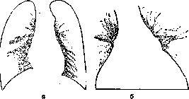
Figure 3. Syphilis of the lung (diagram). Sclerotic-pneumonic process involving the pleura: a - dorso-ventral image; b - in the Fleischner position (tilting the body backward).
and Pulmonary Tuberculosis often causes extensive sclerotic changes even far beyond the limits of the main focal lesions (classic cirrhosis of the lung). In these cases, recognition is sometimes very difficult, and the main support for the diagnosis in all doubtful cases will be the presence or absence of those same Koch's bacilli in the sputum.
V. Einis. X-ray diagnostics. The success of X-ray examination in pulmonary tuberculosis is determined by the fact that it provides a clearer and more accurate representation of the quantity, shape, and location of pathological foci than all other diagnostic methods. It allows for the recognition of a number of forms of tuberculosis otherwise inaccessible to examination. In combination with other methods, it allows one to approach the resolution of the question of determining the nature of pathological-anatomical changes. X-ray examination of the lungs can be considered complete only when both fluoroscopy and a radiograph have been performed. The technique of both methods must be impeccable. As a rule, the exposure time for a radiograph should not exceed 0.2 seconds, since the basis of the shadows of the pulmonary pattern and the lung roots is formed by pulsating arterial vessels. Furthermore, the parts of the lungs adjacent to the mediastinum are shaken by the pulsations of the heart. As a result, with long exposures, small foci of disseminated forms of tuberculosis and other minor changes may not be fixed on the radiograph. By the nature of the shadow of a focus, it is generally impossible to judge its etiology with certainty, and there is no such picture of X-ray changes that would be pathognomonic for tuberculosis. However, in practice, the characteristic and most frequently observed X-ray pictures in tuberculosis often allow the physician to assume the presence of a tuberculous process. If a good radiograph allows one to express an assumption about the prevailing nature of pathological-anatomical changes, it does not provide the possibility to judge the structure of each individual focus. The features of the image of the latter depend on too large a number of factors: the technique of the radiograph, the location of the focus in relation to the film and the focus of the tube, the state of the surrounding tissues, etc. There are lesions that are not detected even on a good radiograph. Such are many fresh disseminated processes, small foci lying in the region of the lung roots, and even quite significant changes hidden by the shadow of thickened pleura or other large foci, etc. A radiograph records only the statics of the tuberculous process, its picture at the moment of exposure. The development of the process in each individual case can be judged only by a series of successive radiographs. A detailed acquaintance with the normal X-ray anatomy of the lungs is absolutely necessary (see Lungs, X-ray diagnostics). Only by insufficient knowledge of the latter can one explain the fact that, under the influence of erroneous views on the development of tuberculosis, a series of signs of initial lung damage was created that did not stand the test at all. We are talking about the "apical veil," "enlargement of peribronchial glands at the lung roots," "stranding from the root to the diaphragm," etc. These symptoms, in combination with corresponding clinical signs, formed the basis of the syndromes of bronchoadenitis (in adults), apicitis, etc. These diagnoses, recently still so frequent, have now almost gone out of use. Isolated uniform darkening of the apex only rarely depends on pathological changes in the pulmonary parenchyma. More often, it is a consequence of the uneven thickness of the skin or muscle coverings of the right and left sides of the upper part of the chest, inaccuracy in the positioning of the patient or the X-ray tube, scoliosis of the cervical spine, thickening of the apical pleura, etc. Stranding from the root to the diaphragm was encountered for many years in fluoroscopy protocols and was interpreted as a sign of tuberculous peribronchitis. In reality, it was usually just a matter of the shadow of normal blood vessels of the lower parts of the lungs. It was overlooked that the vessels of these parts are normally somewhat wider, and that the shadow of the dome of the diaphragm cuts off the peripheral part of the lung field from below. Here, large vascular shadows reach the very edge of the field, unlike other parts where the pulmonary pattern in the marginal zones is almost absent. In cases of congestive phenomena in the lesser circulation, for understandable reasons, the vascular pattern of these same parts of the fields changes first of all. Finally, the shadows of normal or congested vessels of the lung root were constantly mistaken for pathologically altered peribronchial glands. These errors were largely facilitated by the blurriness of the shadow of the lung roots, which was obtained on radiographs with exposures of 1.5-2 seconds or more. The left root is usually, especially in pyknics, partially covered by the shadow of the heart, therefore enlargement of the peribronchial glands was usually found on the right. Of course, the glands of the lung root are sometimes changed in tuberculosis of the respiratory organs in children and even in adults so that they can be detected by X-ray examination, but the resulting pictures are very complex and require great caution in their interpretation. All the listed symptoms of initial pulmonary tuberculosis, just like the lack of clearing of the apex during coughing (Kreuzfuchs phenomenon) and the lagging of one half of the diaphragm during breathing (Williams phenomenon), have lost their diagnostic significance. The smallest of the shadows observed in pulmonary tuberculosis are miliary (see separate table, figs. 1, 2, and 3). They have a size from 1 to 3 mm, from a millet seed to a lentil seed. Their shape is usually round, less often irregular. The shadow of the focus is not particularly intense and is not homogeneous; the outlines are sometimes clear, more often more or less blurred. This type of focus is usually encountered in miliary tuberculosis and in other disseminated forms of tuberculosis. An elementary tubercle with its diameter of 0.05 mm does not give a shadow even on an impeccable radiograph. A miliary focus on a radiograph in tuberculosis thus corresponds to a focus of miliary alveolitis that has captured a group of alveoli, a confluent tubercle, or a miliary tubercle surrounded by a zone of perifocal inflammation. Certain difficulties are presented by the distinction of miliary foci from the point shadows of the cross-section of vessels, places of dichotomous division, and the crossing of the latter. The distinction of vascular shadows is facilitated by the following signs. A circle or oval of a vascular shadow usually lies along the course of the usual strip of the vessel shadow and in its size corresponds quite accurately to the diameter of the latter. The outlines of such a circle are quite even, and the homogeneity of the shadow is complete. Somewhat larger shadows are given by formations of the type of Charcot's peribronchitic nodule and processes capturing an entire acinus or a group of acini of the lung. The most characteristic shadows of these foci have the shape of a triangle or a trefoil. Usually, the edges of the shadow are quite clear, but at certain stages of the development of the tuberculous process or depending on the location of the focus and the technique of the radiograph, they lose this clarity. Since the studies of Graeff and Küpferle, the described type of foci is considered by many to be characteristic of productive tuberculous lesions (see separate table, fig. 4). To a certain extent, this is true, since purely acinous small foci are relatively rare with a predominance of exudative phenomena. Both miliary and acinous foci usually lie in isolation, not merging with neighboring foci. It would seem that with the multiplicity of foci of miliary tuberculosis, their shadows, falling on one plane of the radiograph, should have merged with each other. If, nevertheless, only a relatively small number of individual foci are visible on the radiograph, it is only because only the foci of the parts of the lung adjacent to the film are recorded on it. More distant foci are not captured, being illuminated by secondary rays emitted by neighboring foci and the surrounding lung tissue. With the involvement of a group of acini, the sizes of the foci and their shadows become larger, their shape more bizarre, and the unevenness of the shadow even clearer. With the involvement of a large group of acini, an entire lobule or a group of lobules, the shadows on the radiograph look like snowflakes. The diameter of such foci is 1-2 cm or more. Corresponding to the larger sizes of the foci, their shadow is also more intense than that of the foci described earlier. The central parts of the foci are the darkest; towards the periphery, the intensity of the shadow decreases, and the outlines turn out to be indistinct, blurred. The tendency of the shadows of individual foci to merge with each other is pronounced. Flake-like foci often correspond to pneumonic and pneumonic-caseous processes and are therefore designated by many as exudative or pneumonic (see separate table, figs. 5 and 6). However, it is more correct here too to limit oneself to purely descriptive names. Purely pneumonic foci give the same shadows as foci of cheesy pneumonia, while nodes of focal fibrosis are distinguished by greater clarity and irregularity of outlines. Usually, strips and strands extend from such nodes into the surrounding field, making them look like spiders. The next group of foci in many respects resembles the one just described. These shadows are usually larger, reaching from 1 to 5 cm or more in diameter. The shape of the shadows is quite strictly spherical, the outlines are sometimes very clear, sometimes more blurred. The intensity of the shadow is usually significant. In some cases, the focus seems completely homogeneous, in others, the darkening is uneven. This is, for example, how "early infiltrates" and tumor-like enlarged lymph glands look in tuberculosis (see separate table, figs. 7 and 8). The largest diffuse shadows occupy an entire lung field or its large parts. Their boundaries partly merge with the edges of the field. Most interesting are the changes corresponding to the involvement of lobes of the lung or their significant parts. Radiographs in these cases give a picture very similar to classical lobar pneumonia.
The most characteristic shadows in disease of the upper lobe of the right lung (lobitis dextra superior of French authors) (see separate table, fig. 9) are those that are most typical. The lower boundary of this lobe lies approximately horizontally, i.e., along the path of the X-rays. The boundaries of the other lobes lie more vertically. Thus, the upper lobe of the left lung can be called anterior, and the lower one posterior. In standard dorso-ventral radiographs, the image of the lower lobe of the left lung is superimposed on the image of the upper lobe, and the interpretation of the resulting pictures is difficult. Thus, the topography of the pulmonary lobes partly explains the commonly noted frequency of isolated solid lesions of the upper lobe of the right lung and the rarity of similar lesions of the upper lobe of the left lung. Other shadows do not correspond to the entire lobe, but to some part of it. Usually, in the upper lobes, the sections adjacent to the interlobar boundary are affected (periscissuritis of French authors). On the right, this forms a triangular shadow with the apex at the root and the base in the axillary region (see separate table, fig. 10). On the left, the shadow goes in an expanding band, rising from the root to the outer end of the clavicle. In the lower lobe of the right lung, the triangular opacity usually has its apex at the root, its base on the diaphragm, and one of its sides coinciding with the edge of the mediastinal shadow. All kinds of diffuse shadows are encountered in various forms of pulmonary tuberculosis. The last type is shadows in the form of strands, ribbons, and various networks. In some cases, this is a matter of intensification of the normal pulmonary pattern. The resolution of the question of the boundaries of the norm and the beginning of pathology is especially difficult here. In other cases, the observed changes clearly do not correspond to the normal pulmonary pattern. In addition to pulmonary changes, linear shadows can also be caused by pleural ones (see Pleurisy). Sometimes a linear shadow, upon careful examination, turns out to be composed of individual nodules. Shadows of the described type can occupy the entire surface of the lung fields (bronchitis, etc.) or some parts of them, most often the area between the root and the apex. Some authors consider most reticular shadows to be the normal pulmonary pattern, others consider them a remnant of previously transferred processes, and still others consider them active changes of an initial tuberculous process creeping from the root of the lung into its tissue (see separate table, fig. 11). Probably, most of the strand-like and reticular non-vascular shadows correspond to processes of sclerosis, thickening of the connective tissue framework of the lung. In the scheme presented here, the basis for dividing shadows into groups was mainly their area size, and their outlines and shape were partly taken into account. In recent years, however, attempts have been made to use the varying intensity of shadows. Two goals were set: to obtain an idea of the chemical composition of the focus under study and to determine its approximate depth, and therefore the volume of the lesion. The first goal is more or less convincingly achieved only for calcified foci, which give extremely intense shadows. However, recognition of caseous and non-caseous pneumonia and various other lesions by the intensity of shadows can still be considered impossible. The determination of the thickness of the pathological focus, in the opinion of some authors, can be achieved by comparing the intensity of its shadow with the intensity of the shadow of normal structures: ribs, heart, liver. It is possible to compile an approximate scheme according to which a focus giving a shadow equal in intensity to a rib shadow has an average thickness of 3 cm, with a shadow intensity equal to that of the heart, the focus has a depth of 7.5 cm, and with a shadow corresponding to the liver, the depth of the focus is 15 cm, etc. Knowing further the area of the focus, one can determine its approximate volume. Along with opacities in pulmonary tuberculosis, translucencies can also be detected, both diffuse and limited. Diffuse translucencies, in turn, fall into two subgroups. In the first, which is clearer upon fluoroscopy than in radiographs, the normal, often somewhat intensified, pulmonary pattern is preserved in the area of translucency. This is observed in tuberculous patients with vicarious emphysema. In the second subgroup, a vast zone is occupied by a completely homogeneous translucency. This happens with the accumulation of gas in the pleural cavity (artificial and spontaneous pneumothorax) or in the lung (giant cavities). Sometimes the lower part of the image is occupied here by an intense shadow with an upper boundary that is strictly horizontal in any position of the patient (pneumopleuritis, fluid in a cavity). Limited translucencies are divided into more or less round and tubular ones. The former in most cases correspond to cavities and only very rarely to bullae of marginal emphysema or limited pneumothorax. Sometimes a section of relatively normal pulmonary parenchyma surrounded by pathological shadows can be mistaken for the translucency of a cavity (see separate table, fig. 20). The character of the cavity image depends on a number of factors and, first of all, on the state of the surrounding areas. If the cavity is located in a slightly changed area of the lung field, it usually appears as a light circle or oval with a more or less wide border around it. The central translucency of such a ring-shaped shadow is sharper the greater the depth of the cavity and the less the lung tissue and pleura anterior and posterior to it are changed. On a good radiograph, the translucency of a cavity is by no means homogeneous, because the shadows of normal, and more often pathological, structures projecting here from other layers of the lung are drawn on it (see separate table, fig. 12). The ring-shaped shadow of the wall is characterized by its continuity and theoretically should be visible as a closed ring when examining the patient with different directions of X-rays. However, this is usually hindered by the shadows of normal structures of the chest (ribs, mediastinum, etc.) and pathological changes in its organs (thickening of the pleura, pathological foci in the pulmonary parenchyma, etc.). One sometimes has to search persistently, fluoroscoping the patient in different positions, moving the screen away from him (to increase the size of the shadows), etc. The recognition of cavities is facilitated by the frequent finding of a horizontal level of liquid content in them. Sometimes such a level is the only convincing sign of cavities. That is why the examination of a patient when searching for cavities should not be performed in a lying position; the patient must sit or stand. An important symptom of a cavity is its connection with the root of the lung by a strip of translucency, bounded by two dark walls. Such tubular translucency usually corresponds to a bronchus. Changes in their size during coughing, and in the rarest cases even during normal breathing, have very limited significance in the diagnosis of cavities. False ring-shaped shadows, resulting from the projection onto one plane of strand-like structures of different layers of the lung, decompose into their constituent parts when examined in different directions. Limited bullae of pneumothorax, resembling cavities, are rare and are usually easily recognized by the data of the anamnesis and clinical examination. Even rarer are the mentioned large bullae of marginal emphysema, which are usually located in the marginal zones of the lung. Their diagnosis is very difficult. If a cavity forms in a significantly changed area of the lung, in a zone of pneumonia or caseous decay, it appears as a translucency, as if scooped out with a sharp spoon. The boundaries of such translucencies are often bizarre, as if eaten away. Distinguishing such cavities from zones of normal or emphysematous tissue may encounter difficulties, but is greatly facilitated by parallel clinical examination (see separate table, fig. 6). At present, a number of typical cavity pictures have been identified: "early cavities" (see separate table, fig. 13), "stamped cavities of disseminated tuberculosis" (see separate table, fig. 3), etc. (see Cavities). Besides shadows and translucencies, which owe their origin to changes in the pulmonary parenchyma, X-ray examination in pulmonary tuberculosis captures shadows associated with lesions of the pleural layers (see Pleurisy). In some cases, their pleural origin is quite clear, in others it is determined with great difficulty or remains in doubt. By the quantity and size of various shadows and translucencies, one judges the extent of the lesion; by their character, location, and mutual combinations, one judges the nature of the anatomical changes. The goal of X-ray examination in pulmonary tuberculosis largely consists in identifying such typical pictures. The first manifestation of tuberculosis of the respiratory organs—the primary complex—is characterized mainly by the presence of two foci: pulmonary and glandular. The focus of primary pneumonia usually gives a large, diffuse, not particularly intense shadow. The homogeneity of the shadow is often incomplete. Often, the shadow of the pulmonary and glandular foci initially merges with each other, also absorbing the root of the lung. In most cases, the shadow then decreases, breaking up into its main parts, connected by a more or less wide band. This phase of bipolarity (Redeker) is characteristic, but not mandatory for the primary complex. Subsequently, the shadow of the pulmonary focus usually becomes gradually smaller, its outlines sharper. In conclusion, no visible changes may remain in its place, or a delicate network of induration of the interstitial tissue is formed, or finally, a small (from the size of a millet grain to a cherry) focus of intense opacity.
This is a calcified Ghon focus, the primary affect (see separate table, fig. 15). Changes in the bronchial glands provide clear pictures apparently almost only in the presence of pronounced inflammation in the pulmonary parenchyma adjacent to them (see separate table, fig. 14). Outside of this, the glands are clearly visible either when the foci contained within them undergo the same calcification as the primary affect, or when they are tumorously enlarged. The main clusters of bronchial glands are located at the bifurcation of the trachea, in a place inaccessible to examination during the usual dorso-ventral path of the rays. Small broncho-pulmonary glands of the lung root are included in a complex plexus of vessels and bronchi. Of all the groups of glands, only those adjacent to the trachea on the right, even with a relatively small enlargement, are clearly outlined against the background of the lung field in the form of the already mentioned spherical shadow (see separate table, fig. 8). If one adds that medullary swollen lymph glands do not noticeably block X-rays, it is understandable that soon after the resorption of the inflammatory infiltrate around the glands, their shadow decreases and disappears. As a result, the healed primary complex is recognized by the calcified primary focus in the lung field and similar foci in any of the groups of bronchial glands. More often, however, one of the two constituent parts of the calcified complex is not detected. It should also be remembered that the glandular origin of shadows is absolutely convincing only at the above-mentioned place at the edge of the trachea in the II intercostal space on the right. Rounded and polygonal shadows in the region of the lung root can be a projection not only of blood vessels, but also of pathological formations lying in the lung anterior or posterior to the root. For more precise localization of the foci, it is therefore necessary to resort to examination in the transverse position. Not in all cases does the primary complex end in a cure; sometimes progressive pulmonary consumption develops from it.
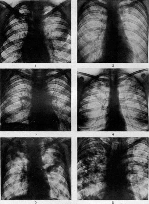
Fig. 1. Acute miliary tuberculosis (at autopsy miliary caseous pneumonia). Figure 2. In both lungs, an eruption
of subacute miliary spread. Figure 3. Chronic benign dissemination in both lungs with a thin-walled cavern on the right. Figure 4. Small foci of productive tuberculosis in the upper sections of both lungs. Figure 5. Flocculent shadows of caseous pneumonia. Figure 6. Flocculent shadows of caseous pneumonia with clearings of small caverns on the right.
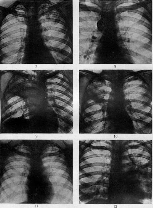
Figure 7. Spherical shadow of an "early infiltrate" under the left clavicle. Figure 8. Calcified glands. Fig. 9. Upper
lobitis on the right with a cavern under the clavicle: beginning of resorption and fibrous transformation. Fig. 10. Shadow of "periscissuritis" in the lower part of the right upper lobe. Fig. 11. Network of perilobulitis in the upper lobe of the right lung. Fig. 12. Cavern with a draining bronchus in the upper lobe of the left lung. Fibro-cavernous changes.
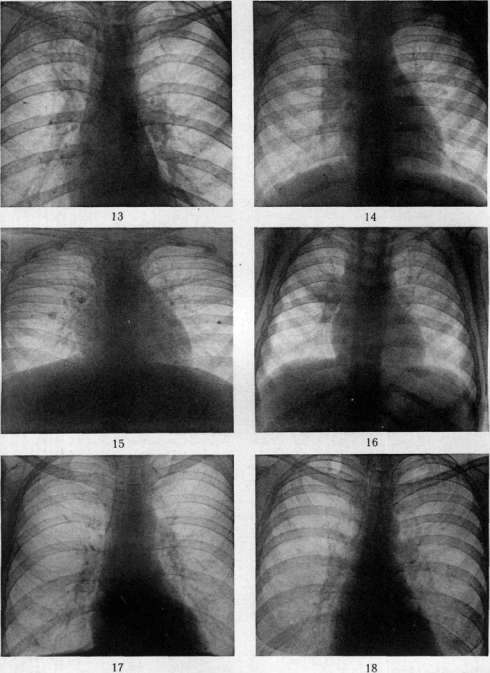
Figure 13. On the right, a subclavicular cavern connected to the root by a draining bronchus; around it are small foci of productive tuberculosis.
Fig. 14. Lesion of the glands of the root of the right lung. Figure 15. Calcified primary complex with a Ghon focus on the right. Figure 16. Perihilar infiltration on the right; linear shadow of thickened interlobar pleura. Fig. 17. Focal fibrosis of the apex of the left lung. Fig. 18. On the left, as well as under the clavicle, are cloud-like shadows of a tubercular infiltrate. To the art. Pulmonary Tuberculosis.
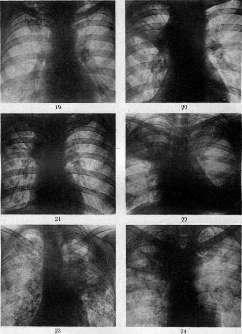
Fig. 19. Under the right clavicle is a round shadow of an infiltrate; outward from it is a round calcified focus. Figs. 20 and 21. Lobitis on the right and its fibrous transformation. Fig. 22. Caseous lobar pneumonia of the upper lobes, pericarditis. Fig. 23. Fibro-cavernous tuberculosis of the upper lobe of the left lung, fibro-productive process of the upper lobe of the right lung. Fig. 24. Fibrous upper lobitis on the right, caseous bronchopneumonia with cavities on the left. To the article Pulmonary Tuberculosis. The pulmonary focus in this case increases, forming ever new flocculent shadows or diffuse foci of the lobitis and periscissuritis type. Upon the formation of cavities, areas of rarefaction appear here, as if scooped out with a sharp spoon. Along with these shadows, or in their absence, the lung field is often seized by a multitude of punctate miliary foci. Such disseminated forms of tuberculosis are described below. Repeated outbreaks of pulmonary tuberculosis in children are characterized, on the one hand, by the appearance of vaguely outlined foci of opacification in the root areas of the lung field, and on the other, by less clear involvement of the glandular apparatus than in primary processes (see separate table, figs. 14 and 16). Often, here too, the shadows take on the character of those observed during infiltration of lobes or interlobar areas. The formation of cavities is a relative rarity; more often, gradual resorption occurs. In the course of the latter, small dense foci and strands appear in place of the diffuse opacification. In tuberculosis of the bronchial glands in children (non-tumor-like), the clearest picture is discovered upon involvement of the aforementioned packet of paratracheal glands on the right. A characteristic representative of hematogenously disseminated forms of pulmonary tuberculosis is acute miliary tuberculosis. While at the very beginning of the disease individual foci are usually too small and are not detected during fluoroscopy and even on radiographs, after a few weeks the situation changes. On a radiograph taken with a sufficiently short exposure, a fairly uniform scattering of small, very numerous, non-confluent, and identically sized foci is discovered (see separate table, fig. 1). Apparently, the formation of such miliary foci usually occurs in different parts of the lungs non-simultaneously. The longer the disease, the larger the individual foci, the more irregular their outlines, and the more uneven their sizes. Individual foci merge at this time with one another, and their distribution becomes less uniform. Often, changes in the upper parts of the lung fields turn out to be more pronounced than in the lower ones. Such pictures characterize subacute forms of miliary tuberculosis (see separate table, fig. 2) and serve as a transition from its acute forms to the group of chronic hematogenous dissemination. In the latter, one encounters, on the one hand, processes whose X-ray representation does not differ in any way from general miliary tuberculosis, while the clinical picture is relatively benign (see separate table, fig. 3). It is precisely cases of this type that served as the basis for sometimes erroneous descriptions of observations on the cure of acute miliary tuberculosis. Indeed, in chronic disseminated forms, one observes the disappearance of some foci and the calcification of others. Sometimes the foci disappear so that no traces remain of them; in other cases, only a part of them remains, more often located in the upper parts of the lungs. The hematogenous origin of such foci is judged by their identical sizes, their greater number, and by the presence of tuberculous foci that appeared simultaneously in organs of the systemic circulation, lymph glands, and pleura. However, multiple uniform eruptions can also be bronchogenic, as is shown particularly demonstratively by observations on the spraying of lipiodol in the lungs during coughing. A decision on the nature of the process in such cases is possible based on an assessment of the entire complex of clinical data. Often, in one or both apices, one finds small-focal shadows, the multiplicity and identical sizes of which compel many authors to recognize a hematogenous path of their origin. Sometimes small rounded petrificates (Simon's foci) are scattered there as well. In other cases, individual clearly outlined foci serve as inclusions in a network of more or less delicate strands (a picture of apical sclerosis) stretching toward the upper pole of the corresponding lung root. Sometimes such a network turns out to consist of individual nodules, giving rise to the thought of an eruption of tubercles along the lymph fissures of the lung stroma (perilobulitis) and of the spread of tuberculosis from the root into the lung field (see separate table, fig. 11). Changes in the pulmonary apices do not essentially differ from those observed in any other part of the lung field. Perhaps here, the involvement of pleural adhesions is more often expressed in the form of individual spots of opacification or in the form of bizarre bands forming the outlines of arcades downward from the second rib. In pneumonic (non-caseous) processes, otherwise called infiltrates, the previously described spherical shadows are encountered anywhere in the lung field, most often in its upper parts. Spherical foci more often lie under the clavicle (Assmann's focus, early infiltrate, subclavicular infiltrate). Their outlines either blur cloud-like into the surrounding field or are so sharp that they resemble a foreign body. Often, the first manifestation of pulmonary tuberculosis on a radiograph is small cloud-like foci, more resembling flocculent shadows in character (Grau's foci) (see separate table, fig. 18). During fluoroscopy, they are easy to miss. All three types of infiltrates (Grau's foci, "early infiltrates," lobitis) can appear either in a field that was completely normal before or in an area containing remnants of previously existing lesions (an indurative field after a pneumonic outbreak or hematogenous eruption, etc.). Finally, pneumonic outbreaks and zones of perifocal inflammation can envelop foci of any of the previously described types in a cloud of shadow. A tuberculous infiltrate, even reaching the size of a lobitis, can be resorbed without leaving behind changes detectable on a radiograph. Sometimes, in its place, there remains a network of strand-like shadows of sclerosis or small, clearly outlined foci of an acinous process, often delimited from normal parts of the lung field by a clear line of thickening of the interlobar pleura. Often, a small and unclear rarefaction appears first in the pneumonic cloud, gradually increasing and acquiring a more definite wall, a rim around it. Before us is an "early" cavity, sometimes with a wide ridge of perifocal inflammation, sometimes with a narrow ring from the period of resorption of the infiltrate, and finally, sometimes gradually decreasing and disappearing. In the latter case, a strip of a small scar more often remains in its place. Caseous tuberculous pneumonia gives, upon X-ray examination, pictures resembling those described for non-caseous pneumonias. It is often impossible to draw a boundary between them on the basis of an isolated radiograph. Lobar pneumonia gives in all cases the same shadow of lobitis, and the difference here is revealed only with the help of other methods of clinical examination or by repeated imaging (or fluoroscopy), which show, in the case of non-caseous pneumonias, a gradual resorption of the shadow or its decrease as a result of processes of shrinkage and scarring. In caseous pneumonias, the dimensions of the shadow do not decrease; only areas of rarefaction appear in it, as if scooped out with a sharp spoon (cavities) (see separate table, figs. 5 and 6), and large flocculent shadows of caseous bronchopneumonia may erupt in the surrounding areas. The picture of such pure bronchopneumonias is somewhat easier to distinguish. Here, the lung field is strewn with intense flocculent shadows, sometimes merging in the upper parts of the field and rarer below, sometimes densest at the roots and rare at the periphery. With multiple small cavities, characteristic pictures are created, described by French authors under the name of the bread crumb type. Pictures of pseudo-lobar lesions often arise, differing from true lobar ones by the presence of individual smaller flocculent shadows at the periphery of the lobe. Pronounced tuberculous cirrhoses are encountered in three main forms: extensive lesions occupying large parts of the lung field entirely, large- and small-focal fibroses, and finally, tree-like shadows often permeating the entire thickness of the lung. For all these types of lesions, the presence of displacements of adjacent organs is equally characteristic (see separate table, fig. 23). The light strip of the trachea and the shadow of the large vessels of the heart are displaced toward the scarring process, bending like a bow. The shadow of the heart is shifted there as well, simultaneously rotating. Taut pleuro-pericardial adhesions give angular outlines to its contours. The shadows of the vessels of the lung root are displaced. Thus, in cirrhosis of the upper lobe of the right lung, the right root rises to the level of the left or even higher, and the shadows of the vessels, usually winding from the root to the diaphragm, are pulled taut, straightening out completely. The same phenomena of shrinkage affect the bony skeleton, bringing adjacent ribs closer together and making their direction more vertical. Sometimes even scoliosis of the spine occurs.
As a result of extensive pleural changes, uniform shadows cover large areas of the lung field, and adhesions to the diaphragm lead to the closure of the pleural sinuses and disfigurement of the diaphragm dome. Diaphragm movements are impaired. The third characteristic feature of cirrhosis is the frequency of emphysematous lucencies in the surrounding areas. When, for example, a lobe of the lung is affected, it shrinks, while the adjacent one enlarges, encroaching upon it and correspondingly becoming more lucent. In focal fibrosis and in tree-like strands corresponding in their location to the vessels and bronchi of the lung, the lucency covers the surface of the entire lung field or both lung fields, creating a picture of tuberculous emphysema of the lungs. In a number of cases of pulmonary cirrhosis, there are shadows not of one type, but various combinations of them, e.g., focal and strand-like shadows side by side. The very nature of cirrhosis shadows was mentioned earlier. Caverns, which are frequent in cirrhosis, are characterized by thick walls, often angular due to the action of the same cicatricial traction. Giant caverns, occupying the areas of entire lung lobes, accompany cirrhosis more often than other types of changes. The variegation of the pictures becomes even greater due to the congestive intensification of the lung pattern and the formation of more or less characteristic rounded or tubular bronchiectatic lucencies. Along with the described pure types of tuberculous lung lesions, mixed pictures are also very common. The most important of these corresponds to chronic cavernous consumption or ordinary pulmonary tuberculosis (phthisis vulgaris). In this variegated picture, elements of all the previously described forms are mixed. The upper sections of the lung fields are most often occupied by shadows of focal fibrosis, lucencies of thick-walled caverns, and the diffuse haze of pleural thickenings. Almost always there are displacements of the trachea, large vessels of the heart, and the lung root. Below are large or small flocculent shadows, often corresponding to bronchogenic foci of simple or caseous pneumonia (see separate table, fig. 24). In the phase of a pneumonic flare-up of the process, individual clearly defined foci are enveloped by a diffuse, vague shadow. The picture is further complicated by the presence in some areas of clearly defined small foci, a network of sclerotic changes, or lucencies of vicarious emphysema. If pleural changes play a major role in all the described pictures, they acquire independent significance in various pleurisies (see). The most characteristic pictures of the main forms of pulmonary tuberculosis as they appear on radiographs were described above. However, cases are not rare where qualitative diagnosis of tuberculous changes is very complex, and even more often one has to be content with establishing the predominant nature of the lesions. Attempts at qualitative diagnosis by means of fluoroscopy should generally be rejected. It is a gross error to make a diagnosis of pulmonary tuberculosis based on data from an isolated X-ray examination, since all the pictures described here can also be observed in other diseases. In deciding the question of the tendency of the development of the tuberculous process, a single X-ray examination helps relatively little. Still, pictures of an early infiltrate, an infiltrate with a cavern, flocculent foci, etc., allow one to speak about the activity of the process. Much more is provided by a comparison of images produced with strictly identical technique repeatedly at known intervals of time. In cases of progression of the process, the number of shadows increases, their sizes become larger, belts of cloud-like shadow appear around them, caverns are formed, etc. In opposite cases, the shadows may disappear, decrease in size, and increase the clarity of their outlines. At the same time, ring-shaped shadows become smaller, their walls thinner, and sometimes the lucency disappears completely or is replaced by a narrow strip of shadow (scar!). Thus, serial images, and in extreme cases also fluoroscopy, provide extremely valuable objective data for determining the course of the tuberculous process. Such serial studies are also very important for evaluating the effect of various therapeutic interventions and especially for managing collapse therapy. X-ray examination helps to determine the nature of changes in the lung subject to surgical intervention, the location of the main pathological foci in it, the state of the second lung, etc. These data largely determine the establishment of indications for one or another type of intervention and the methodology for it in each given case. In relation to artificial pneumothorax, X-ray examination also helps, though not with complete certainty, to determine the location of pleural adhesions and to choose a site for puncture. After the introduction of the first portions of gas, a light, structureless strip appears at the edge of the lung field. If, despite fluoroscopy of the patient in different directions and in the phase of full exhalation, this strip is not visible, one can still manage to notice sharp changes in the character of cardiac pulsations. When gas passes between the lung and the heart, the normal smooth movements of the heart are replaced by jerky jolts, similar to the flapping of a sail. Subsequently, periodic examinations are performed. The location of adhesions, the maximum dimensions of the bubble, the degree of mediastinal displacement, the formation of mediastinal bays, etc., are determined by fluoroscopy immediately after insufflation. The state of the collapsed lung and the opposite one, and the minimum dimensions of the bubble are best examined before the next insufflation, when the gas bubble is smallest. The correct choice of intervals between insufflations and the amounts of gas introduced, as well as the correct management of artificial pneumothorax in general, is possible only with good X-ray control. All other types of collapse-therapeutic interventions—independent and auxiliary—exeresis of the phrenic nerve, cauterization of adhesions according to Jacobaeus, thoracoplasty, etc., require the same X-ray control. The latter is especially difficult with thoracoplasty in relation to the lung on the operated side. X-ray examination in a number of cases helps to distinguish pulmonary tuberculosis from other chronic diseases of the respiratory organs (see Lungs, X-ray diagnosis). Unlike malignant tumors of the lungs, pulmonary tuberculosis on serial images is characterized not by a steady and relentless increase in the size of shadows, but by a change of diverse periods. Pathological shadows sometimes increase in their size and number and often acquire a blurriness of outlines, then decrease and sometimes become more clearly defined, and then finally do not change at all for a long time. If, with a lung tumor, changes usually remain unilateral for a long time, then with extensive tuberculous lesions they are found in the second lung field as well. Diaphragm paresis is a rarity even with extensive tuberculous lesions and is very common with lung tumors. In bronchogenic cancer, the bronchus often becomes impassable for Lipiodol soon after the onset of the disease, while in tuberculosis it retains a free lumen. In lobar tuberculous processes, the dimensions of the affected area decrease compared to normal, whereas in tumors, except for the rarely encountered scirrhus, they increase. The main difference between syphilis of the lungs and tuberculosis boils down to the fact that anti-syphilitic treatment usually leads to the rapid disappearance of the pathological shadows of the former and does not affect the shadows of the latter origin. The frequent location of shadows in the root zone in elderly syphilitics is atypical for tuberculous processes in this age group. Unlike bronchiectasis and other bronchial lesions, tuberculosis usually gives greater changes on X-ray examination than could be expected from physical examination data. In tuberculosis, as a rule, changes in the upper sections of the lung fields predominate, while in bronchial diseases—in the lower ones. However, both syphilis and malignant tumors can produce pictures indistinguishable from those observed in pulmonary tuberculosis, and bronchitis and bronchiectasis can be caused by tuberculosis itself. Diagnosis is especially complex with the simultaneous involvement of the lungs by tuberculosis and some other process. Of particular practical importance is the sometimes complete similarity of X-ray pictures of pneumonic forms of tuberculosis and pneumococcal and influenza pneumonias. The shadows of the latter can completely imitate 'early' infiltrates, hilar infiltrations, etc. A single, and often even a repeated X-ray examination, provides perhaps no reference points here for the diagnosis of the affliction. In recent years, X-ray examination of large groups of the population has been spreading more and more widely for the rapid and cheap detection of pulmonary tuberculosis in them. This path is very useful for examining those being drafted into the army, when hiring for large new construction projects, when monitoring persons meeting with tuberculous patients in everyday life and at work, etc. Fluoroscopy allows excluding cases of pronounced pulmonary tuberculosis more reliably and faster than all other methods of examination. More detailed data are provided by images on film or special paper.
F. Mikhailov. T. Prognosis. Prognosis for pulmonary tuberculosis is extremely difficult to establish. The protracted nature of the disease, numerous, for the most part suddenly arising flares with subsequent, sometimes very long intervals make establishing a prognosis for a more or less long period sometimes completely impossible. Depending on the nature of a newly arisen metastasis (e.g., formation of a cavern at the site of an old calcified focus with seeding through the bronchial system or a hematogenous metastasis on the soft membrane of the brain during a sluggishly flowing tuberculosis of the bronchial glands, etc.) the process can, from being fully compensated and by all signs completely quieted, immediately become violently progressive and incurable. This instability particularly characterizes chronic forms of pulmonary consumption. Despite all the complexity of the interaction of conditions determining the course and development of the process, one can, however, note a number of moments characteristic of individual phases of the development of the tuberculous process, which to a certain extent allow one to assess the prognostic prospects for a given patient undergoing a given phase of the process. Thus, the phase of the primary complex in the overwhelming majority of cases resolves quite successfully and ends with the complete liquidation of active phenomena. The remaining phenomena of allergy, intoxication caused by the presence of Koch's bacillus in the organism, and reduced resistance to various irritations in those infected with tuberculosis manifest themselves mainly in childhood, the more sharply the fresher the infection and the younger the organism; by the period of maturity, in most cases, these signs also cease to manifest, and insignificant traces of a once-existing infection (petrificates, fibrosis, pleural adhesions, etc.—see above) are discovered mostly as an accidental finding during mass examinations in completely healthy, able-bodied people. In early childhood, less often at the age of maturation, and even more rarely, but still, as observations of recent years show, quite often in adulthood as well (about 200-250 such cases pass through the Moscow Tuberculosis Institute annually), cases are observed where the primary complex does not undergo self-healing, but immediately after emergence or after some time after a lull begins to show tendencies toward the growth of foci, toward the formation of lymphogenous and hematogenous metastases and generalization of the process. If in such cases it is not possible to localize the process (artificial pneumothorax, general treatment, etc.), then quite soon the disease acquires a malignant course, passing into generalized tuberculosis with violent dissemination or giving large foci of caseation with decay (see above—Clinical picture). Sometimes the process smolders for a long time, giving from time to time trifocal reactions or being accompanied by hematogenous spread of bacilli and the formation of small, rapidly healing metastases (protracted generalization—protrahierte Durchseuchungsperiode, Schürmann), but in such cases, the formation of large foci in organs (kidneys, bones, etc.) can sharply change the prognostic prospects for the worse. The course of hematogenous forms of tuberculosis is especially capricious and unpredictably variable (see above). The emergence of a cavern, if it is impossible to quickly liquidate it with therapeutic interventions (see above, as well as Caverns, Artificial pneumothorax, Thoracoplasty, etc.), is a moment that sharply worsens the prognosis, and all the physician's attention must be directed toward preventing this almost fatal turn in the course of the tuberculous process. The appearance of tuberculous metastases in the larynx is just as serious in prognostic terms; tuberculous involvement of the intestine darkens the prognosis even more, and the appearance of symptoms of amyloid degeneration of organs (in cases with prolonged suppuration: abscesses, empyemas, etc.) is completely bleak. Finally, even when the tuberculous process quiets down, the remaining cirrhotic changes and pleural adhesions in the lungs, as well as changes in the liver, kidneys, heart, and other organs that developed as a result of prolonged intoxication, can have a significant influence on the functions of organs important for life (first of all, the cardiovascular system), the patient's working capacity, and even viability, and thus significantly darken the prognosis. If, therefore, establishing a final or long-term prognosis is difficult or even sometimes impossible, then with systematic observation of the patient and careful consideration of all clinical, social, and everyday data, one can at any given period, based on the entire complex of facts, determine the prognostic prospects with fairly high accuracy. In this regard, some data from clinical-laboratory examination acquire very serious significance. 1. Age-related moments have no small significance for assessing prognostic prospects. In early childhood, the transitional period, and from 45 to 55 years of age, severe, acutely proceeding flares are more often observed, and the prospects for the outcome of a disease that has manifested during these periods of life must be determined with special restraint and caution. Also, a moment that worsens the prognosis is the disease in people from families burdened with tuberculosis, who have already lost a number of their members to this disease. 2. A number of diseases have long been known as provocateurs of tuberculous flares, and a tuberculous patient contracting one of these diseases can sharply worsen the course of the process. Especially serious is the disease in early childhood with measles, pertussis, or scarlet fever. The role of typhus and influenza is less clear, since very often the diagnoses of these diseases are made erroneously during acute flares of tuberculosis, and thus a false impression is created as if the disease preceded the tuberculosis. Chronic lung diseases, conioses, and plastic pneumonias do not have a clear provoking influence on the tuberculous process, but the increased incidence of active forms of tuberculosis in such tuberculous patients still does not allow one to exclude their significance in worsening the prognosis. Even more complex and confused are the interrelationships of syphilis and malaria with tuberculosis. Along with a course that is completely non-reactive on the part of tuberculosis, cases of a definitely malignant turn of the process are observed after the addition of one of these diseases to chronic tuberculosis. The antagonistic influence of arthritism on tuberculosis must be evaluated very cautiously, as cases are observed where, despite pronounced phenomena of arthritism, the tuberculous process progresses and invalidates the person. 3. The patient's appearance often provides rich material for a prognosis: swollen facial features with a fleeting bright flush, full bright red lips, eyes with a veil—the typical appearance of a patient with an acute exudative-caseous process—together with a sharp, characteristic for this form smell of sweat, difficult to eliminate even with careful hygiene, immediately create an impression of a very serious situation for the patient, despite even a relatively good state of nutrition and general well-being. The appearance of a cirrhotic patient in a period of cardiac decompensation (dyspnea, heavy cough, cyanosis, earthy complexion) and a patient in the terminal phase (sharpened features, mummy-like expression, sharp emaciation, dry, peeling skin, etc.) is equally characteristic. 4. The patient's weight in itself does not yet play a role; what is important is the difference between the weight at the moment of examination and the weight during a period of full health. Even more important is the tendency of the weight to increase or decrease. A drop in weight without visible causes during good nutrition should attract the physician's attention. Asthenics usually give lower weight indicators against standards. In such cases, the state of general nourishment can be determined by examining the skin fold according to Bunak (the fold in the lumbar region, taken between the index finger and thumb, should not be thinner than 1-1.5 cm). 5. Temperature. With the exception of chronic forms of cavernous consumption and destructive chronic forms of tuberculosis of the bones, and tuberculous lesions of the skin, the presence of normal temperature indicates a quieting and stabilization of the process. Flares of the process are always accompanied by temperature rises. The temperature during severely proceeding diffuse pneumonic and hematogenously disseminated processes takes the form of high fever with various forms of temperature waves (see above). The duration of this wave and its amplitude are to a certain extent directly proportional to the severity of the course and the gloominess of the prognosis. The speed of the fall of the wave and the transition to normal temperature during the implementation of therapeutic measures and regimen corresponds to a certain extent to the tendencies of the process toward quieting, and with the persistence of an afebrile state, it can conditionally serve as a sign of the possibility of a favorable outcome. Sometimes, however, even after a multi-month febrile period, a favorable and stable turn and lasting recovery can occur. The measurements of temperature simultaneously in the armpits on both sides recommended by Sternberg can in some cases help to detect more active foci on the side of the higher temperature (check the thermometers). 6. The pulse for the most part corresponds to the temperature and is very rapid during high fever. Constant tachycardia and instability of the pulse, which becomes sharply rapid even with small movements, excitement, etc., in cases of tuberculous diseases proceeding at the moment of observation without fever, acquire independent and unfavorable prognostic significance. With such a gap between the temperature and pulse curves, one has to expect new and severe flares and metastases. 7.
Sweating, if there are no other causes for its occurrence and if it accompanies a clearly expressed focal or diffuse tuberculous process, is also a sign that worsens the prognosis. 8. Hemoptysis in itself, if it is not very profuse and does not recur very often, does not worsen the prognostic outlook, but a complication with aspiration pneumonia can catastrophically change the course of the process and therefore, as a rule, hemoptysis is considered prognostically a grim symptom. 9. Sputum in the presence of tubercle bacilli and elastic fibers always indicates tissue breakdown and the danger of the formation or presence of cavities. Thus, the appearance of tubercle bacilli and elastic fibers is always considered an indicator of worsening prognostic prospects. Although cases of prolonged bacillary excretion with a stationary state of tuberculous foci are known, in the overwhelming majority, progression of the process is observed with increasing disability over 3-4-5 years. Generally speaking, the number of bacilli in the field of vision is not of serious significance; however, with the correct performance of sputum analysis (see), the constant detection in the sputum of a multitude of bacilli and elastic fibers with the preservation of their alveolar structure indicates ongoing destruction; conversely, a sharp decrease in bacilli to single bodies in the preparation, their disappearance for a more or less prolonged time (facultative bacillary excretors) indicate the development of fibrosis, the localization of areas of breakdown, and the possibilities of a favorable outcome. Persistent disappearance of elastic fibers and tubercle bacilli, not detectable even upon culture and inoculation into a guinea pig, allows one to outline prospects for the cure of the process (given other favorable signs) the more confidently, the longer this state lasts. The very amount of sputum (daily, without saliva) is determined to a significant degree by the state of the bronchi and upper respiratory tract, but with ongoing breakdown, it usually increases, and with the healing of foci, it decreases and sometimes disappears completely. Thus, measuring the daily amount of sputum also helps to substantiate the prognosis of a given case. The protein content in the sputum also increases to a certain extent in parallel with the intensity of the breakdown processes. The remaining bacterial flora does not have decisive prognostic significance. But still, the presence of a large number of pathogenic microbes in the sputum should attract our attention (danger of mixed infection). 10. The appearance of protein and parenchymal elements in the urine signals only acutely occurring processes or far-advanced chronic processes, when the prognostic prospects are already clear. The appearance of tubercle bacilli in the urine is of enormous significance (collect urine for a day, centrifuge the sediment, make a culture to isolate the strain, repeat the examination three times), indicating, upon the appearance of blood and protein, damage to the renal tissue. This fact makes the prognosis immediately very serious and raises the need to decide the question of appropriate therapeutic measures. The appearance of the Weiss urochromogen reaction (see Weiss reaction) or the diazo reaction (see) is noted already in the period of a far-advanced, malignantly occurring process and only complements an already bleak prognostic symptom complex. 11. Stool. The presence of tubercle bacilli in the stool in adults with destructive pulmonary processes adds nothing to the data already usually available. Detection of blood according to Gregersen, with the exclusion of meat for 3-5 days and the absence of bleeding from the stomach or lungs, indicates ulcerative lesions of the intestines and is a factor worsening the prognosis. In small children who do not expectorate sputum, tubercle bacilli in the stool indicate the presence of breakdown in the lungs (verify by examining gastric mucus taken on an empty stomach) and play the same role as the detection of tubercle bacilli in the sputum. 12. Blood provides extremely valuable data for establishing a prognosis. It is necessary first of all to note the significance of the erythrocyte sedimentation rate (ESR). With the exception of rare cases, ESR indicators are normal only in the absence of signs of an active process. If there are no other causes for the acceleration of the ESR besides tuberculosis, then this acceleration speaks for the activation of the process. One can be guided, as a rule, by ESR indicators to evaluate the effectiveness of therapeutic measures and to determine the tendency of the process toward subsidence or progression. An indispensable condition is the repetition of the ESR performance to compare a series of observations. Systematic observation and evaluation of the activity of the process are impossible without the ESR. The Daranyi, Costa, and some other reactions, proposed in their time, are less simple and clear than the ESR and have been supplanted by it in the USSR. Hyperleukocytosis in the absence of other causes speaks for the presence of large exudative-caseous acute pneumonias and the presence of cavernous breakdown. A shift of the neutrophil group to the left speaks for the active character of the process, and the degree of the shift usually corresponds to the freshness of the flare-up. Lymphocytosis with a small shift and a leukocyte count close to normal speaks for the appearance of reparative phenomena. Finally, an increase in monocytes as a rule corresponds to the moment of the formation of new foci (this is especially clearly seen in fresh hematogenous eruptions), and a drop in their indicator with unfavorable indicators of other groups (shift to the left and neutrophilia, lymphopenia) speaks for the onset of a phase of unfavorable course of the process (experimental and clinical observations of N. A. Shmelev at the Moscow Regional Tuberculosis Institute). Thus, not being specific for pulmonary tuberculosis, these hematological studies, with the exclusion of other diseases leading to analogous changes, help in establishing a prognosis for tuberculosis. The detection of tubercle bacilli in the blood as a sign of the presence or possibility of hematogenous dissemination, as well as the isolation of other microbes from the blood, are, of course, a factor that makes the prognosis, if not more severe, then in any case more uncertain and unstable. 13. Many do not recognize the prognostic significance of tuberculin reactions, however, the observations of Ravich-Shcherbo and Kholtsman allow their indications to be used among a number of other criteria. Favorable significance is held by bright, sometimes hyperallergic reactions with exudative phenomena, but with a short course. They are observed in active processes, but with good resistance of the organism. Unfavorable significance is held by hyperallergic prolonged reactions (active, acutely occurring processes with a tendency toward generalization) and sluggish, late, and prolonged hypoallergic reactions in chronic processes with poor reactivity of the organism. Serological reactions (complement fixation with Besredka antigen, etc.) still cannot be used for prognosis. 14. X-ray diagnostics in itself does not provide grounds for a prognostic evaluation of a case, but in combination with the remaining data, it plays a large role, giving a vivid characterization of the pathomorphological changes, the extent of the process, and on serial films or sketches of fluoroscopy data, registering the changes occurring in one direction or another. As the most general rule, it should be noted that the prospects are the more favorable, the older the foci (fibrosis, calcifications), the smaller they are in size and the less numerous, and, conversely, the fresher the foci (infiltrative, perifocal reactions, pneumonic foci), the more diffuse and larger they are, and the greater their number, the grimmer the prognostic prospects. 15. The degree of reduction in vital capacity (spirometry) and the breath-holding time (according to Stange or other methods), indicating the size of the lesion of the pulmonary tissue, adhesions of the pleural layers, emphysema, and disturbances in the compensation of the cardiovascular system, is a serious objective prognostic criterion. A decrease in these indicators by more than 50% against standards, in the absence of a tendency toward increase, is a serious prognostic sign of a far-advanced, difficult-to-cure process, in which the use of active therapy (in particular, surgical) is not indicated. 16. As treatment progresses, the prognosis may change depending on the effect of therapeutic measures. Thus, for example, a rapid drop in temperature during bed rest or upon the induction of artificial pneumothorax, the disappearance of bacilli and radiological signs of a cavity after collapse therapy measures, a persistent increase in weight with a decrease in local tuberculous changes, etc.—all these are more or less reliable signs of a turn for the better, and the more stable they are, the longer they hold, the more reliable the prospects for a good outcome become. But great caution in formulation is necessary on the part of the physician, since it is sometimes enough for the appearance of, for example, exudate during artificial pneumothorax or another complication for a sharp, sometimes irreparable deterioration to occur in the course of the process. Thus, the assessment of the long-term results of our measures in the treatment of tuberculosis and the establishment of a more or less firm prognosis upon the completion of treatment should be carried out after at least 2 years of observation, and only those cases should be considered firmly cured where, after 2 years of the regime of life and work usual for the given person, no signs of activation of the process are noted. 17.
All other things being equal, in cases with prospects for a favorable outcome, the prognosis depends to a significant degree on the behavior of the patient, his ability to consciously and precisely follow medical instructions, and the conditions of his daily life and labor.
V. Holzmann. VI. Treatment. The treatment of tuberculosis makes very great demands on both the physician and the patient. The chronic, for the most part, course of the disease with periods of flare-ups, alternating with intervals of remission of the process, sometimes for years, requires from both the physician and the patient much patience, persistence in carrying out the necessary therapeutic measures, and skillful maneuvering depending on the stage of development of the process at a given period, the living and working conditions of the patient, and other circumstances. Especially important is the continuity of the plan and the unity of therapeutic guidelines when the patient passes from one physician to another. In cases with a protracted course of an active process, it is necessary to organize the patient's treatment so as to concentrate therapeutic measures at the most necessary moment, and in periods of remission, to grant the patient the maximum freedom possible according to his condition in organizing his work and rest. The constant consciousness of the need to carry out these or those prescriptions of the physician bores the patient, begins to irritate him, and may lead to a decision to stop treatment altogether and leave medical supervision. It depends on the tact and experience of the physician whether he will be able to keep the patient within the framework of a cheerful, active mood and preserve in him the will to fight for health, without turning the patient into a fearful person constantly obsessed with the thought of the dangers awaiting him. No less important is the education of the members of the patient's family, friends, and work colleagues in a conscious and sympathetic attitude toward the treatment measures being carried out in the given case. All these fundamental prerequisites are provided in the organization of the Soviet tuberculosis dispensary, where, through the combined efforts of the physician, the nurse, and the social activist, it is possible to fully resolve both the unity of the treatment plan and the constant influence on the patient's will and the behavior of those around him in a direction that guarantees the best implementation of therapeutic instructions. We still do not possess methods of treatment that eliminate the causative agent of the disease and thus any possibility of its further manifestations. All our therapeutic measures are aimed at making the patient's organism most resistant to the spread of the process and to the influence of Koch's bacillus. More precisely, we strive to create conditions that inhibit the development of already emerged tuberculous foci, the spread of Koch's bacillus throughout the organism, and their multiplication in the foci. The encapsulation of the focus, the development of connective tissue in it, and the resolution of exudative phenomena are conditioned by the active activity of the reticulo-endothelial system; an increase in the activity of this system is achieved by a number of general influences on the organism (see below). The works of Gavasch (Central Tuberculosis Institute, Moscow) clearly demonstrate the great role of monocytes in the processes of healing the focus (phagocytosis). Experimental works by Shmelev (Regional Tuberculosis Institute, Moscow) speak of the same thing. Model (Moscow) proved that the multiplication of Koch's bacillus ceases upon the enrichment of the nutrient medium with capillary-active substances (salts of fatty acids, protein breakdown products, lecithin, and many others) and upon the alkalinization of the medium beyond the optimum pH necessary for the growth of Koch's bacillus. The indicated experimental data provide a solid theoretical foundation for empirically tested methods of general therapy for tuberculosis. In this direction, we can have a number of highly effective measures that fully justify the currently universally accepted position on the curability of tuberculosis, especially if treatment is started in a period when the process is still fresh and little spread, and is conducted skillfully in accordance with the individual indications of the given case. The basis of the treatment of tuberculosis in all its manifestations is the hygienic-dietetic regimen. Its elements—rest during periods of exacerbation, rational nutrition, hardening, and physical training as the active phenomena subside—were tested long ago, even by ancient medicine, and have passed through a number of centuries without significant fundamental changes. Recently, the credit for the particularly thorough and concrete development of the details of the hygienic-dietetic regimen is attributed to Brehmer (80s of the 19th century) and his successor Dettweiler, and in terms of application in a therapeutic setting and for therapeutic purposes, a great deal of graduated labor has been done by Wolff (Reiboldsgrün, Germany) and Paterson (England), who about 30 years ago carried out a great deal of work on the propaganda of so-called occupational therapy in tuberculosis sanatoriums. The hygienic-dietetic regimen itself can be carried out in the patient's home environment if the necessary objective conditions are present, but practice shows at every step that even with all the objective conditions present, both the patient and those around him very often allow such violations of the regimen in the home environment that reduce its therapeutic value to zero. Therefore, in cases where treatment is started for the first time in periods when it is especially important to carry out the regimen with all consistency, it is necessary to place the patient in a special tuberculosis hospital of the hospital or sanatorium type and keep the patient there until his condition allows him to switch to a lightened regimen, feasible in the patient's home life. The main tasks of the hygienic-dietetic regimen are: a) shifting the process from the phase of progression, flare-up, to a period of the most stable remission possible, b) raising the patient's nutrition if it has suffered during the exacerbation, c) hardening the patient, strengthening his neuro-psychic sphere through a system of physical therapy procedures, d) cultivating in him the necessary hygienic habits and self-discipline in caring for himself. Since in the overwhelming majority of cases the treatment of tuberculosis begins during a period of exacerbation, with elevated temperature, the first stage of the hygienic-dietetic regimen is aimed at eliminating fever and shifting the process to an afebrile course. The best means for eliminating fever turns out to be bed rest. Therefore, in all cases with persistently elevated or unstable temperature that shows large fluctuations, treatment begins with placing the patient in bed and completely forbidding any movements. In well-organized tuberculosis hospitals, with sufficient staff, it is necessary to provide the patient with such care that he is spared even the need to walk or go to the toilet. Where, due to local conditions, this is associated with difficulties (general wards, etc.), and the patient's condition allows for small movements, one can make concessions and allow the patient to perform his morning toilet outside the bed and in the toilet. The patient's bed must be comfortable, equipped with a good mattress and a bedside table, allowing the patient to eat, read a book, etc., in bed. It is especially important that the mattress and blanket sufficiently protect the patient from drafts and that the bed temperature be even, corresponding to the 'comfort zone'. Under such a strict bed rest regimen, the patient must remain all the time until the temperature drops to normal or insignificant subfebrile figures with a small (less than 1°) amplitude. If temperature measurements over 5-7 days have shown a persistent disappearance of fever, bed rest is replaced by room rest: the patient is allowed to sit in an armchair, first during meals, then for reading and needlework, and is allowed to walk around the room and go out onto the balcony attached to it. If these movements do not cause an increase in temperature, walks along the corridor of the medical institution and going out to the dining room and common rooms are allowed. Finally, if the patient's movements inside the building and his stay out of bed are gradually brought up to 3-4 hours without a temperature reaction, the patient is allowed walks in the fresh air, first after breakfast and evening tea for 15 minutes, then, in the absence of a temperature reaction, their duration is increased daily by 15-20 minutes until the stay outside the building fills all hours free from treatment, meals, and sleep. The speed of transition from one regimen load to another must be determined strictly individually. The criteria are the indicators of the activity of the process (see above—clinical, prognosis), primarily temperature, pulse, weight. Any scheme is unacceptable, regulating the regimen without direct conversation with the patient, based on reports from a nurse or reviewing diaries, is unacceptable. The slightest symptom of fatigue, adynamia, or increased intoxication must be taken into account as a signal requiring a slowing down of the pace of regimen loads. It is better to move forward more slowly than to be forced to return back during a new flare-up. The regimen must be regulated and transitions to new loads must be made with particular caution during periods with unstable weather (spring, autumn). If the patient tolerates a one-hour walk (4 km on flat terrain or with a slight 7 degree incline uphill) without reaction, then he can be allowed to participate in games that do not require sharp movements and running, and to work in workshops. And here, of course, the same strict control is necessary. Whatever the patient's loads during treatment, he retains as a mandatory minimum the prescription to be in bed from 10 p.m. to 8 a.m. and to spend 1.5-2 hours in absolute rest after dinner (on the veranda or in the ward with open windows). These requirements, at the slightest opportunity, should also be carried out after the patient is discharged from the medical institution. Simultaneously with the rest regimen (Liegekur), the fresh air regimen (Freiluftkur) also begins.
In the patient's room, depending on the climate, time of year, and weather, a window or transom is opened, if possible, around the clock. In regions with severe winters and winds, where it is impossible to regulate heating so that the temperature with an open transom is not lower than 13-15°C, the transom must be closed from time to time. The staff must keep an unceasing watch over this. In the warm season, in the south, the bed with the patient can be rolled out onto a balcony or veranda even for the whole day, provided that the design of the verandas ensures the necessary protection of patients from possible rain, snow, and sun, and that the necessary care for bedridden patients on the veranda is possible (toilet, feeding, etc.). Maximum attention should be paid to the construction of verandas at tuberculosis hospitals, and they should ensure that all patients can stay in the fresh air. Abroad, windows have been designed in a number of institutions that slide into the wall, thus opening wide access to air. Individual ingenious attempts to construct sliding windows resembling screens have also been implemented in our country (Kalinin Institute of Physiotherapy, Department of Bone Tuberculosis). For protection from the sun's rays, a roof or awning is necessary; in the ward, light, washable curtains at the windows. As the patient's strength and stability increase, the influence of flowing air is intensified by prescribing air baths (see Aerotherapy). The third element of the hygienic-dietary regimen is skin care and the exercise of its neuro-regulatory system, which ensures the proper activity of sweat glands and skin capillaries and the sensitivity of their reaction to fluctuations in the ambient temperature. This is achieved, firstly, by regular baths, which ensure the cleanliness of the skin and the proper functioning of its pores. Baths can also be given to febrile patients if their strength allows them to stay in the bath for a few minutes and if quick and skillful service by the staff is ensured. For weak patients, baths are replaced by hygienic rubdowns with water at room temperature with alcohol, cologne, etc. Secondly, starting with daily rubdowns, which are mandatory for every patient, patients must go through a system of hydrotherapeutic hardening procedures (see), depending on the equipment of the institution and their condition. Finally, the concluding link in the complex of hardening is sun baths. They are prescribed when there are clear signs of the process subsiding and in the absence of phenomena of fresh metastasis (for methodology and details, see Heliotherapy). The organization of rational nutrition for a tuberculosis patient is also one of the basic conditions of the hygienic-dietary regimen. The often observed lack of appetite or its inconsistency, combined with weight loss and the inadequacy of the digestive apparatus of the tuberculosis patient, make the establishment of a proper diet for tuberculosis patients extremely complex, especially in large medical institutions, where financial limits and the need to feed a large number of patients simultaneously greatly complicate the organization of an individual diet corresponding to the habits and inclinations of a given patient. Any attempts to create a diet that has therapeutic significance for the tuberculosis process [Sauerbruch-Herrmannsdorfer salt-free regimens: 90.0 protein, 200.0 fat, few carbohydrates; and Gerson: 40 purine-free protein, no more than 100.0 fat, many carbohydrates in the form of fruits, berries, fruit and vegetable (carrot) juices] have not yielded positive results. The work of L. M. Model, who subjected the basal and intermediate metabolism in various forms of tuberculosis to in-depth study, the real balance of daily food consumption in connection with the institution's regimen and therapeutic procedures, and the inhibition of the growth of tubercle bacilli under the influence of protein fractions and fatty acids, allow for an approach to constructing a rational food ration for tuberculosis patients. The average level of real consumption of adult patients with active pulmonary tuberculosis, studied by L. M. Model at the Shafranovsky resort sanatorium, is as follows: Food consumption: Proteins 173.2, Fats 114.1, Carbohydrates 438.7, Calories 3,580. Percentage of caloric value: 19.8. Maximum consumption: 214.7, 148.3, 606.0, 4,514. Minimum consumption: 131.2, 97.1, 307.6, 2,772. From this, it is evident that a tuberculosis patient consumes more protein and more fat than a healthy person (according to data from the Central Statistical Administration, the urban population of the USSR in 1925 consumed about 115 g of protein, 70 g of fat, and 515-516 g of carbohydrates; 3,239 calories, of which 14.6% were protein, 20.1% fat, and 65.3% carbohydrate). Protein reserves are necessary for the tuberculosis patient both for reconstructive processes and as a factor inhibiting the growth and reproduction of tubercle bacilli; fat reserves, by enriching the organism with capillary-active substances, also apparently exert a powerful inhibitory influence on the reproduction of tubercle bacilli (Model). Based on the data of his research, Model proposes the following composition of the food set as a percentage of the total caloric value: Products: Model, Norms of the People's Commissariat of Health, 1931. Fats: 42.6, 48.9. Dairy products: 5.9, 4.9. Meat, fish, poultry, eggs: 8.1, 3.8. To ensure this set, a set of products in grams is necessary: White bread 300, Black bread 300, Wheat flour 60, Potato flour 12, Macaroni 25, Various cereals 50, Legumes 10, Potatoes 400, Various vegetables 600, Melons and watermelons 50, Berries and fruits 100, Dried fruits 30, Sugar 90, Milk 600.
Related articles
Mentioned in
Cite this page
“Pulmonary Tuberculosis.” Soviet Medical Encyclopedia. English translation of Bolshaya Meditsinskaya Entsiklopediya, 1st ed. (Moscow, 1928–1936), ed. N. A. Semashko. https://sovietmedicalencyclopedia.pages.dev/article/pulmonary-tuberculosis/