Thoracoplasty
Historical document, translated for reference. It reflects medical knowledge of the 1920s–30s and is not medical advice.
Summary
Thoracoplasty is a surgical procedure on the chest wall to mobilize it and create new conditions affecting lung, pleura, or heart function. It was developed in the late 19th century and found particular application in treating tuberculosis by collapsing lung cavities.
Encyclopedia article (1928–1936)
THORACOPLASTY (the term was introduced into surgery by S. Spengler in 1890), an operation applied to the chest wall with the aim of mobilizing it and creating new conditions that affect the function of the lungs, pleura, or heart. The increased pliability of the wall is created by partial or complete removal of ribs, their cartilages, clavicle, and sternum, or by removal together with the ribs of the soft tissues of the intercostal spaces and parietal pleura. In the first case, the method is performed without opening the pleura and therefore can be considered as extrapleural, while in the latter, radical wall plasticity belongs to the intrapleural methods. In relation to the lung, extrapleural T. is applied as a method that collapses it similarly to artificial pneumothorax (see) and has an influence similar to it. The chest wall, having lost its elasticity along the line of the removed ribs, gives way to the elastic tension of the adjacent lung tissue. The lung collapses and reduces its respiratory excursions. The changed dynamic conditions contribute to the collapse of pathological cavities contained in the lung and weaken their stretching during breathing, thanks to which the absorption of breakdown products of lung tissue is limited, destructive phenomena are reduced, and scarring of the affected area occurs. This influence is the reason why T. has found wide application in cavitary-productive forms of tuberculosis of the lungs. Historically, however, the idea of affecting pulmonary disease by operative intervention on the chest wall was first proposed by Freund, but for other, opposing motives. After studying degenerative changes in the costal cartilages in certain diseases, Freund attributed congenital anomalies of these cartilages and their hardening to the causal relationship with the occurrence of apical pulmonary tuberculosis and pulmonary emphysema and expressed in 1859 the idea of the possibility of curing them by resection of the cartilages. By excising the cartilage of the 1st rib, he assumed to achieve thereby the expansion of the pathologically narrowed ring and the elimination of the excessive rest favorable to the development of the tuberculous process at the apex during breathing. Freund also considered such restoration of respiratory movements possible in pulmonary emphysema by applying the indicated plastic operation to other costal cartilages. Developing his idea in a 1901 work, he substantiated it on the grooves at the apex of the lungs from the depression of the 1st rib in apical tuberculosis noted by pathologist Schmorl and the curvatures and narrowings of the apical bronchi by Birch-Hirschfeld. Freund's theoretical considerations caused great activity in the development of the question of the influence of the shape and movements of the chest wall (ribs and cartilages) on pulmonary processes, but the explanations he proposed for this influence, i.e., the causal role of the found changes in the lung, were rejected by most researchers (Morriston-Davies, Ulrici and others). The operation proposed by Freund in relation to emphysema was first performed on a patient in 1908 by Hildebrandt on the proposal of Kraus; while for tuberculosis, the first publication of 2 cases of Freund's operations occurred in the same year and belongs to Seidel. See Rubashev in his dissertation (1912) was able to collect 72 cases for Freund's operation. Diterikhs (1923) cites 9 French cases before 1914, 9 German cases and 2 of his own. At present, for pulmonary emphysema, this type of thoracoplasty still retains its relative indications; but for the treatment of pulmonary tuberculosis with the current settings of collapse therapy, it is insufficient and has only historical significance. The correct path to understanding the collapsing significance of T. and its healing effect for pulmonary cavities was first indicated by Quincke. In 1888, he expressed the position that a cavity is maintained in the lung because it is stretched: "to close it, it needs to be compressed." To this period also belong the first attempts by Quinke with the help of T. to achieve the closure of pulmonary cavities, and since then the application of this method begins to develop and the success of its effect in tuberculosis is reinforced by reports from Spengler (1890), Turban (1899), Landerer (1902), Bier (1904), Mikulicz and Garre (1904) and others. Resection of ribs progressively increases in its extent. Performed according to Quincke's proposal above the cavity, T. under the influence of the striving for a greater collapse effect begins to be performed also at a distance from the cavity. Spengler already performs T. from a periscapular incision and removes ribs from III to VII behind, bringing the resection at the spine to the costal angle. The developing doctrine of artificial pneumothorax since 1882 and especially its growing popularity since 1894 after the international congress in Rome accumulate material clarifying the mechanism of the influence of collapse therapy on the course of pulmonary tuberculosis and specifying the requirements for T. The introduction into the clinic of the radiological method discovered in 1896 sharply changes the conditions for studying the clinic of tuberculosis and gives the key to understanding the cases of insufficient effectiveness of artificial pneumothorax and the causes of failures of T. These possibilities were utilized by Brauer. After his works on improving artificial pneumothorax, he expanded the ideas of his predecessors and published in 1906 his method of extensive extrapleural T. Recognizing the insufficiency of limited measures, Brauer set himself the task of achieving by operative means the same complete collapse of the entire lung as is obtained with artificial pneumothorax. In the operation he proposed, he demanded complete subperiosteal removal of all ribs, complete "decostation" (Entknochung) of the chest wall, using in his idea the method of Schede, which was used in extensive empyemas. The operation was first carried out by Friedrich. The first patient he operated on gave a favorable and lasting effect, however, subsequent cases convinced of the excessive severity of this type of T. Of the 27 patients operated on by Friedrich, 8 people died within the first 20 days after the operation (Quincke), while the survivors with difficulty tolerated the postoperative period. The soft pliable chest wall deprived of ribs created severe dyspnea due to paradoxical breathing (retraction during inspiration and bulging during expiration), trembling of the diaphragm, mediastinum, and pendulum-like movement of air in the chest cavity. The severity of the operation, its aforementioned negative sides prompted the search for new operative paths. In 1911, Wilms and independently of him in 1912 Sauerbruch published new methods of extrapleural T. Excision of 10-11 ribs in them was performed from a paravertebral incision (Fig. 1). In the pillar-like paravertebral thoracoplasty of Wilms (Pfeilerresection), the ribs were removed in two moments from behind on a very small extent 3-4 cm long, and in case of an insufficient effect being revealed, in the 3rd moment, resection of the costal cartilages was added in front along the sternum. In the more radical method of Sauerbruch, it was proposed to remove also paravertebrally, predominantly in 2 sessions, significantly larger sections of ribs - 8-10-15-20 cm long, depending on the required degree of collapse and localization of the process; the lower ribs are longer, the upper ones are shorter. In the first operations, removal of 1 rib was not considered obligatory, in the subsequent ones it was recognized as an integral part of T. Both methods - of Wilms and Sauerbruch - in the beginning of their proposals had many similar features and were based on the same idea - to make the operation easier by performing it in several stages and, using the most advantageous place for excision of the rib in terms of effect, namely its very posterior part (Fig. 1), to eliminate the possibility of obtaining too pliable a chest wall and the severe aforementioned complications that occurred after the Friedrich-Brauer operation. Some divergences in the details of the operation (length of the excised pieces, the necessity of removing the X rib, which affects the elevation of the diaphragm, whether to start from above or below, etc.) in the practice of both authors after a series of mutual concessions to a large extent smoothed out. Not being satisfied, however, with the effect of thoracoplasty of Sauerbruch and even more so of Wilms and taking into account the negative sides of his first proposal together with Friedrich, Brauer in 1914 published a new modification of his method - subscapular T. Its basic requirements aimed 1) to weaken the excessive traumatic nature of the intervention of the first modification, 2) to strengthen the collapse by removing a much greater extent of ribs than that given by the Sauerbruch-Wilms operation, 3) to use the scapula as a shield, pressing and fixing during breathing the rib-deprived chest wall. Of the last three proposals - of Wilms, Sauerbruch, and Brauer - at present the operation of paravertebral T. according to the method of Sauerbruch in its final modification, i.e., with removal of the 1st rib and resection of ribs depending on the length, localization of the cavity, its volume and extent of the process, enjoys the greatest popularity. Under the influence of such extending over the entire chest cavity subperiosteal T., the lung, having received weakening of the traction from
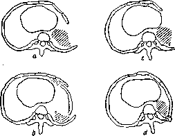
Figure 1. Graphic scheme of Gourget, illustrating the advantages of removing ribs from behind paravertebrally (d) compared to lateral (a-c).
side of the chest wall, due to its elasticity, collapses, the ends of the resected rib shift (the peripheral one descends) and approach each other; at the same time, the diaphragm rises due to the resection of the I rib. After restoration from the preserved periosteum, the rib becomes shorter, i.e., the thoracic cavity and the diseased lung contract throughout the entire resected area, contracting irreversibly. In view of this, life has naturally set the task that with limited pulmonary damage, apply T. not over the entire chest, but only within the affected area of the lung, in order to preserve normal excursion of respiratory movements in the healthier parts. Such partial T. may be required over the lower, more often one upper, lobe of the lung, where in most cases the greatest damage is concentrated. However, with upper T., there is always a threat of aspiration pneumonia for the lower lobes, regarding which there is still no complete unanimity at present. Some authors, starting with Wilms, consider the fear of aspiration exaggerated, others insist on its danger (Sauerbruch and others). In order to reduce this risk, combined types of T. have been created: plastic surgery with basal pneumothorax [proposed by Jessen], plastic surgery of the upper half after preliminary phrenicectomy for compression of the lower lobe, etc. To the category of combined T. can also be added T. of the lower parts with a plug over the upper lobe. However, despite the very real possibility of aspiration with upper T. without reducing its risk by any measures on the lower lobes, in recent years the application of single upper T. is beginning to gain greater popularity. Review
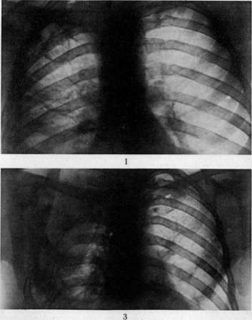
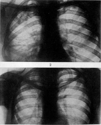
Figure 2. [I rahoi: ro roi pni perhushchnaya kaverna at the level of the upper two ribs. Fig 3. The same case after total thoracoplasty, complete disappearance of the cavity. The same case after 1 1/2 months after the first phase of thoracoplasty: the cavity has significantly narrowed. A lasting effect for 5 years shows: no cavity, no VC and elastic fibers, full working capacity. Figure 4. Apical cavity over the upper 4 ribs.
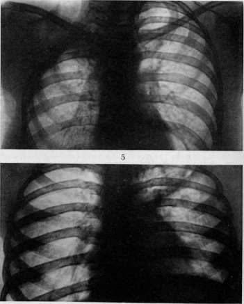
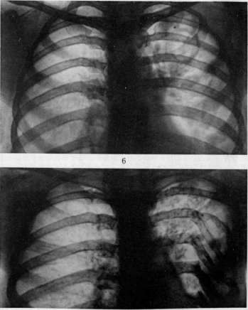
Fig. 5. The same case (see fig 4) after upper thoracoplasty (5 ribs removed): disappearance of the cavity and collapse of the lung; lasting effect; no PC and elastic fibers. Figure 6. Thoracoplasty as a final method after a complete effect from phrenicectomy; X-ray before the operation; cavity at the level of IV V ribs on the left. Fig. 7. The same case after a month after phrenicectomy; the cavity is smaller, but there are VC and elastic fibers. Fig 8. The same case. After 3 years after thoracoplasty from IV to VIII ribs, lasting effect
clinical effect; no cancer. no elastic fibers; full working capacity. However, the question of this cannot be considered finished, as observations from individual institutions speak against ignoring the possibility of complications from aspiration, especially in cases of very large cavities and with a large amount of sputum. The relative severity for weak patients is not only of complete T., but also of limited upper, which requires removal of no less than 7 ribs, the presence of contraindications from the other lung in the limitation of the cavernous process only by the very upper parts of the lung created the need to limit T. to one apical region. Under the influence of this request, the American surgeon Coffey published in 1929 his method for applying in these cases the removal of a piece of the first rib from an anterior supraclavicular incision with the simultaneous transection of the scalene muscles proposed earlier by the surgeon Sato, together with the removal of the thoraco-abdominal nerve and n. thoracici longi. This type of partial T., combined with removal and at the same time limited in extent, attracted the general attention of phthisiatricians, but however did not receive sufficiently widespread distribution. In our Union, it is being propagated by the Sukhumi doctors Bodungen and Antelava who developed it. On the basis of his experimental research on the approach to the I rib and joint clinical observations with Bodungen, Dr. Antelava somewhat modified Coffey's method (see below) and, convinced of its insufficiency, supplemented it with the resection of II and III ribs from the anterior incise he proposed. The main property of extrapleural T. to reduce the volume of the thoracic cavity prompted soon after the introduction of paravertebral T. into operative practice to extend it to purulent pleurisy for reducing the pleural cavity in open forms of chronic and more recent empyemas (residual cavities in non-healing fistulas) and closed (in tuberculous purulent pleurisy) in combination with washing with rivanol, weak iodine solutions 1:10,000 etc. This method of T., being successful also in relation to empyemas, especially when using 2-3 stage operations, replaces the methods of Simon, Küster, Estlander, Subbotin and others in more extensive cavities (see Pleurisy), but is not always sufficiently effective in very old empyemas with very strongly developed adhesions. In these cases, a more radical intrapleural type of T. comes into its own, in which the pleural rib adhesions are removed together with the ribs over their entire length, empyemas. To this category of T. belongs the operation proposed by Schede in 1890 (see below) and other methods that seek to reduce the traumatic nature of Schede's operation and the dangers arising from it by limiting the extent or dividing it into several sessions (modifications of Friedrich, Helferich, Beck, Krause and others.).-Among the varieties of T. should be included the extensive resection of ribs in the heart area proposed by Brauer for adhesions of the pericardium with the anterior chest wall that hinder cardiac activity. The operation bears a widespread but not entirely correct name 'cardiolysis,' or thoracolysis praecardiaca. It is necessary to mention that paravertebral T. has also found application for the purpose of correcting the shape of the chest wall itself and the curvature of the spine in scoliosis (see). Indications and contraindications to T. Any kind of T. comes into its own when it is impossible to apply artificial pneumothorax or when it is insufficiently effective. Yielding to artificial pneumothorax in the strength and gentleness of its action, extrapleural T. occupies the most prominent place as an operative method for collapsing the lung. Most often T. is performed for tuberculosis of the lungs, chronic empyemas and residual cavities in empyemas, but it also has indications when it is necessary to collapse cavities in the lung that have arisen after various pathological processes (echinococcus, drained lung abscess etc.). However, clinical experience with the application of T. in bronchiectasis has revealed the insufficiency of the effect obtained from T. After more or less brief success, the severe symptoms of bronchiectasis usually return. Large cavities with completely collapsed lung give indications for operations of the Schede type. In pulmonary tuberculosis T. has in general indications in cases of productive cavernous process and mainly in its fibro-cavernous type when the other lung is healthy or in a stable state of a quiescent or productive process (see separate table, figures 1-8). Total T. should be applied in widespread tuberculous processes extending over large areas of lung tissue and in large residual pleural cavities. Limited lesions give rise to a choice between T., phrenicectomy (see.), plombage and thoracocautics according to Jacobaeus, used in cases of insufficient effect from artificial pneumothorax and in cases of partial pulmonary adhesions (see-Thoracoscopy). In such cases, one should strive to apply partial T. and especially of the combined type with artificial pneumothorax, phrenicectomy etc. Cavities of the upper lobe of the lung, located in the posterior 'dead angle' (at the heads of the ribs), may give indications for extended T. by additional excision of the transverse processes and disarticulation of the heads of the ribs, according to the proposal (1929-1932) of Proust, Maurer and Rolland. Very limited apical cavities give grounds for applying the Coffey-Antelava operation, but can be successfully and more easily collapsed by paravertebral upper T. with the above-mentioned modification. For cavities located anteriorly medially ('sternal'), anterior T. is applicable, which is practiced as the 3rd stage in case of insufficiency of paravertebral T. and can be performed by the method of Wilms, Brauer, Sauerbruch or Antelava. 'Hilar', i.e. located close to the hilum of the lung, cavities are not eliminated by paravertebral T. even in its most widespread form and therefore are contraindications to such operative intervention. The introduction since 1927 of bilateral artificial pneumothorax has led to attempts to expand the indications for T. in bilateral processes. However, observations on this kind of intervention cannot be considered finished. If combined T. on one side with artificial pneumothorax on the other apparently gives grounds for cautious application, then the still few attempts to apply T. on both sides do not yet give the right to any conclusion.-Contraindications to T. are: 1) cachexia and severe general weakness and intoxication; 2) active process in the other lung; 3) exudative form of tuberculosis; 4) specific ulcerative lesions of the intestine; 5) kidney lesions; 6) insufficiency of the cardiovascular system. Relative contraindications are: age over 45 years, ulcerative processes of the larynx, bronchiectasis, hilar cavities. Technique of T. The operation in all types of T. is performed with the patient lying on the healthy side with the upper part of the body elevated. The overwhelming majority of authors perform it under ether anesthesia. The latter is especially urgently indicated in pulmonary-pleural diseases. Despite the extensive extent of the operative field, paravertebral anesthesia of the intercostal nerves or by Vishnevsky's method by infiltration into each intercostal space can provide complete anesthesia, and the operation is well tolerated even by weaker patients. This deep anesthesia can be carried out immediately after the skin-muscle infiltrative anesthesia along the line of the proposed incision or after the incision of the skin and the superficial layer of muscles, making injections paravertebrally into the intercostal spaces under direct vision. Finally, the third general question for all types of T. is the question of the periosteum. Although some authors insist on its excision in order to prolong the period of maximum possible collapse of the lung, most surgeons do not do this, as experience speaks against this: quite sufficient collapse of the lung is obtained even while preserving the periosteum; it is only necessary to observe the proper length of the ribs to be removed. Extrapleural T. according to Friedrich-Brauer (figure 2). With a large horseshoe-shaped incision, the ribs from II to X are exposed in their entire length. The anterior vertical part of this incision begins at the level of the II rib, turns posteriorly at the VI-VII rib and connects with the horizontal part above the IX-X rib with the posterior vertical, which descends along the spine, beginning at the level of the III spinous process. Along the line of this incision, with one stroke of the knife, all layers from the skin to the ribs are cut through, after which the entire outlined skin-muscle flap together with the scapula and the serratus anterior muscle is separated from the chest wall and with the patient's arm strongly raised, the flap is retracted upward and outward. After stopping the bleeding, they proceed to the subperiosteal resection of the ribs, starting from the lowest one and exposing each rib with a longitudinal incision of the periosteum from the head to the costal cartilage. The ribs are first transected at the anterior end, and then at the posterior end, as close as possible to the transverse process.
The stumps of the ribs are carefully rounded with forceps (to prevent neuralgic pain). To complete the operation, Friedrich recommends excising all the intercostal muscles along the length of the removed ribs together with the remaining periosteum and to resect the nerves high up. These additions, however, are not considered mandatory by all, and some authors even consider them harmful (Küttner). The skin-muscle flap is placed in position, and the wound is sutured in layers. In front and behind, at the deepest parts of the wound, to prevent the easy formation of hematoma, a rubber drain is inserted for 2-3 days. An aseptic dressing is applied, over which, for some reinforcement of the now excessively yielding wall, transverse strips of adhesive plaster are placed from the sternum to the spine. The arm is bandaged to the trunk on a sling. The postoperative period is conducted with the patient in a semi-sitting position, with all measures to combat the unfavorable consequences of the Friedrich-Brauer method described above. Paravertebral thoracoplasty of the subscapular region (Brauer's operation).
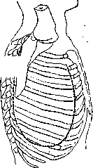
Figure 2. Incision of soft tissues according to Friedrich-Brauer.
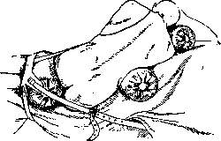
The modification of this operation described in 1914 by Becker (Becker) at Brauer's suggestion consists in reducing the wound surface and a more conservative attitude toward the components of the chest wall. The ribs are removed subperiosteally through two incisions—a posterior and an anterior. The posterior one is made paravertebrally along the edge of the scapula, and from it, pieces of ribs from X to II are resected as close to the heads as possible. After this, the patient, who has been lying on the healthy side, is turned onto the back, and an anterior incision of the soft tissues is made along the anterior axillary line. After resection from this incision, the anterior parts of the ribs are freed from under the scapula with the help of special retractors designed by Brauer, forming a tunnel through the periosteal sheath for the intermediate parts of the ribs (the part left un-resected between the excised posterior and anterior portions). The pieces of ribs thus freed are pulled out from under the scapula, and the latter, having lost its firm support, can then easily, in the form of a bolster, be pressed in and appose the collapsed lung. The intervention can be performed in one session or, depending on indications, divided into 2-3 or more sessions. The operation is completed by suturing the muscles and skin in layers, with the area of the chest wall freed from ribs being fixed with strips of adhesive plaster over an aseptic dressing. Paravertebral thoracoplasty according to Sauerbruch. The operation is mostly performed in 2 stages. In the 1st stage, 6-7 of the lower ribs are removed, in the 2nd—4-5 upper ones. With sufficient strength of the patient and good anesthesia, it is possible to remove all ribs in one session. If thoracoplasty is preceded by phrenicectomy, it is possible to limit oneself to removing 8-9 ribs. The interval between stages is usually set at one month. In some cases, with good strength of the patient, the 2nd stage can be performed after 18-20 days. The length of the pieces of ribs to be removed depends on the location of the cavity and the extent of the pulmonary process. Over the lobe containing the cavity, the longest pieces are resected, and conversely, in the area of the healthiest part of the lung, shorter pieces of ribs are excised. The patient is placed on the healthy side with the upper half of the trunk elevated. The position should be as comfortable as possible, in no way forced. A roll is placed under the side, slightly arching the operated side, which helps to spread the ribs to facilitate their resection. Under the buttock region, support is provided either in the form of a roll in a loop of a towel supporting the buttocks (the loop is fixed in the front part of the table) or a metal clamp fixed to the edge of the operating table (Fig. 3). The patient's head is placed on a pillow in a slightly inclined position toward the chest.
Figure 3. Position of the patient according to Sauerbruch.
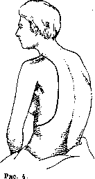
For the greatest spreading of the scapulae, the arms are abducted forward, and the patient is given a pillow rolled up like a bolster to embrace. The skin incision is begun at the level of the V-VI spinous process, three transverse fingers laterally from the spine, which corresponds to half the distance between the abducted scapula and its spinous processes. For longer pieces of ribs to be excised, it is more advantageous to make the incision closer to the spine than to the scapula (so that the scar does not fall on its edge). The line of incision is carried parallel to the spine to the IX rib, from where it bends arcuately forward and outward, crosses the X rib, and ends on the axillary line. The skin, muscles (m. trapezius, latissimus dorsi, part of rhomboidei) are immediately incised down to the ribs. According to Sauerbruch's suggestion, it is more advisable to begin the resection with the X rib, then quickly shorten the IX-V ribs, and after that, excise the XI rib. The length of the pieces to be extended is 8 to 15-20 cm, depending on indications. When freeing the posterior parts of the ribs and spine from the periosteum, the teeth of the long back muscles are carefully and meticulously separated. A careful attitude toward them eliminates the risk of subsequent scoliosis (Brauer). If it is possible to continue the operation and complete it in one session, the other ribs are also removed. To perform this 2nd phase of the operation, the upper thoracoplasty incision according to Sauerbruch is extended. The muscles (mm. trapezius, rhomboideus, serratus post., sup.) cut simultaneously with the skin are strongly pulled forward with a hook together with the freed scapula. Starting from the lower one, the ribs are resected one after another over a length of 6-8 cm up to and including the II rib. Throughout the operation, during coughing movements, the assistant counteracts the bulging of the lungs by pressing with a gauze tampon. To remove the I rib, the incision is extended again, this time bending it arcuately over the shoulder forward to the anterior edge of the m. trapezius. By this fractional production of the incision, Sauerbruch seeks to reduce the severity of the operation, limit blood loss, and the nerve injury received from the sudden incision of tissues along the entire length of the chest from top to bottom (Fig. 4). With the upper, scapula-encircling incision, two arteries are usually intersected and ligated: aa. cervicalis superficialis and transversa colli. With very strong spreading by hooks and pulling outward of the edge of the wound (mm. trapezius and levator scapulae) and especially the scapula in the depth of the wound, the I rib can be seen up to the subclavian artery and brachial plexus, if the posterior scalene muscle (attached to the II rib) and the middle one are first cut and pulled upward (Fig. 5). According to Sauerbruch's requirement, the first rib is resected over a length of 3 cm from the transverse process to the Lisfranc tubercle. The anterior scalene muscle is cut at the very edge of the rib. After all ribs are removed, the vessels are ligated, the wound is toiletted, and catgut sutures are placed in layers on the muscles and silk sutures on the skin. Into the depth of the wound, down to the bed of the I rib, a rubber drain is inserted for 2-3 days to prevent hematoma. Aseptic dressing; 2-3 spiral strips of adhesive plaster for fixation from the healthy shoulder to the anterior abdominal wall. A gauze dressing with the arm bandaged to the trunk. In the concluding part of the described type of thoracoplasty, the most difficult part is the resection of the I rib. The depth of the wound in the method of incision according to Sauerbruch creates a substantial obstacle for calm operating by sight and by
Incision according to Sauerbruch.
Figure 5. Approach to the I rib according to Sauerbruch: 1 and 2—subclavian vascular-nerve bundle; 3—I rib.
Figure 6. Straight-line incision according to Eipov.
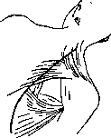
of access to the rib. Meanwhile, the best effect of T. is obtained by resection of the largest piece of the I rib, and for some localizations of cavities, it is necessary to remove its entire extent from the head to the cartilage (Graf, 1925). Significant relief of conditions can be obtained by some modification of the Sauerbruch operation method (Esipov). First, the skin-muscle incision continues upward without inclination along the shoulder forward, parallel to the spine to the level of C VI-VII (Fig. 6). By cutting the short horizontal fibers of m. trapezii, the elevation with the scapula of the freed upper parts of its levator scapulae is facilitated (Esipov, Nalivkin). Second, immediately after cutting the skin-muscle layer, exposing and anesthetizing the ribs, the anterior serratus muscle's (m. serratus anticus) tooth from the II rib is subperiosteally shifted, unlike others very close to the spine and more than all other teeth hindering good elevation of the scapula and opening of the I rib (Esipov) (Fig. 7-8). In this modification, the upper T. sharply changes the conditions of access to the I rib, making it well visible not only to the operating surgeon but also to the surrounding observers of the operation and giving the possibility to manipulate with simpler instruments, with short beaks (Fig. 9). Paravertebral thoracoplasty according to Wilms (Pfeiler resection) is characterized by shorter sections of resected ribs - 3-4 cm from 2 incisions: posterior and anterior. In the original version of the operation, from a longitudinal skin incision along the spine, 3 transverse muscle incisions were made along the fibers at the level of II, V and VII ribs. The beginning of resection from the VIII rib, VII and VI from the lower muscular cleft, then V-IV ribs were removed from the middle and III, II and I ribs from the upper. After 34 T., the Wilms method took the following form: a) for limited processes of the upper part of the upper lobe: 1 session - paravertebral resection from VII to I rib, 2nd session (conditionally, if insufficient) - parasternal resection of cartilage from I to V rib, 3rd session (also conditionally) - parasternal resection of the clavicle, so that its outer remainder in collapse would press with its medial end against the clavicular notch of the sternum, being held above by the sternoclavicular muscle and below by the subclavian. b) In case of lower lobe involvement: 1) 1st session - paravertebral resection from VIII-IX ribs to I, 2nd session - parasternal resection of 5-6 cartilages; the 2nd or 1st session is split into two: the upper 6 ribs are resected short,
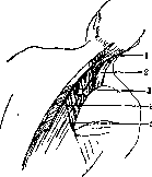
Figure 7. Relationship of Esipov's straight incision to paravertebral muscles: 1-m. splenius; 2-m. levator scapulae; 3-m. rhomboideus; 4-m. trapezius; 5-line of incision.
in the second step - VII, VIII, IX ribs on a large extent (10-15 cm). The operation, if necessary, is completed by a third step, in which
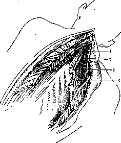
Figure 8. Muscle and vascular relationships in the method of approach to the I rib according to Esipov: 1-m. levator scapulae; 2-m. scalenus post.; 3-brachial plexus; 4-upper 2 teeth of m. serratus ant. shifted by a retractor.
the second step - VII, VIII, IX ribs on a large extent (10-15 cm). The operation, if necessary, is completed by a third step, in which
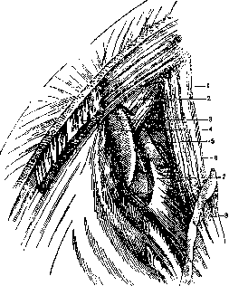
Figure 9. Exposure of the I rib according to Esipov: 1-m. levator scapulae; 2-m. scaleni med. et post.; 3 and 4-m. rhomboideus; 5-I rib; 6-brachial plexus; 7-subclavian artery; 8-m. serratus ant.
not earlier than 6-8 weeks after the first, the sternal cartilages are resected from the front. The operation of Koffi belongs to the category of interventions on the I rib, performed from a supraclavicular incision (Fig. 10-11). The approach to the rib is made from an incision that is carried along the bisector of the angle between the sternoclavicular muscle and the clavicle. Deepening into the soft tissues, it is necessary along the way to intersect m. platysma, ligate v. jugularis externa, intersect m. omohyoideus, ligate and cut aa. and vv. transversae colli and scapulae. Perform a phrenic exeresis, separate from the rib m. scalenus anticus and medius, expose and cut passing along the edge of the latter muscle n. thoracalis longus to enhance the collapse of the lung, thereby causing paralysis of m. serratus anticus, and finally, taking on a loop a. subclavia and the brachial plexus and moving aside with a hook the edge of m. trapezii and scaleni post., proceed to the subperiosteal removal of the I rib. The latter is excised for a length of 3 cm at the posterior edge of the bed of the subclavian artery and at the transverse process of D1. The Koffi method was modified and supplemented by the Soviet surgeon Antelava, who, excluding from the operation the cutting of n. thoracalis longi, added the wide extrapleural apicolysis proposed in 1929 by

Figure 10. Koffi incision. The approach to the rib is made from an incision that is carried along the bisector of the angle between the sternoclavicular muscle and the clavicle. Deepening into the soft tissues, it is necessary along the way to intersect m. platysma, ligate v. jugularis externa, intersect m. omohyoideus, ligate and cut aa. and vv. transversae colli and scapulae. Perform a phrenic exeresis, separate from the rib m. scalenus anticus and medius, expose and cut passing along the edge of the latter muscle n. thoracalis longus to enhance the collapse of the lung, thereby causing paralysis of m. serratus anticus, and finally, taking on a loop a. subclavia and the brachial plexus and moving aside with a hook the edge of m. trapezii and scaleni post., proceed to the subperiosteal removal of the I rib. The latter is excised for a length of 3 cm at the posterior edge of the bed of the subclavian artery and at the transverse process of D1. The Koffi method was modified and supplemented by the Soviet surgeon Antelava, who, excluding from the operation the cutting of n. thoracalis longi, added the wide extrapleural apicolysis proposed in 1929 by
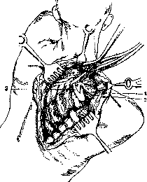
Lauwers when removing the I rib and added resection of the rib (Fig. 12). Anatomical studies by Antelava proved that the Koffi operation is easier to perform in the brachymorphous and mesomorphic types of the superior thoracic aperture and encounters greater difficulties in dolichomorphous ones (Fig. 13), since the posterior part of the I rib in them is covered by the brachial plexus and it is almost impossible to isolate the subclavian artery, which together with the plexus must be moved forward and medially. Thoracoplasty in empyemas. Of the plastic operations for empyema, the most radical, but at the same time more severe operation is T. according to Schede (Fig. 14) (see Pleurisy). The method according to Schede (intrapleural T.) with the patient on the healthy side. With a large horseshoe-shaped incision penetrating through all layers, the skin-muscle flap is outlined corresponding to the entire extent of the empyema. Its anterior vertical begins at the IV rib, at the lower edge of the pectoralis major muscle, the posterior one - at the level of the upper edge of the scapula, the lower horizontal - at the X rib along the axillary line. All muscles, including the serratus anterior muscle, are separated from the ribs. After freeing, the skin-muscle flap together with the scapula is pulled upward with hooks with the arm strongly abducted and raised upward. After stopping
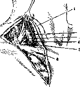
Figure 11. Koffi-Antelava incision: 1-phrenic nerve; 2-m. scalenus ant.; 3-brachial plexus; 4-a. subclavia; 5-v. subclavia; 6-clavicle.
the bleeding, subperiosteal resection of ribs should be performed on the entire extent from X to II. The next moment is the freeing of a new, deep, rib-less flap from adhesions and its mobilization. For this purpose, one penetrates into the pleural cavity or by lengthwise incision along the intercostal space expands a fistula or immediately cuts out a flap through the thickness of the intercostal spaces and periosteum along the edge of the first skin-muscle flap, starting from behind. Along the way, the intercostal vessels are ligated and the nerve trunks are cut. The operation is completed by the final freeing of the flap from adhesions, toilet of the wound and suturing with nodal stitches the mobilized second and first flap applied to the lung to complete contact. Rubber drains are inserted into the depth of the wound along the edges of the anterior and posterior vertical incisions. An aseptic dressing, over which
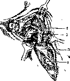
Fig. 12. Removal of the I rib according to Koffi-Antelava: 1-brachial plexus; 2-I rib; 3-clavicle; 4-stump of the anterior part of the I rib.
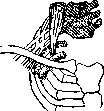
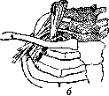
Fig. 13. The two extreme types (a and b) of the superior aperture of the chest cavity according to Antelava. Sticky plaster strips are applied diagonally and transversely to fix the chest wall. The arm is bandaged tightly to the trunk with a Dosage bandage. Some authors open the pleura and fashion a flap without performing subperiosteal resection of the ribs or resorting to it partially, since this is required for pressing the flap to obliterate the cavity. The severity of the extensive operation of Schede, which historically served as the prototype for subsequent proposed methods of T., can be alleviated by performing it in several stages. For better mobilization and to give greater plasticity to the flap, Zudek suggested performing one of these sessions in the same way as Beck and Krause perform extensive resection of the scapula; Helferich used an H-shaped incision in the prone position (I), excised the ribs with periosteum and removed the entire thickness of the deep layer of the wall. Technique of paravertebral extrapleural thoracoplasty in empyemas. T. for empyemas according to Sauerbruch's method is also performed in one or several sessions, as in pulmonary tuberculosis. It is only necessary to begin the operation with upper T. for more favorable drainage of the pus. To avoid infection of the wound cavity from the fistula, the fistulous opening should first be isolated by two semilunar incisions and sutured along the edge over the fistula, covered with a gauze tampon. The ribs are removed above and below the fistula, with special care in separating them from the periosteum around the fistulous tract. To avoid opening it, especially in cases where the fistula is surrounded by a bony ring, a piece of bone near the fistula can be left untouched, cutting the rib behind and in front of the fistula (Yesipov). Postoperative regime. The postoperative period after T. often proceeds with various severe phenomena for the patient, especially after one-stage T. To eliminate and alleviate them, the following methods should be used. More or less pronounced difficulty in breathing depends on the loss of fixation of the lung to the chest wall in the resected area. Shortness of breath is significantly relieved by the application of sticky plaster or by the Brusger and Sauerbruch method of sewing in strips of bandage rubber that stretch on inspiration and collapse on expiration. The patient's arm should be well bandaged to the trunk, but circular turns of the bandage should not tightly constrict the other side of the chest. The patient is placed quietly in bed in a semi-sitting position with support under the head and under the buttocks to prevent slipping. For the first 1-2 days, in case of painful coughing, narcotic agents are used, later they are contraindicated and replaced by expectorants. Cardiac activity is maintained with camphor, infusions of glucose and saline solution, in more severe cases - by administering digitalis. In cases of shock, morphine is administered. An increase in temperature in the first days may be due to intoxication of the body by products of a specific nature; often the reason lies in the development of a hematoma. With high temperature rises, early dressing and careful examination of the wound are necessary in terms of possible infection. At the first signs of it, partial removal of the sutures, drainage, and in case of certainty of the development of suppuration, urgent radical measures are required. Immediately after the operation, the amount of sputum decreases, patients forcibly suppress coughing, and retention of expectoration may be a causative factor for increased temperature. It is necessary for the physician to fight this by timely switching to expectorants and suggestion to the patient. Drainage from the wound with normal temperature is removed on the 2-3rd day. In empyemas, if drainage was not inserted during the operation, it is reinserted into the fistula (with great care to avoid rupture of the fistula wall) at the first signs of retention of pus in the pleural cavity (increase in temperature), usually on the 2-3rd day. The bandaged arm is freed on the 7-12th day, after which active movements with it are permitted. The patient is usually allowed to get up after 3-4 weeks, in some cases it is possible to allow walking as early as the 10th day. Outcomes of T. The material for T. in tuberculosis is usually cases of ineffective artificial pneumothorax. Under these conditions, in the practice of all authors, the success of T. depends largely on the correctness of the indications, the experience and technique of the operating surgeon. Brauer defines the outcomes by the following ratios: 1/3 of cases gives complete clinical recovery, 1/3 - improvement (relative working capacity) and the last third - unsuccessful outcomes and death. According to the largest statistics of Sauerbruch, recovery is noted in 33%, improvement in 27%, while mortality amounts to 25%, of which 12% is related to the operation and 13% to the long-term result. A combined statistics of various authors in 740 cases gives 34% clinical cures, 28% improvement and 38% unsuccessful T. (including mortality). Calculation of long-term results of T. over 10 years of work (1923-33) of the Moscow Regional Tuberculosis Institute led to the following data on 158 T.: 1. A total of 105 T. were performed for pulmonary tuberculosis. Of these, recovery occurred in 45 people = 48% (full working capacity), improvement - 22 people = 24% (tubercle bacilli at times); no change - 6 people = 6%. Death - 21 people = 22%. No information - 11 people. Of this number, complete T. was performed on 57 patients: recovery - 24 people = 52%, improvement - 13 people = 28%. Death - 9 people = 20%. No information - 11 people.
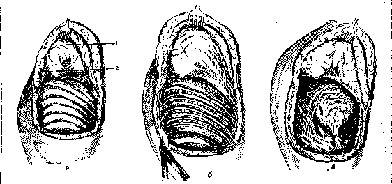
Fig. 14. Various phases (a, b, c) of the Schede operation: 1 - the turned up skin-muscle flap; 2 - the protruding lower angle of the scapula.
i
I) Inc. 1 and 2. Visible Hfcpea Гаракоскм.....utri ll ten рнлнлос пережигание тшка легочной ьерхушки до н мосле иисдоцик. JЕодтнн\тдн и рис, I Рис, 3. I [лаетялчатип. кроеного пветаn fiornrun сосудами тяж. Нсрхлнн доля, c каверной подтянута к стенке, Рис, 4, Тяж пережжен. Ле; самостоятельно. Рис, 5. hcr.icoGpainuli тик c ch.tmhjm иптяженнем легкого и конуса париетальной иленры. Фор ча тяжа, наиболее опасная и верхняя доля после пересечения тяжа I рис. 2) сильно сократились. Легкое сократилось Кчльтн wpojwTi.....т, K|Ч)потСчспие осгленчшм*<ц, ,.,-j н смысл* ипгожшютн миелин легочной ткани. Рисунок 6. i ГоепЕнШни пережигание, Нерсжжеля нанжеткоА и не игральная нленрп. Легко*, осооСоднишнсь от lice, сойетсиноп мост......остью потягннйег и истончает тяж при тдльиеГпнсм пережигании его. 1*нс h Тнж пережжен. Легкое птнпло. сократилось. Индид скноль дни клади ни пи на стоике-след устраненного статення. Рисунок B. Дна верку шечкьи нлпетиичншх тяжп. Pac У. Пристеночная культя после пережигания сильно кровоточила (струен). Осглнонкп кровотечения прижатием холодного клутири, Рисунок HI. Пластинчатый эаяавесчлтын складчотин тяж. У оснонрння- проснечнипюшне пудирькн на поверхности легкого {oedema ЪиИоэит). При прорыве и* йипвет спонтваный ллс&чоторакс K ст. TopahotrnmitH «18
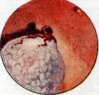
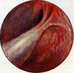
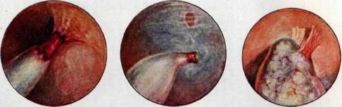
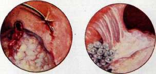
Partial upper T. in 34 patients. a) With artificial pneumothorax on the same side - 22 people. Recovery - 12 people = 54%. Improvement - 5 people = 28%. No change - 1 = 28%. Death - 4 = 18%. b) Without artificial pneumothorax - 12. Recovery - 6 = 50%. Improvement - 4 = 33%. Death - 2 = 17%. With upper T. without basal artificial pneumothorax, aspiration pneumonia occurred 3 times; in one case with fatal outcome on the 16th day after the operation. Partial lower T. - 14 patients. Recovery - 3 people = 22%. No change - 3, worsening - 2 = 35%. Death - 6 people = 43%. 2. A total of T. was performed for empyemas (after spontaneous pneumothorax and in residual cavities) - 43. Recovery - 7 = 20%. Significant improvement - 7 people = 20% (fistula remains, but full working capacity). Improvement - 10 = 29%. Death - 11 = 31%. No long-term information - 8 people. 3. T. for bronchiectasis - 7. No change - 2, slight improvement - 2. Remote death - 3. 4. T. for abscesses of the lung: recovery - 1, improvement - 1, death after 2 months - 1. Regarding functional results, despite extensive dissection of the scapular muscles, movements of the shoulder girdle are fully restored (fig. 15). Scoliosis never occurs with careful and careful work on the long muscles of the back at the time of isolating the posterior angle and end of the ribs. The chest wall caves in significantly, most strongly with complete T., due to shortening
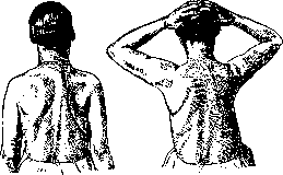
Fig. 15.
of the ribs in length and the downward displacement of the entire chest cavity. Cavities in tuberculosis should be collapsed in the outcome. Only in this case does complete recovery occur with the disappearance of all signs of the disease.
Related articles
Mentioned in
Cite this page
“Thoracoplasty.” Soviet Medical Encyclopedia. English translation of Bolshaya Meditsinskaya Entsiklopediya, 1st ed. (Moscow, 1928–1936), ed. N. A. Semashko. https://sovietmedicalencyclopedia.pages.dev/article/thoracoplasty/