Thoracoscopy
Historical document, translated for reference. It reflects medical knowledge of the 1920s–30s and is not medical advice.
Summary
Thoracoscopy is a method for examining the pleura and lung during artificial pneumothorax, first proposed in 1911 by Swedish physician Jacobaeus. It was initially used for evaluating lung and pleural surfaces, then adapted for adhesiotomy to improve artificial pneumothorax effectiveness.
Encyclopedia article (1928–1936)
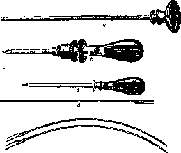
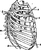
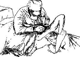
Thoracoscopy, a method for examining the pleura and lung during artificial pneumothorax. First proposed in 1911 by the Swedish phthisiologist Jacobaeus, initially exclusively for evaluating the condition of the lung surfaces, pleura, and the entire cavity, and then in 1913 for burning adhesions of the lung that hindered the full effectiveness of artificial pneumothorax (see below). The first attempts at T. were performed by Jacobaeus using a cystoscope, which was later modified by him according to local needs in thoracoscopy. T. received its greatest development and popularity in Germany after substantial changes to the instrumentation and technique were made by Unferriht, Ulrici, Kremer, and Gross in 1916-22. In the USSR, the method began to be used in 1929. At present, T. is performed using the instrumentation shown in Fig. 1. The straight thoracoscope (a) is inserted into the chest cavity through the cannula of a trocar (b), which is punctured under local anesthesia in the intercostal space and equipped with an automatically acting valve, which limits the entry of air into the pleural cavity when the trocar stylet is removed and allows T. to be performed during pneumothorax in negative phases of intrapleural pressure; the examination is performed in the same way as during cystoscopy. However, the ovoid shape and size of the pleural cavity enhance the shortcomings of the thoracoscope inherent in optical instruments of the cystoscope type. Perspective is particularly distorted, and to a greater extent one must account for deviations of the image in the reverse direction with certain relationships of the endoscope axis to the object being examined. Nevertheless, with skill, T. makes it possible to perform a very clear examination and manipulation in the pleural cavity, bringing the optical 'eye' of the thoracoscope closer to or farther from the place where the research or operation is applied by inserting and withdrawing it in the trocar cannula and rotating it on its axis (see color table, figs. 1-10). T. is used primarily for tuberculosis of the lungs and pleura as a preliminary stage or simultaneously with thoracocautery according to Jacobaeus. Thoracocautery according to Jacobaeus consists of burning adhesions of the lung to the pleura under the control of the thoracoscope without opening the chest cavity by incision. The purpose of the method is to eliminate, under visual control, strands that hold tuberculous cavities in a stretched state and prevent their collapse during treatment with artificial pneumothorax. The operation is performed under novocaine in the presence of an air bubble in the pleural cavity and consists of 2 moments: 1) puncture of the chest wall with a trocar for the thoracoscope to examine existing adhesions, and 2) insertion of a new trocar for the introduction of a galvanocauter (the complex thoracoscopes proposed by Mendel and Kremer, which include both optics and cautery similar to catheterization cystoscopes, have not found wide application in practice). The galvanocauter according to Unferriht has a straight or curved shape (fig. 1d), accordingly a straight or curved trocar cannula is used, or even better, a spiral flexible cannula according to Unferriht, suitable for any curvature. The long dimensions of the cautery make it possible to burn strands at different levels of the lung from one puncture point. Some strands, however, are more advantageously eliminated from specific puncture points (fig. 2). The greatest freedom of movement is provided by the placement of both trocars in which the thoracoscope and cautery cross in the cavity at an angle close to right. The overwhelming majority of adhesions can be eliminated by inserting one instrument from the axillary area at the edge of the pectoralis major muscle and the other at the edge of the latissimus dorsi muscle in the II-III-IV intercostal spaces (fig. 3). The presence of multiple adhesions, upper and lower, sometimes requires 2 punctures for the cautery at different levels. With multiple or
Figure 2. Diagram of Dill and Kre-
mer: t-opening for the thoracoscope; ft-opening for the cautery.
Figure 3.
very short, mostly apical, dangerous due to their anatomical position strands, or during prolonged operations, it is rational to perform the burning in 2, 3 or more sessions, between which artificial pneumothorax is applied. For performing thoracocautery, the following is required: 1) the presence of an air bubble sufficient for movement of the instrument in the cavity without risk of damaging the lung, 2) bringing the intrapleural pressure to 0 or low negative or positive figures (-2, -4) and 3) a clearly performed radiograph. Contraindications are generally severe condition, severe shortness of breath, very persistent cough, fresh increasing exudative pleurisy, pleural adhesion, very short (less than 1 cm) strands. Bilateral artificial pneumothorax with good compensation is not a contraindication to thoracocautery. The technique of thoracocautery is very delicate, requiring good knowledge of local topography, attention and care in work. The duration of the operation usually ranges from several minutes to 1-2 hours and depends on the nature of the structure of the strands. The latter may consist of connective tissue with rich or poor inclusion of fatty tissue, vessels, sometimes with greater or lesser content of lung tissue, into which the wall of the marginal cavity may also be drawn. The strands themselves may appear as thin or thick plates, threads, cords, the thickness of which can reach the diameter of a thumb. These varieties may be the source of difficulties for
\!Л
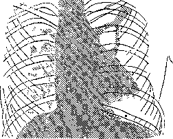
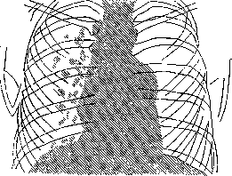
Figure 4. Diagram of a hanging cavity before burning the strand with cautery, the reason for the impossibility of rapid work. In addition, considerable delays are caused by the need to stop burning to remove the smoke that obscures the field of vision from the cavity using the cannula. For this purpose, there is a suction device that reduces time loss, proposed by Ulrici. The postoperative period is conducted with strict bed rest. Resumption of artificial pneumothorax should begin 7-15 days after thoracocautery. A successful result of the operation manifests in the coming weeks: the cavity collapses (figs. 4 and 5), BK, elastic fibers, sputum disappear, temperature drops, blood composition rapidly improves. COMPLICATIONS: 1) The most serious complication can be bleeding during or after cautery, when the patient is already in the ward. It is possible with unsuccessful burning of a strand too close to the chest wall (branch of intercostal vessel) or to the lung (lung parenchyma or a. bronchialis on its surface). Often the strands themselves are richly vascularized and bleed easily. This complication is not often encountered, varying among different authors from 0 to 3-5%; usually bleeding is venous and stops easily on its own. In cases of arterial re-. it is recommended to press the strand or its stump with the cautery sleeve and stop the bleeding by pressure or to cauterize the bleeding vessel with the cautery. A preventive measure is the use of red, not white, loop heating of the cautery. Maurer almost completely eliminated the complication of bleeding by proposing a device that coagulates the vessels of the strand before burning with diathermy. 2) The danger of burning the lung, # cavity wall, pericardium,
Figure 5. Disappearance of the cavity after burning the strand (diagram). * subclavian or azygos vein and others is prevented by caution in procedures and precise orientation in topography. In addition to these complications, shock, acute shortness of breath with rapid increase in intrapleural pressure before the operation, spontaneous pneumothorax (Unferriht-3 cases, Dill and Kre-mer-2 cases, Stoyko-1 case, Yesipov-1 case) may occur. A very frequent complication (50-70%) in the postoperative period is subcutaneous emphysema, which usually disappears very quickly, and an increase in temperature to 38-39°. The latter usually falls after 6-15 days if not associated with pleural infection. Very often after cautery, pleural exudate is observed. Its frequency varies greatly - from 16% (Dill and Kremer) to 72% (W. Liidke). Exudate most often arises from irritation and with this etiology disappears after 8-10 days. Delay in resorption may be associated with infection, which can come from the lung stump, in severe cases - from damage to lung parenchyma, and sometimes from the wall of the cavity itself. Tuberculous exudative pleurisy may occur when burning strands containing tuberculous nodules and recognized by most authors as not suitable for cautery. The final result of thoracocautery is usually good. With strict selection of cases for cautery, it reaches 75% (Pomplon), with a bolder attitude toward cautery, namely when performing thoracocautery in 80-85% of thoracoscopies, the method gives 60-62% success, i.e., conversion of ineffective pneumothorax into full, effective (Unferriht, Dill and Kremer, Stoyko, Yesipov and others.).
Related articles
Mentioned in
Cite this page
“Thoracoscopy.” Soviet Medical Encyclopedia. English translation of Bolshaya Meditsinskaya Entsiklopediya, 1st ed. (Moscow, 1928–1936), ed. N. A. Semashko. https://sovietmedicalencyclopedia.pages.dev/article/thoracoscopy/