Cannula
Historical document, translated for reference. It reflects medical knowledge of the 1920s–30s and is not medical advice.
Summary
A cannula is a hollow tube used in medical practice for various purposes including introducing substances into the bloodstream, measuring blood pressure, irrigating and draining fluids from closed cavities, and connecting internal organs to the external environment. They are made from various materials including metals, rubber, and glass, and come in different specialized forms for different medical procedures.
Encyclopedia article (1928–1936)
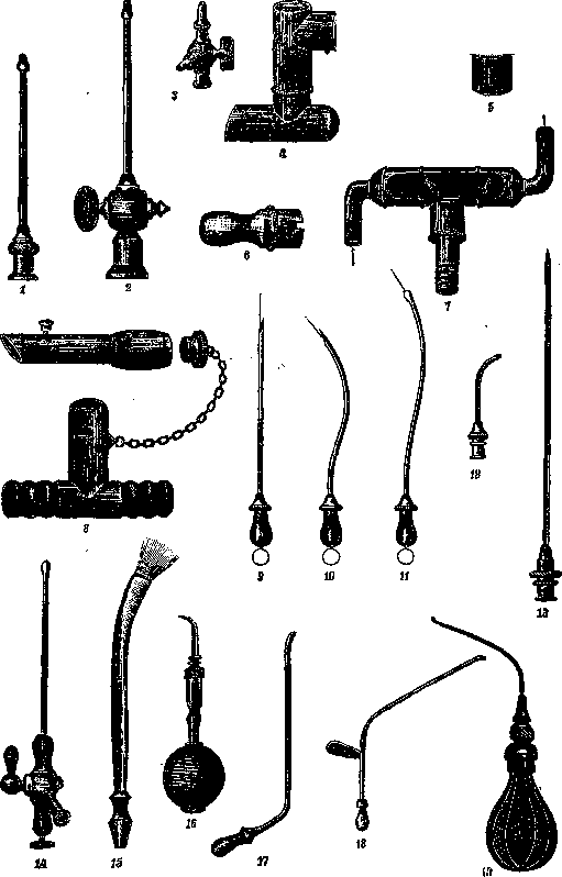
CANNULA (from Latin canna - tube), a hollow tube, the form of which varies greatly depending on its particular purpose. C. is used for introducing various substances into the circulatory system, for recording blood pressure in animals, for irrigating and draining fluids from closed cavities, and for connecting internal organs and cavities with the external world. C. are made of platinum, gold, silver, and other metals, as well as of hard rubber, celluloid, and glass. For a n a t. works, metal C. with a pear-shaped expanded blunt end (for fixing the vessel wall on it) are used. The central end of the C. has threads, which allows it to be screwed onto a syringe; this makes it possible to introduce liquid under high pressure. In addition to simple C. (fig. 1 and 2), there are more complex ones with taps and side tubes (fig. 3).-For physiol. works, C. of various sizes are used, which do not differ in principle from C. for anat. works. For artificial respiration, C. of Francois-Franck (fig. 4 and 5) are used. In addition to them, there are simpler ones (fig. 6). C. of Verdin (fig. 7) is used for studying exhaled gases in dogs. Tracheal C. differ in many ways from those used in humans (fig. 8).- For introducing therapeutic liquids into the circulatory system and for venepunctures, C. with sharp and blunt ends are used. The advantage of the former is that in the case of well-expressed veins, such cannulas can be used without preliminary venesection. Cannulas used for this purpose are a straight or S-shaped hollow tube. Blunt C. at the peripheral end have a small expansion that allows fixing the vessel wall on it. The central end has a more significant expansion, onto which a rubber tube connecting the C. with an infusion apparatus or syringe is fitted (fig. 9, 10 and 11).-For subcutaneous infusions, C. with sharp ends are used. In addition to C. connecting with a syringe via a rubber tube, there are C. that can be put on or screwed directly onto a syringe (fig. 12).-For
CANNULA II
CANNULA III
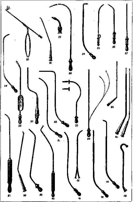
spinal anesthesia and lumbar punctures, C. with a mandrel are used; the latter is well fixed due to its special design. For this purpose, cannulas of Bier (fig. 13) are most commonly used. For venous anesthesia, Minz proposed a C. that allows regulating the introduction of anesthetic substance through a side tap (fig. 14).-In ophthalmic practice, in addition to C. suitable for syringes, a Terson cannula (fig. 15) is used for irrigating the conjunctival sac; for irrigating the lens capsule - a Kuhnt cannula (fig. 16). In diseases of the ear, throat, and nose, the following C. are used: for the tympanic cavity - Hartmann C. of silver or hard rubber (fig. 17); von Stein with plates for fixation (fig. 18); Politzer with a balloon (fig. 19); Milligan with a handle for fixation (fig. 20), thanks to which the C. can be fixed very firmly, which frees the p-nt from sometimes very painful sensations caused by the movement of the C. For introducing boric acid into the tympanic cavity, Bezold C. (fig. 21) are used. Frey cannula is used for irrigating the attic (fig. 22). For trial punctures of the maxillary cavity from the lower nasal passage, von Stein C. (fig. 23) are used. With these same C., injections can be made, both through the lower and middle nasal passages. For the same purposes, Ki llian C. (fig. 24), Sprenger C. with a trocar (fig. 25) and Chiari C. (fig. 26 and 27) are used. For irrigating the maxillary cavity, Hartmann C. (fig. 28), Eicken C. (fig. 29), Killian C. (fig. 30) are used. For irrigation from the alveolar process - Hartmann C. (fig. 31), Frankel C. (fig. 32), Ankidinov C. (fig. 33); the latter C. is provided with a silver drain for maintaining the perforated opening in an open state. The flexible C. of the same author (fig. 34) can be used for irrigating the frontal sinuses, etc. For irrigating the sphenoid cavity, Herzfeld and Grünwald C. (fig. 35 and 36) are used. C. of these same authors serve for irrigating the frontal cavity (fig. 37 and 38). For the same purpose, Hartmann C. (fig. 39), Panas C. (fig. 40), Killian C. (fig. 41), Bresgen C. (fig. 42) are used. Also used for irrigating the frontal cavity are the sharp Killian C. (fig. 43) and his C. for the ethmoid cavity (fig. 44). For the nasopharyngeal space, Fischer C. (fig. 45), Schwarze C. (fig. 46), Fröltsch C. (fig. 47), Vascher C. (fig. 48) are used. For injecting carbolic solutions into the tonsils, Taube-Heubner C. (fig. 49) is used.- On cannulas used in tracheotomy, see Tracheotomy. In urological practice for irrigating the urethra, the following are used: glass cannulas according to Janet (fig. 50 and 51) (the latter may be with or without a tap); Tuffier C. (fig. 52); Spitzer C. with double flow (fig. 53); Piccinini C. (fig. 54); self-closing Piccinini-Gentile C. - glass (fig. 55); Kiefer C. - metal with double flow (fig. 56). In female urethritis, special celluloid Fritsch C. suitable for each Praetz syringe are used for injections. In gynecological practice, a special C. for tamponade is used; this C. is screwed onto a Courant syringe, at the end it is provided with a notch that helps during tamponade; on the side there is an opening for the outflow of liquid (fig. 57); with the help of a syringe, the introduced strip of gauze is impregnated on the spot with a disinfecting solution. For vaginal puncture purposes, Flatau proposed a special C. It consists in that on a curved C. a button in the form of an ellipse is put on; the latter is so peculiarly drilled and cut that when pressed on soft tissue it springs. The button is put on the end of the C. in such a way that it completely covers it. When pressed on a denser body (tumor), the C. passes through the button, which itself moves along the C. tube (fig. 58). For punctures of the pelvic cavity through the rectum, ordinary curved sharp C. that fit tightly on a syringe are used (fig. 59).
F. Yanishevsky.
Cannulas (physiological)—tubes applied to various parts of the gastrointestinal tract on one side for collecting contents from its higher parts, and on the other side for introducing various substances into specific parts of this tract. Metal tubes that can be closed with plugs are most convenient for this purpose; for collecting contents, cannulas (or the same thing) fistula tubes with a larger diameter are used; for introduction, with a smaller diameter. The application is performed according to general surgical rules. After appropriate preparation, the animal is narcotized, the operative field is prepared, an incision is made in the abdominal wall, and the necessary areas are located. Then a purse-string suture is applied corresponding to the disk of the fistula tube, an incision is made in the middle of the suture, into which the tube is inserted, and the suture is tightened so that the mucosa does not protrude outward. In essence, the application of temporary supporting sutures is not required; it is sufficient to close the wound and pull the fistula disk by placing a gauze tampon between the skin and the upper disk, which must be changed more often due to tissue swelling. The fistula tube is much more securely fastened if it is brought out not through an incision but through a corresponding trocar puncture. For this purpose, tubes with a screw-on disk are needed, which is screwed on after the tube is brought to the proper place (Fig. 61). Care must be taken that the disk cannot unscrew, because then the tube will go into the lumen of the intestine. Figure 60 shows a standard fistula tube for the stomach of relatively large diameter. Usually they are made of silver I or nickel silver. Tubes of the same size with a screw-on disk are used for trocar insertion. In addition, Pavlov considers it necessary to have a set of collapsible cannulas (Fig. 62) in case the cannula falls out or (which happens more often) the upper disk breaks, causing the tube to fall inside. When using a trocar, an incision must be made with a knife, otherwise the skin turns out in the wound and contaminates it. For intestines, tubes of smaller diameter are usually used and brought out through a trocar, so that with one incision several tubes applied in different areas can be brought out. Prof. London somewhat modified the shape and size of the intestinal tubes for better isolation of different segments of the intestine; for greater reliability, he introduces a balloon, which is inflated, and thus individual areas are isolated. Recently, London introduced a new form of cannula, borrowing it from the form of vasostomy cannulas (see Vasostomy). The essence is that a narrow tube with ears at the end is secured to the serous membrane of the intestine without opening the lumen by sewing on the aforementioned ears. With the help of a thin needle and syringe, desired substances can be introduced into the intestine as needed. The advantage is that in this case the intestine is not opened. Recently, Babkin applies a T-shaped tube to the esophagus in the neck. Outside of the experiment, the tube is closed with a plug, during the experiment the lower opening is closed with a corresponding plate, and food falls out of the upper opening.
Related articles
Mentioned in
- Anatomical Instrumentarium
- Anelia Spritz
- Autopsy Instruments
- Blood Pressure
- Bronchography
- Decapitation
- DRAINAGE (is used for removing fluids from body cavities,)
- Drip Infusions
- Gabastou Method
- George Crile
- Gerota Method
- Instillators
- Interplantation
- Laryngostomy
- Operations on Animals
- Syringes
- Tachography
- Thoracoscopy
- Vasopuncture
- Vasostomy
Cite this page
“Cannula.” Soviet Medical Encyclopedia. English translation of Bolshaya Meditsinskaya Entsiklopediya, 1st ed. (Moscow, 1928–1936), ed. N. A. Semashko. https://sovietmedicalencyclopedia.pages.dev/article/cannula/