Vasostomy
Historical document, translated for reference. It reflects medical knowledge of the 1920s–30s and is not medical advice.
Summary
Vasostomy is a method for creating permanent access to deep blood vessels, developed by E.S. London and his colleagues in 1919. This technique allows for repeated blood sampling from specific vessels in healthy animals, enabling research on intermediate metabolism and the fate of various chemical substances in organs.
Encyclopedia article (1928–1936)
Vasostomy, or angiostomy, is a method for creating permanent access to deep-lying blood vessels. This method, which allows at any moment to take from a healthy animal blood flowing from any organ, as well as arterial blood, has proven very valuable for studying intermediate metabolism and the fate of various chemical substances entering a given organ with the blood. The method of vasostomy was developed by E.S. London and his students. The first experiments were conducted in 1919 on the portal and hepatic veins. The vein was brought from the depth of the abdominal cavity, sutured to the abdominal wall, and a metal cannula was attached to it, through which the vein was punctured with a syringe needle at the necessary moment and blood was taken for analysis. Subsequently, this technique was modified in the sense that the vessel was left as much as possible in its natural position; only the portal vein is fixed by being sutured to the vena cava (see Figure 1), while the other veins remain completely undisturbed. The essence of the method lies in the fact that near the desired vein in the dog a special cannula made of hard, inflexible metal with two lugs on one end is sewn in. These lugs are not sutured to the vessel itself but to some immobile point near it so that the opening of the cannula faces the vessel. When operating on the portal vein, the cannula is sutured to the vena cava, which has been previously sutured to the portal vein. It is also sutured to the vena cava when applying the cannula to the v. renalis and v. lumbalo-suprarenalis at their mouths. When applying the cannula to the v. lienalis and v. pancreaticoduodenalis, it is sutured to the portal vein at their mouths (see Figure 2). The lugs
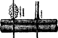
Figure 2.
of the cannulas become covered with strong elastic connective tissue, which holds the cannula in place. To increase the animal's ability to form such tissue, London recommends performing a preliminary operation two weeks before the main operation, consisting in opening the abdominal cavity and performing some minor manipulation in it, for example, in ligating and cutting off a small branch of the splenic vein. Such preliminary operations significantly increase the chances that the cannula will become covered with dense connective tissue and remain in place. The other end of the cannula is brought out through a special incision in the abdominal wall. The cannula is selected of such a length (from 6 to 16 cm) that the free end protrudes 2-3 cm. It is possible to prepare dogs with several cannulas; thus, in London's laboratory there are dogs that, in addition to an intestinal fistula, have cannulas in three blood vessels: the portal, hepatic, and renal (see Fig. 3) veins. To take
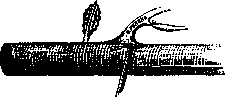
Figure 3.
blood, a needle (usually made of gold or iridium platinum) is inserted into the cannula, with the cannula held in such a way that after puncturing the vein, the needle runs along the length of the vein, not across it. Blood flows freely through the needle, and in this way up to 300-400 cubic cm of blood can be obtained. The dog does not even notice the moment of the puncture and remains indifferent to the blood being taken. At present, more than 30 works have been published, the data of which were obtained by means of the V. method. Almost all of these works came from London's laboratory. The topics of these works are diverse: intermediate nitrogen and carbohydrate metabolism, the fate of fats and cholesterol in intermediate metabolism, purine metabolism and the formation of uric acid, the equilibrium of hydrogen and hydroxyl ions, metabolism during fasting; the effect of low atmospheric pressure, pharmacological studies on the fate of quinine and iodine, the effect of insulin and adrenaline, the fate of inorganic phosphorus, specific metabolic secretions of internal organs - liver, spleen, adrenal glands, intestines, etc. The method of vasostomy is being improved year by year in London's laboratory. His employees (N. Kochnevoy and L. Rabin-kov) have developed the V. method also with respect to rabbits.
N. Vereshchagin. Vasostomy, Vasotomy. Vasotomy is the operation of sectioning the vas deferens, proposed by Voelcker and Belfield for the treatment of inflammation of the seminal vesicles (see Vesiculitis). The operation is performed under local anesthesia. An incision in the skin of small size (4-5 cm) along the course of the vas deferens at any point along its length. After freeing the deferens from surrounding tissues, it is brought out into the wound in the form of a loop. Along the length of the deferens, its wall is incised down to the lumen, through which a thin, blunt cannula is inserted, and through it the seminal pathway is irrigated. The irrigating fluid is discharged into the urethra. The deferens can be left for a longer or shorter time on a gauze strip in the open skin wound for repeating injections. To prevent the edges of the deferens wound from healing until the need has passed, a cannula or a metal probe used for the lacrimal duct, or a thread-like elastic bougie, should be left in its lumen. When injections are stopped, the deferens is lowered into the wound, and its incision is closed by the granulations of the skin wound. In those cases where the nature of the primary process (tuberculous epididymitis, purulent or tuberculous orchioepididymitis) requires its exclusion, vasotomy is replaced by vasostomy, which differs from vasotomy in that the deferens is cut. The central end is brought out into the angle of the skin wound.
Related articles
Mentioned in
Cite this page
“Vasostomy.” Soviet Medical Encyclopedia. English translation of Bolshaya Meditsinskaya Entsiklopediya, 1st ed. (Moscow, 1928–1936), ed. N. A. Semashko. https://sovietmedicalencyclopedia.pages.dev/article/vasostomy/