Portal Vein
Historical document, translated for reference. It reflects medical knowledge of the 1920s–30s and is not medical advice.
Summary
An anatomical and physiological overview of the portal vein (vena portae) from the 1st edition of the Soviet Great Medical Encyclopedia. It details the vein's origin, course, variations, tributaries, and the collateral circulation pathways (hepatopetal and hepatofugal) that develop under pathological conditions.
Encyclopedia article (1928–1936)
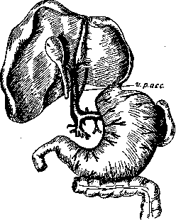
PORTAL VEIN (vena portae), collects venous blood from the stomach (except the cardia), intestines (except the lower segment of the rectum), pancreas, spleen, and gallbladder, and carries it to the liver (see plate, Fig. 1). Formed by the small veins of the aforementioned organs, the trunk of the portal vein begins behind the pancreas, posterior and to the left of the superior part of the duodenum, and runs toward the porta hepatis within the thickness of the hepatoduodenal ligament (posterior to the hepatic artery and bile ducts; Fig. 1. Variations of the portal vein: see plate, Fig. 2; 1—v. portae; 2—v. mesenter. sup.; 3—v. lienalis; 4—v. mesenter. infer.; 5—v. coronaria ventr. sup. (Valker). at the porta hepatis, the portal vein divides first into two branches, and then, gradually branching, breaks up into capillaries in the right and left lobes of the liver. From here, collected into more or less large hepatic veins (vv. hepaticae), the blood enters the general venous system (vena cava inferior). The length of the portal vein trunk ranges from 2 to 8 cm, depending on sex and age; with increasing age, the trunk of the portal vein elongates, which can be explained by the flaccidity of tissues associated with the aging of the body; in old age, the trunk of the portal vein is shorter, which is probably due to shrinkage resulting from atrophic processes; in women, the portal vein is thinner and longer. In general, the portal vein is formed by the superior and inferior mesenteric veins (vv. mesenterica sup. and inf.), the splenic vein (v. lienalis), and the left gastric vein (v. coronaria ventriculi superior), but there are significant variations in the order of their fusion, which can be grouped into 3 types (Valker; see Figure 1)—the portal vein is formed by the fusion of: 1) the superior mesenteric vein and the splenic vein, where the inferior mesenteric vein may empty into a) the superior mesenteric vein or b) the splenic vein; 2) the superior mesenteric vein, the splenic vein, and the inferior mesenteric vein; and 3) the left gastric vein, the superior mesenteric vein, the splenic vein, and the inferior mesenteric vein. The largest trunk is the superior mesenteric vein. Its length ranges from 0.5 to 5 cm; it usually accompanies the branches of the artery of the same name and is situated with the latter between the two layers of the mesentery of the small intestine. Its main trunk runs upward and along the attachment of the root of the mesentery, located to the right of the artery; on its way, it receives venous branches from the small intestine and the proximal part of the large intestine. Reaching the pancreas, it passes behind its head and here merges with the inferior mesenteric vein and the veins of the stomach. More frequently (predominantly in men), it is formed by the fusion of the superior mesenteric vein proper and the inferior, less frequently by the fusion of three trunks of intestinal veins into one, and (predominantly in women) by the fusion of many venous trunks. A characteristic feature of the intestinal veins is the formation
by them of a series of arches, with their convexity facing the intestinal loop, and these arches can be arranged in several rows (arches of the first, second, etc. order). The greatest number of them occurs in the section of the small intestines, the smallest in the section of the large intestines. In some cases, the arches are very weakly expressed, especially at the edge of the intestine, and then individual venous branches, which do not form new arches, depart from the main arch to the intestinal loop. Therefore, the entire venous system of the intestines can be divided into two types: 1) plexiform (looped) and 2) branching.—The superior pancreaticoduodenal vein (v. pancreatico-duod. sup.) runs along the inner edge of the upper portion of the vertical and upper horizontal part of the duodenum and empties most often into the left gastric vein (v. coronaria ventr. sup.) or into the gastrocolic vein, and sometimes into the inferior mesenteric vein, the middle colic vein, and directly into the portal vein. The inferior pancreaticoduodenal vein (v. pancreatico-duod. infer.) anastomoses with the preceding one, is located along the inner edge of the duodenum, and collects blood from the lower portion of the vertical and lower horizontal part of the duodenum; sometimes it is double and empties into the superior mesenteric vein or into one of the
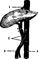
Figure 3. Diagram of an Eck
fistula:
1—v. hepatica;
2—fistula; 3—v. portae;
4—v. cava infer. intestinal veins (vv. intestinales). The posterior pancreaticoduodenal vein (v. pancreatico-duodenalis posterior), located on the posterior surface of the duodenum, collects blood from this area and the adjacent part of the pancreas and empties into the portal vein; in its absence, it is replaced by one or several branches of the superior pancreaticoduodenal vein. The veins of the small and large intestines have the same names as the arteries and accompany them, widely anastomosing with each other, most often forming arches in two rows (branching and looped types).—The splenic vein (v. lienalis) is formed from several roots coming from the hilum of the spleen and from the fundus of the stomach; passing along the upper edge of the pancreas, it receives small veins and behind its head empties into the inferior mesenteric vein or the portal vein.—The inferior mesenteric vein (v. mesent. inf.) is formed on the rectum from the venous plexus and, then receiving branches from the sigmoid flexure, descending colon, and the distal part of the transverse colon, and passing under the head of the pancreas, empties into the superior mesenteric vein, having previously received the splenic vein.—The left gastric vein (v. gastrica s. coronaria ventric. sup.) runs from left to right in the layers of the lesser omentum from the lesser curvature of the stomach and empties into the portal vein. The site of entry varies; most often it is located at a distance of 1.0–1.5 cm from the angle formed by the entry of the splenic vein. Collateral circulation in the portal vein system depends on anatomically pre-existing collaterals and their physiological adequacy. Anastomoses are mostly congenital, but under normal conditions they do not manifest themselves; only when circulation in the portal vein system is obstructed do they expand compensatorily. Anastomoses are divided into: 1) hepatopetal—connecting the portal vein with its hepatic branches, and 2) hepatofugal—diverting portal vein blood into the bed of the superior or inferior vena cava, bypassing the liver. The former can replace the portal vein, but only if the circulatory obstruction is caused by an obstacle in the portal vein itself. If the obstacle lies in the hepatic veins (for example, in atrophic cirrhosis of the liver), then hepatopetal anastomoses are useless. In these cases, hepatofugal anastomoses can come to the rescue, but it is obvious that they cannot fully replace the portal vein, because by diverting blood into other beds, they deprive it of the natural filter that is the liver. Hepatopetal anastomoses include the accessory portal veins of Sappey (veines portes accessoires), which occur as: 1) branches coming from the hepatogastric ligament, 2) small veins from the gallbladder
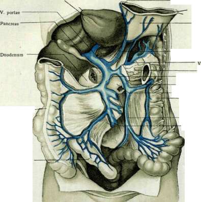
V. coronaria ventriculi Ventriculus V. coron. ventriculi lienalis Pancreas V. mesenterica sup. V. mesenterica inf. Angulus duod.-jejunal. Colon descendens Colon ascendens V. mesenterica inf. Mesenterium Intestin. ileum
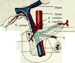
Vesica fellea Ductus choledochus Duodenum Caput pancreatis V. lienalis Pancreas mesenterica inf. A. mesenterica sup. V. mesenterica sup. Fig. 2. The portal vein within the thickness of the hepatoduodenal ligament (modified diagram, after Rouvière).
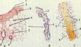
Figure 1. The portal vein system (after Rouvière).
Figure 3. A—structure of a vein: 1—endothelial layer (intima); 2—middle muscular layer (media); 3—adventitia; 4—surrounding loose connective tissue (adipose); 5—small vein; 6—its corresponding artery; 7—arteriole. B—small vein—venule [2], composed of capillaries (1). C—vein of the pia mater: 1—endothelium (intima); 2—thin adventitia (no muscular layer); 3—blood inside the vessel; 4—tissue of the pia mater. To the article Veins, portal vein. ... of the gallbladder, anastomosing with its veins, 3) veins coming from Glisson's capsule of the liver, 4) veins from the peritoneum via the lig. suspensorium hepatis, and 5) from the umbilical region also via the lig. suspensor. and, partly, through the obliterated cord of the v. umbilicalis. These veins (according to Kiernan) empty into the site of division of the portal vein. In addition, Walker described vv. portae accessoriae propriae, which in the form of 1, 2, 3, or more small trunks arise either from the portal vein itself or from one of its hepatic branches, usually at some distance from the hilus of the liver, sometimes at the level of, but more often above, the inflow of the v. coronaria ventriculi superior (see Figure 2), and, having reached the liver, disappear into its parenchyma. They are found in 4% of cases, have a caliber of 0.25 to 1.0 mm, and a length of 1.0 to 2.5 cm. Hepatofugal anastomoses include the veins of the preperitoneal and retroperitoneal tissue: "cava-portal anastomoses," since through them a connection is established between the systems of the v. portae and v. cavae. Every organ of the abdominal cavity, except the pancreas, has extra- and intraorgan anastomoses with the portal vein. The former are located outside the organ, in the extraperitoneal tissue, can dilate relatively freely and, if necessary, divert blood from the portal vein system into the v. cavae system. Intraorgan ones are characterized by low capacity and have no practical significance in this sense. Most frequently observed is an anastomosis between the v. ileo-colica, v. appendicularis, and v. spermatica dextra in the number of 2–3 and up to 6 branches (Torkacheva); a direct connection of the branches of the v. ileo-colica and v. appendicularis with the cava inferior is possible, as well as a connection of the appendiceal veins with the v. sacralis media, which usually empties into the v. iliaca communis sinistra. Blood is also diverted into the inferior vena cava from the mesenteric veins, mainly from the hemorrhoidal plexus of the lower segment of the rectum via the vv. haemorrhoidales mediae, haemorrhoidales inferiores, pudenda interna, and v. hypogastrica, and partly through the v. pudenda externa and v. femoralis. Acute insufficiency of the collateral portal circulation experimentally (upon ligation of the portal vein) manifests as hemorrhagic infarction of the intestines, with abundant hemorrhage into the intestinal lumen, leading to the rapid death of the animal. With a gradual increase of obstruction in the portal vein, the collaterals open up, become physiologically sufficient, and the animal recovers. In humans, with slowly increasing symptoms of circulatory obstruction in the portal vein system, hepatofugal anastomoses take on a certain role in the collateral circulation, expanding significantly, which can be seen, for example, on the veins of the abdominal skin in the umbilical region, sometimes forming a tortuous network (caput Medusae). However, both hepatopetal and hepatofugal anastomoses may prove to be both anatomically and functionally insufficient, as evidenced by the development of ascites (see). An attempt to establish a wide communication between the portal vein system and the v. cava was made by Eck, who proposed, by creating an anastomosis between the portal vein and the v. cava with ligation of the former above the site of its application, to exclude the liver from the portal circulation ("Eck's fistula," see Figure 3). However, animals after this operation quickly die with symptoms of clonic and tonic convulsions, which, according to Nencki, is caused by poisoning with ammonium carbamate entering the general circulatory system, due to the exclusion of the liver, in an unaltered form (not processed into urea), which, however, is disputed (Pal). The conditions of the portal circulation are of great importance in the circulation of the liver, as are the functional sufficiency of the right heart and the influence of nervous stimulation (Pal). In humans, Eck's operation has been successfully performed (in modification) by Bogoraz, Krestovsky (suturing the peripheral end of the transected v. mesenterica superior into the v. cava inferior), and Rosenstein (Rosenstein—anastomosis between the portal vein and the v. cava, without applying a ligature to the former above the anastomosis). The embryonic development of the portal vein is determined by the course of development of the primary vitelline veins. The unpaired trunk of the portal vein arises where the vitelline veins approach the liver and connect by two ring-shaped transverse anastomoses embracing the duodenum (sinus annularis of His); as a result of the right limb of the posterior anastomosis and the left limb of the front anastomosis becoming obsolete, the unpaired trunk of the portal vein is formed, spirally embracing (from left to right) the duodenum and receiving blood from the yolk sac and the intestinal canal, and subsequently—with the reduction of the yolk sac—only from the intestinal canal and the developing pancreas and spleen. With the cessation of placental circulation at birth, the v. umbilicalis and ductus Arantii (hepatic circulation, see Liver) obliterate and transform: the former into the lig. teres hepatis, the latter into the lig. venosum. On the basis of comparative anatomical data, one can conclude about the features of the portal vein system of higher organized animals and, in particular, of humans: the higher the comparative anatomical stage, the more isolated from the rest of the venous system is the portal vein system, and the smaller the number of its roots becomes. In humans and other higher animals, the portal vein is formed by 2, 3, or 4 branches. In birds, it usually connects by an anastomosis with the vena cava (Jacobson's anastomosis); in amphibians and reptiles, there are two hepatic-portal systems: one corresponds to the portal vein of higher animals, the other receives blood from the lower extremities, urinary bladder, ventral abdominal wall, gallbladder, and heart. In bony fishes, the portal vein connects with the veins of the swim bladder and at the posterior end of the body communicates with the v. caudalis (see also Veins). Pathology. Inflammatory diseases of the portal vein are observed quite rarely. In terms of their pathological-anatomical nature and origin, its acute purulent and chronic inflammations (pylephlebitis) present no special features compared to inflammatory processes of veins in general (see Veins). The uniqueness of the process is determined by the exceptional conditions of the portal circulation and the peculiarities of its participation in the general circulation. Suppurative pylephlebitis (pylephlebitis suppurativa s. purulenta) is either the result of the transition of a purulent process from neighboring tissues or is caused by the entry of an embolus from a purulently disintegrating thrombus or by spread per continuitatem. In the latter cases, the sources are purulent and inflammatory processes in the region of the tributaries of the portal vein: ulcers of the stomach and intestines, various diseases of the latter (colitis, typhlitis, paratyphlitis, appendicitis, proctitis, dysentery, hemorrhoids), as well as suppuration in the cellular tissue of the abdominal cavity and processes in the mesentery itself. Likewise, diseases of the organs served by the portal system, and of the pelvic organs in women, can cause pylephlebitis, and if a purulent embolus is carried into the liver, it can cause the formation of abscesses in it. Pylephlebitis is the most common cause of thrombosis and occlusion of the portal vein (pylethrombosis). Thrombosis can also be caused by slowed circulation (marantic thrombus), compression of the lumen of the portal vein from the outside by a tumor or of its intrahepatic ramifications in cirrhosis, or (rarely) by parasites penetrating into the lumen (Distomum haematobium). In case of insufficiency of the collateral circulation, thrombosis of the portal vein, depending on the level of occlusion, entails significant disorders, manifesting in a prolonged course as damage to the hepatic parenchyma (atrophic processes), and in the impossibility of collateral circulation—as catastrophic changes in a number of organs (stomach, intestines, spleen, pancreas).—Portal circulation, see Liver.
Related articles
Mentioned in
Cite this page
“Portal Vein.” Soviet Medical Encyclopedia. English translation of Bolshaya Meditsinskaya Entsiklopediya, 1st ed. (Moscow, 1928–1936), ed. N. A. Semashko. https://sovietmedicalencyclopedia.pages.dev/article/portal-vein/