Coccyx
Historical document, translated for reference. It reflects medical knowledge of the 1920s–30s and is not medical advice.
Summary
The coccyx is the lowermost part of the human vertebral column, consisting of 4-5 fused vertebrae. It forms a synchondrosis with the sacrum and serves as attachment points for muscles and ligaments.
Encyclopedia article (1928–1936)
Coccyx (os coccygis), the lower, terminal part of the human vertebral column. It is homologous to the tail skeleton of animals. It consists of 4, very rarely 5 vertebrae (vertebrae coccygeae), gradually decreasing in size from top to bottom, giving the entire coccyx a triangular shape (figs. 1 and 2). Its broad base connects with the sacrum, while the apex is freely directed downward. On the first coccygeal vertebra, one can distinguish the vertebral body (corpus vertebrae), poorly marked transverse processes (processus transversi), and two horns (cornua coccygea). The latter extend from the posterior surface of the coccyx and correspond to the superior articular processes. In the II vertebra, it is sometimes also possible to distinguish a hint of transverse processes. The remaining vertebrae form rudimentary round bodies of spongy bone tissue. All coccygeal vertebrae, except the first, in old age, especially in men, fuse together; the V vertebra, if present, is also often fused with the vertebra above it. The coccyx forms a synchondrosis (symphysis) with the sacrum, containing an intervertebral disc with a small central cavity. - Ligaments of the coccyx: lig. sacro-coccygeum articulare, s. breve - connects the coccygeal horns with the sacral ones; lig. sacro-coccygeum laterale - connects the crista sacralis lateralis with the processus transversus of the I coccygeal vertebra; lig. sacro-coccygeum anterius - passes along the anterior surface of the sacro-coccygeal joint; lig. sacro-coccygeum posterius superficiale and lig. sacro-coccygeum posterius profundum - along the posterior surface of the sacro-coccygeal joint (fig. 3). - To the coccyx is attached a paired, quadrangular, thin coccygeal muscle (musculus coccygeus; fig. 4). Starting from the spina ischiadica of the ischial bone, the coccygeus muscle extends to the lateral edges of the lower sacral and upper coccygeal vertebrae, intertwining here with the posterior bundles of the m. ilio- et pubo-coccygei (levator ani) and, together with the latter, tenses and elevates the muscular diaphragm of the pelvis, receiving innervation from branches of the sacral plexus. In front, at the beginning of the coccygeus muscle, is located the coccygeal plexus (plexus coccygeus). It is formed by the anterior branches of the coccygeal, V sacral, and partly IV and III sacral nerves, anastomoses with the lower end of the sympathetic trunk, supplies the internal organs with nerve fibers, and gives rise to three to five anococcygeal nerves (nervi ano-coccygei), which pierce the coccygeus muscle and, directed backward, spread out in the skin of the coccygeal area. - The coccyx and the soft tissues immediately adjacent to it are supplied with blood from branches of the middle sacral artery (a. sacralis media; fig. 5), which originates from the abdominal aorta above its division, descends downward along the median line, in front of Lv, the sacrum, and the coccyx, and ends under the latter as the 'coccygeal gland,' or 'coccygeal plexus' (glomus coccygeum). Blood drains through the corresponding vein (vena sacralis media). The coccyx is sometimes subject to traumatic effects: contusions, dislocations, fractures, and gunshot wounds. The first three types of damage occur almost exclusively as a result of falling onto the hard prominence of the coccygeal area. Dislocations can occur forward and backward. The latter are rarer and may occur as a result of birth injuries and pressure from tumors developing in the anterior sacro-coccygeal area. Symptoms of traumatic injuries consist of sharp local pain, increasing when sitting and walking, tenesmus, and in dislocations and fractures, in addition, mobility and displacement of the injured bone, which is
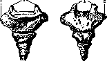
Figure 1.
Figure 2.
Figure 1. Coccyx from the front. Figure 2. Coccyx from the back. 1-cornua coccygea; 2- processus transversus. round bodies of spongy bone tissue. All coccygeal vertebrae, except the first, in old age, especially in men, fuse together; the V vertebra, if present, is also often fused with the vertebra above it. The coccyx forms a synchondrosis (symphysis) with the sacrum, containing an intervertebral disc with a small central cavity. - Ligaments of the coccyx: lig. sacro-coccygeum articulare, s. breve - connects the coccygeal horns with the sacral ones; lig. sacro-coccygeum laterale - connects the crista sacralis lateralis with the processus transversus of the I coccygeal vertebra; lig. sacro-coccygeum anterius - passes along the anterior surface of the sacro-coccygeal joint; lig. sacro-coccygeum posterius superficiale and lig. sacro-coccygeum posterius profundum - along the posterior surface of the sacro-coccygeal joint (fig. 3). - To the coccyx is attached a paired, quadrangular, thin coccygeal muscle (musculus coccygeus; fig. 4). Starting from the spina ischiadica of the ischial bone, the coccygeus muscle extends to the lateral edges of the lower sacral and upper coccygeal vertebrae, intertwining here with the posterior bundles of the m. ilio- et pubo-coccygei (levator ani) and, together with the latter, tenses and elevates the muscular diaphragm of the pelvis, receiving innervation from branches of the sacral plexus. In front, at the beginning of the coccygeus muscle, is located the coccygeal plexus (plexus coccygeus). It is formed by the anterior branches of the coccygeal, V sacral, and partly IV and III sacral nerves, anastomoses with the lower end of the sympathetic trunk, supplies the internal organs with nerve fibers, and gives rise to three to five anococcygeal nerves (nervi ano-coccygei), which pierce the coccygeus muscle and, directed backward, spread out in the skin of the coccygeal area. - The coccyx and the soft tissues immediately adjacent to it are supplied with blood from branches of the middle sacral artery (a. sacralis media; fig. 5), which originates from the abdominal aorta above its division, descends downward along the median line, in front of Lv, the sacrum, and the coccyx, and ends under the latter as the 'coccygeal gland,' or 'coccygeal plexus' (glomus coccygeum). Blood drains through the corresponding vein (vena sacralis media). The coccyx is sometimes subject to traumatic effects: contusions, dislocations, fractures, and gunshot wounds. The first three types of damage occur almost exclusively as a result of falling onto the hard prominence of the coccygeal area. Dislocations can occur forward and backward. The latter are rarer and may occur as a result of birth injuries and pressure from tumors developing in the anterior sacro-coccygeal area. Symptoms of traumatic injuries consist of sharp local pain, increasing when sitting and walking, tenesmus, and in dislocations and fractures, in addition, mobility and displacement of the injured bone, which is
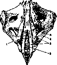
Figure 3. Ligaments between the coccyx and sacrum. 1- processus transversus; lig. sacro-coccygeum post. prof. (2) et superfic. (3); 4-lig. sacro-coccyg. lat.; 5- apex ossis sacri.
recognized by
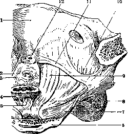
Figure 4. Right m. levator ani. 1-symphisis oss. pubis; 2-urethra; 3-trigon. urogenitale; 4-rectum; 5-m. recto-coccygeus; 6-os sacrum; 7-m.coccygeus; 8-m. ilio-coccygeus; 9-m. pu-bo-coccygeus;i0-arcus tendin.m.levat.ani;//- arcus tendin. fasciae pelv.; 12-v. dorsal, penis.
the area of the coccyx onto a hard prominence. Dislocations can occur forward and backward. The latter are rarer and may occur as a result of birth injuries and pressure from tumors developing from the anterior sacro-coccygeal area. Symptoms of traumatic injuries consist of sharp local pain, increasing when sitting and walking, tenesmus, and in dislocations and fractures, in addition, mobility and displacement of the injured bone, which is
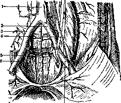
Figure 5. Arteries of the pelvis: 1-aorta abdom.; 2-a. iliaca comm.; 3-a. sacralis med.; 4-ureter; 5-a. sacral, lat.; 6-a. hypogastr.; 7-a. iliaca ext.
The diagnosis is made by palpation from the skin side and from the rectal side. The latter is also often damaged (the coccyx being dislocated forward, its fragments). Reduction is achieved by direct pressure on the displaced parts with the fingers, for which one of them is inserted into the rectum, and the others manipulate from the outside. It is more difficult to keep the reduced coccyx, resp. its fragments, in place. A thick-walled drainage tube inserted into the rectum for several days and strips of adhesive plaster applied to the skin behind the coccyx can help with this. Subsequent treatment consists of bed rest, warm (up to 40°) sitz baths, and gentle massage. Sometimes, despite treatment, persistent pains remain, known as coccygodynia (see). In such cases, resection of the coccyx is indicated, performed through a vertical median incision, sometimes with an additional transverse incision above the base of the coccyx. Gunshot wounds of the coccyx are observed mainly during wars and constitute about 1% of all gunshot wounds of the pelvis. In such cases, the surgeon's main attention is not drawn to the coccyx itself, but to the simultaneously damaged internal organs (rectum, bladder, etc.).-Of infectious diseases, only tuberculosis of the coccygeal bones is of interest, but it is also rare, occurring according to the type of carious lesions of flat bones. In case of involvement of the posterior surface of the coccyx, a cold abscess is detected very early and precisely at the site of the carious focus. When the anterior surface is affected, the abscess is detected much later; it is located to the side or below the coccyx or around the anal opening. Rupture of the abscess into the rectum is possible. Tubercular fistulas can be confused with pericoccygeal fistulas originating from dermoid cysts. The diagnosis is often only established after finding hairs in the fistula discharge, which constitute the characteristic content of the dermoid. In the presence of a fistula, its direction and the localization of the primary focus can be determined by injecting contrast substances such as bismuth salicylate, lipiodol, iodipin, etc. into the fistula, followed by an X-ray. Of particular interest are tumors of the coccyx. They are rare but show great morphological diversity. On the anterior surface of the sacrococcygeal area, lipomas, lymphangiomas, and teratomas occur. On the posterior surface-meningocele, as well as lymphangiomas and teratomas. Meningocele originates from the sacral canal opening and represents a protrusion of the meninges in the area of the sacrococcygeal cornua resembling a cystic tumor. Meningocele is often associated with lymphangioma or teratoma, but never with lipoma, which is the direct opposite of sacrolumbar tumors, which are often combined with lipomatous, cavernous, and cystic formations (Bergmann). More common than other coccygeal tumors are cystic and cavernous lymphangiomas without meningocele. Teratomas of the coccygeal area show great diversity. They may contain elements of all three embryonic leaflets (tridermomas). Upward, coccygeal tumors never extend above the upper edge of the gluteal fold. The skin over the tumor is sometimes normal, but often atrophic, red, and sometimes ulcerated. During straining, e.g., when crying, the tumor tenses. Palpation is painless. Tumors developing in front of the coccyx displace it backward and may cause compression of the rectum. In differential diagnosis, it is necessary to exclude hernias of the intestine and bladder. The growth of the tumor is slow in some cases (lipomas), rapid in others (cystosarcomas). Diagnosis of the various forms of tumor before surgery is very difficult. The prognosis is serious even for benign tumors, since the soft tissues over the tumor easily (due to the proximity of the anal and urethral openings) become contaminated, infected, inflamed, suppurate, and ulcerate. This in turn easily leads to general sepsis. The mortality rate of newborns with coccygeal tumors is about 90% in the very first days. As a rare exception, patients survived to old age. Any coccygeal tumor should be surgically removed. The greatest technical difficulties in excision are presented by anterior tumors. It is best to dissect them after preliminary resection of the coccyx through a T-shaped incision. If even under these conditions it is not possible to bypass the upper pole of the tumor, the tumor is severed as high as possible and the remnants are cauterized with a thermocautery (Steinthal).-Besides tumors in the strict sense of the word, peculiar formations, so-called tail appendages, are also found in the area of the coccyx. They can reach a length of 10 cm. Virchow distinguishes among them 1) complete tails, consisting of an increased number of coccygeal vertebrae, 2) soft, boneless tails, connected to the spine by rudimentary tissue, 3) tail-like skin appendages arising from inflammatory processes occurring between the egg membranes and the upper surface of the embryo. All types of tail appendages require surgical excision. The technique here is much easier, and the prognosis is much more favorable than in coccygeal tumors.
M. Fridiand. CORTEX (lat. cortex), cortical substance, the peripheral part of certain organs, differing from the rest of the centrally located part (called the medulla) in its color, density, and structure; anatomically and functionally, the cortex and medulla are closely connected to each other. Cortical substance (lat. substantia corticalis) is distinguished in the kidneys, adrenal glands, ovaries, brain, and thymus gland.-The kidney cortex, or renal labyrinth, forms the superficial part of the kidney 5-7 mm thick, extends to the sides to the edges of the renal sinus, and in addition gives off processes (Bertini's columns) between the pyramids of the medulla. Its color is reddish-brown, the appearance is uniform, slightly granular.-The adrenal cortex is dense, of a rich yellowish or brownish-yellow color; it covers the medulla, which has a significantly smaller mass.-The ovarian cortex represents the most important part of them and contains glandular tissue in the form of egg follicles with eggs.-In the thymus gland, the cortex is the darker, peripheral part of the main lobes-rich in cells and blood vessels.-The cerebral cortex is gray in color, up to 5 mm thick, covers the periphery of the brain, dipping uniformly in a layer into all its convolutions.
Related articles
Mentioned in
Cite this page
“Coccyx.” Soviet Medical Encyclopedia. English translation of Bolshaya Meditsinskaya Entsiklopediya, 1st ed. (Moscow, 1928–1936), ed. N. A. Semashko. https://sovietmedicalencyclopedia.pages.dev/article/coccyx/