NERVE FIBERS
Historical document, translated for reference. It reflects medical knowledge of the 1920s–30s and is not medical advice.
Summary
Nerve fibers are long processes of nerve cells located in the central and peripheral nervous systems. They can be formed by axons or dendrites of nerve cells, with or without myelin sheaths, and their structure varies between central and peripheral locations.
Encyclopedia article (1928–1936)
NERVE FIBERS, long processes of nerve cells, located in the central and peripheral nervous systems. In most cases, nerve fibers are formed by the axons of nerve cells (see), but in some cases by long dendrites (dendrites of unipolar or bipolar sensory nerve cells). The axons of nerve cells passing through the gray matter of the brain, in many cases, do not show special sheaths, but upon entering the white matter of the brain, they all connect with special cellular or syncytial formations, which are usually called the sheaths of N. f. The larger part of N. f., especially those originating from the central nervous system, already in the white matter of the brain has a sheath consisting of a special lipoid substance, myelin or nerve medulla, which is what the white matter of the brain owes its color to. These fibers * are called myelinated or medullated N. f. Most N. f. that make up the sympathetic nervous system do not have this sheath and are called non-medullated or, after the name of the author who first described them, Remakian (Remak; 1838). The structure of medullated N. f. inside and outside the brain is somewhat different, and therefore morphologically central nerve fibers are distinguished from peripheral ones. Peripheral medullated N. f. Their thickness is very diverse; in humans it ranges between 1-16 microns. Apparently, the thicker ones belong to larger nerve cells, the thinner ones to smaller ones. According to Schwalbe, the thicker ones are at the same time the longest. Younger N. f. are thinner than older ones. Along the axis of N. f. runs its main part - the axis cylinder, representing, as stated above, the process of a nerve cell. In a freshly split and unfixed N. f., the axis cylinder has the appearance of a fairly wide, somewhat flattened cylinder [see separate table (p. 607-608), figure 1]. On fixed preparations, it is significantly thinner, as it apparently shrinks due to its high water content. In the so-called vital staining of N. f. with methylene blue, the axis cylinder is stained homogeneously but unevenly. The most intense staining is obtained by the parts of the axis cylinder located on both sides of the nodes of Ranvier (see below). When viewed in the dark field (ultra-condenser), the axis cylinder appears optically empty. The axis cylinder consists of 1) neuroplasm, 07 or axoplasm (Waldeyer), or perifibrillar substance (Bethe) and 2) neurofibrils. Each part is visible only with appropriate treatment. 1) Neuroplasm is a continuation of the plasma of nerve cells and has its properties. According to some researchers, it is independent; others consider it to be one whole with the plasma of the myelin sheath, and give the entire plasma of the internodal segment the common name myeloaxostroma. In the form of thin layers, it is located between the neurofibrils and in addition forms the outer marginal zone. Outside the axis cylinder, a special thin layer was previously described, which bears different names: axolemma (Axolemma), inner sheath (innere Scheide), Mauthner's sheath (Mauthnersche Scheide), cortex of the axis cylinder (Axenzylinder-rinde; Schiefferdecker), inner neurilemma (Boveri). Most researchers at present consider this layer an artificial formation obtained as a result of the shrinkage of the axis cylinder. 2) Neurofibrils, axofibrils are a continuation of the fibrillar network of the nerve cell (see). Just as with the latter, the controversial question - whether there are anastomoses between individual fibrils - has not been resolved. Opinions also differ regarding the significance of each part of N. f. for the conduction of excitation. Apathy, Bethe, Heidenhain, Bielschowsky consider only neurofibrils as conducting elements, Leydig, Wolff, Strasser - only the plasma, Cajal, Retzius, Marinesco attribute significance to both parts. The medullated sheath covering the axis cylinder (see separate table, figure 3), or myelin sheath contains a mixture of lipoid substances, which has been named myelin. When viewed in the fresh state and on preparations fixed with substances that do not dissolve fats, the myelin sheath appears homogeneous and exhibits the phenomenon of uniaxial double refraction. On cross-sections, the medullated sheath has a radial, comparable to the appearance of a wheel (Radspeichenstruktur), or ray-like (Sonnenbildchenfigur) structure. It is stained with special stains for lipids (Ciaccio, Schmidt, Dietrich, Sudan III - pale orange-pink color, with osmium - gray, gray-black). It is intensely stained with hematoxylin according to Weigert. When treated with fat-dissolving substances (alcohol, ether, chloroform), myelin dissolves, and the myelin sheath takes the form of a delicate network, called the neurokeratin network or skeleton (Ewald, Kühne). This network has nothing to do with keratin, can be detected with special stains (Altmann, Snesarev). Some authors consider it an artificial formation, others accept it as crossbars formed by the protoplasm of Schwann cells; new Russian works (Snesarev, Mogilnitsky, Dukhnikova) recognize the neurokeratin network as an independent paraplasmic substance. The homogeneous structure of myelin is characteristic of a nerve that has not lost its excitability. A N. f. damaged in some way and having lost its excitability changes the character of the myelin sheath. Myelin is then detected in the form of droplets or lumps of various sizes and shapes (Cajal). Further destructive processes of the myelin sheath are expressed in the transformation of myelin lipids into neutral fat (see Wallerian degeneration). Accordingly, when N. f. is damaged, its neurokeratin skeleton also changes. In the degeneration of a nerve fiber, its thin network turns into large, irregularly shaped cells and finally completely disappears. The neurilemma or Schwann sheath is a thin, transparent, structureless sheath covering the entire surface of N. f. Under it is a thin layer of cytoplasm, which in places forms accumulations. In thick fibers, such accumulations are more numerous and contain more plasma. In the largest accumulation of plasma, closer to the middle of the segment, is located an oval or round nucleus. In thin fibers and in those poor in plasma, it is flatter, has the shape of an ovoid and lies, indenting into the myelin. From the superficial layer of cytoplasm, strands of cytoplasm extend, anastomosing with each other and penetrating into the depth of the myelin sheath to the axis cylinder. In the cytoplasm, with appropriate staining, special types of inclusions are visible: 1) so-called tγ-granules (tγ-granula; Reich) [see separate table (p. 623-624), figure 11]; they look like very small hooks and are stained metachromatically with thionin in carmine-red color. 2) Round bodies or grains of various sizes, staining like myelin - β-granules (Reich), or Elzholz bodies. 3) Droplets of various sizes, giving microscopic fat reactions. The amount of these inclusions changes with age and in various pathological conditions. Apparently they are products of metabolism or degeneration (see below pathology of N. f.). Medullated N. f. at certain and mostly equal distances receive characteristic constrictions. At such constrictions, myelin is absent, while the Schwann sheath is not interrupted. These sharply defined constrictions are called "nodes of Ranvier". The parts of the Schwann sheath in the interval between two nodes contain in mammals one nucleus and are called internodal segments; their length in thick fibers reaches, according to Schaffer, 1 mm; in thin N. f. it is smaller. At present, it can be considered established that the previously practiced division of sheaths into Schwann and myelin is incorrect, since the myelin sheath is only the inner part of the Schwann cell (Nemilov, Boeke, Heringa, Mollendorff). The Schwann cell (figure 1) represents a formation of cylindrical shape. Its outer part is represented by 1- Ti
т ж
щ
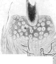
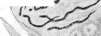
Figure 1. Structure of the vagus nerve fiber of a dog: a - neurilemma; b - myelin; c - axolemma; d - axial cylinder. Figure 2. Regeneration of a nerve fiber. Formation of a bundle of neurofibrils in the ribbon fiber. Figure 3. Myelinated fiber with stained axial cylinder. Figure 4. Calcification of a nerve cell. Figure 5. Motor plate on striated muscle. Figure 6. Nerve plexus around the hair follicle of human skin - large cells of sebaceous glands and small nuclei of the hair follicle epithelium are visible. Figure 7. Neurinoma. (Figure 1 - after Stern, figs. 2, 3 and 6 - after Rakhmanov, fig. 4 - from Spilmaier, fig. 5 - after Voeke). See also: Neurinoma, Nerve fibers, Nerve cells, Nerve endings. 609 with a thin layer of protoplasm with a thickening around the nucleus, while the inner part, adjacent to the axial cylinder and enclosing it, represents a protoplasmic network, in the loops of which are drops of myelin [see separate table (art. 623-624), figures 6 and 12]. The crossbars of this network are so thin that the myelin sheath appears completely homogeneous under the microscope. If the myelin is heavily stained, then along the course of N. f. deep funnel-shaped slits become visible, extending from the surface of the Schwann cell right to the axial cylinder. These spaces are called Schmidt-Lanterman's incisions and divide each segment into 'cylinder-conical' sections. Inside these slits, Golgi and Rezzo
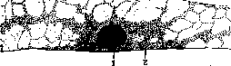
Figure 1. Schwann cell (according to Nemilov): 1-nucleus; 2-cytoplasm. nico) described visible with Golgi staining thin threads, spirally or ring-like surrounding the axial cylinder. Their significance at the present time is not yet sufficiently clarified. It is possible that the notches represent thicker crossbars of Schwann cells (Nemilov). When N. v. are treated with silver and gold salts, a variety of patterns arise, which, as is now established, are for the most part artificial formations. These include the 'Ranvier crosses', obtained at the place of the same-name nodes. They occur because when the nerve is treated with silver, it penetrates inside the nerve trunk in the area of the nodes and is adsorbed more quickly and easily here. During the reduction of silver, it is deposited at the place of entry (a black line perpendicular to the axial cylinder) and in the parts of the axial cylinder closest to the node (a line perpendicular to the previous one). These also include the so-called dark stripes, arranged in a ladder pattern across the axial cylinder, especially near the Ranvier nodes. They are called the lines, or stripes of Fromann. At strong magnification, a granular structure is noticeable in them. It has been established that these lines arise due to the uneven deposition of silver salts, and the entire phenomenon proceeds similarly to the formation of Liesegang rings. At the nodes, the axial cylinder sometimes appears thickened like a club in both adjacent segments; these are the so-called biko-niches swellings; their significance is unclear; it is not excluded that this is some kind of physiol. or pathol. phenomenon, but at present they have no significance for the normal and path. histology of N. v. The central medullated N. v. in some details differs from the peripheral one. The question of the existence of a neurilemma on it has been raised repeatedly. Ranvier and Kölliker denied its existence, Cajal recognized it. It can be considered that glial cells form a protoplasmic layer covering the myelin sheath and giving rise to crossbars penetrating into the thickness of the myelin, forming a plasma reticular basis, in the loops of which the myelin is located. According to the latest data (Cajal, Flechsig, Dogel, Belynovsky) myelin in the central nervous system also forms interruptions, the glial rings of Held, analogous to the Ranvier nodes, but they are closer to each other than in peripheral fibers; the axial cylinder at these nodes is devoid of medulla for a greater extent and in this place is covered by a special, apparently plasma formation, the 'cement' cylinder (Cajal). The medulla in general has the same properties as in the peripheral fiber, but on cross-sections it does not have a clearly expressed radial structure. As a rule, p-granules are not found in the plasma of central fibers. Non-medullated, or Remak's nerve fibers are found in the central nervous system, as well as in large numbers in the peripheral nervous system. In the latter, they can be detected in nerve trunks of spinal origin and in nerve trunks of the autonomic nervous system. The once widespread view that postganglionic fibers of the autonomic nervous system are non-medullated must be abandoned. Postganglionic fibers for the most part have a medullated sheath. This sheath is extraordinarily thin and may be overlooked with insufficiently thorough research (Langley). In thin nerve trunks of the autonomic nervous system, medullated fibers lose their medulla before they reach their terminal apparatus, why such trunks for the most part are non-medullated or mixed. The axial cylinders of non-medullated nerve fibers are usually thinner than those of medullated ones. Sometimes on the axial cylinders, uniformly arranged, clear-shaped thickenings are noticed, the so-called varicosities. Such varicosities in normal conditions are found mainly in the area of the nerve ending, while varicosities along the course of the nerve trunk should be considered either an artificial formation or in some cases a pathological phenomenon. There are no inclusions in the plasma of non-medullated N. v.; formations similar to Ranvier nodes and definite boundaries between cells are also not noted. The sheath of non-medullated N. v. is built of elements that have a great resemblance to the Schwann cells of medullated N. v. Only the myelin-containing inner layer and the Ranvier nodes are absent. Since the boundaries between individual Schwann cells are not detected, the sheath of non-medullated N. v. is called the Schwann syncytium. The nuclei of the Schwann syncytium lie either on the periphery of the fiber, to the side of the axial cylinder, or lie in the center, and then the bundles of neurofibrils diverge, surrounding the nucleus from all sides (Schultze). In the organs innervated by the autonomic nervous system, the Schwann syncytium has a special character. Bundles of neurofibrils, before reaching their place of termination in the working organ, diverge, continuing to be covered by the protoplasm of the Schwann syncytium. These nodal points often have a star-shaped or three-rayed shape. With strong staining of the syncytium protoplasm, these areas have the appearance of multipolar cells. A number of authors recognize these elements as nerve cells ('interstitial cells of Cajal'). At the present moment, it can be considered established that many non-medullated N. v. contain several axial cylinders covered by one common Schwann sheath. This type of structure is called 'cable' (Martin Heidenhain). According to Leontovich, in organs provided with smooth muscles, in glands, in the connective tissue basis of the skin, the strands of the Schwann syncytium often anastomose with each other, forming true protoplasmic networks ('conducting plasmodium', according to Stern). Inside these networks run axial cylinders, which according to the opinion of most authors do not anastomose with each other. Such formations have been repeatedly described as 'nerve networks' and have served as a source for a number of physiol. concepts, especially regarding the innervation of the heart. The question of 'interstitial cells', 'nerve networks', 'protoplasmic networks' cannot yet be considered finally resolved. The significance of the sheaths of N. v. to this day remains insufficiently clarified. Held considers that the sheaths of N. v. represent a continuous protoplasmic path, predetermining the course and development of nerve fibers (see Neuron theory). Of particular importance here are the experiments of Harrison, who succeeded experimentally in obtaining in an animal nerve trunks completely devoid of sheaths, by removing the corresponding rudiment of the medullary tube. He also succeeded in seeing in a living animal the migration of ectodermal elements of the medullary tube and their settling on the axial cylinders. This phenomenon has now been reproduced in tissue cultures outside the organism. It follows from this that the connection of the axial cylinder and the Schwann syncytium is a secondary formation, arising at the moment of embryogenesis. But from the moment this connection was established, the axial cylinder and its sheath represent a morphol. and physiol. whole. This viewpoint was naturally alien to the cell theory, which represented tissue only as a sum of cellular elements; this same viewpoint was also adopted by the neuron theory, representing the elements of the sheaths as a case put on the axial cylinder. Meanwhile, pathol. histology of N. v. shows that when N. v. are affected, both the axial cylinder and the Schwann cell are involved to an equal degree, that is, N. v. reacts as an organic whole. - A whole series of theories has been advanced regarding the physiol. significance of the sheaths of N. v.; they were ascribed 'protective', 'insulating', 'nutritive' and other properties. None of these theories at present has serious grounds. However, there is no doubt that in the processes of exchange of N. v. Schwann elements play a very large role. Both medullated and non-medullated N. v. can branch along their course, usually dividing dichotomically. The division of medullated fibers occurs only at the Ranvier nodes. Before the formation of the terminal apparatus, the medullated N. v. loses the myelin sheath and for some distance is non-medullated. For N. v. in general, the connection of them into bundles is characteristic. Along the course of bundles of nerve fibers, the transition of individual N. v. and their groups from one bundle to another can occur, and cases are possible where two N. v., entering one bundle, diverge in it in opposite directions. From this it follows that the composition of bundles along
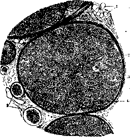
Fig. 2. Cross-section of a cerebrospinal nerve* 1-fat cells epineurium; 2- perineurium; 3-endoneurium; 4- cross-section of nerve fibers.
changes and that in a bundle fibers can pass conducting irritation in different directions. Outside the central nervous system, N. v. unite into bundles and are connected by connective tissue, thus forming peripheral nerve trunks (figure 2) (see Nerves). Age-related changes* of N. v. Just like nerve cells, N. v. at the time of human birth are not finally differentiated. Postembryonic development of nerve fibers continues for a considerable period. A number of nerve pathways in the central nervous system do not have myelin sheaths at birth. Flexigom discovered the patterns according to which the development of myelin in nerve pathways proceeds. It turned out that phylogenetically younger pathways are covered with myelin much later (see Myelination, Brain). For peripheral N. v., cable structure is characteristic (see above) of many myelinated N. v. Buke was able to show that the nerve fibers of the tongue of a newborn mouse are all built according to the cable type and only later differentiate into myelinated fibers containing one axial cylinder each. The appearance of myelin in peripheral N. v. in the postembryonic period has not yet been studied. There are indications that the fibers of the vagus nerve in the cardiac plexuses of a newborn are still devoid of myelin (Lazovsky). P a t. h o l o g i c a l c h a n g e s of N. v. occur both in the axial cylinder and in the sheaths covering it, but with various diseases of the nervous system, these elements can be affected to varying degrees. For example, in a number of neuritis, the axial cylinders react to injury with characteristic lateral outgrowths (Exkreszenz), the appearance of club-shaped thickenings, varicosities, sometimes of giant size, fragmentation, etc. The cytology of Schwann's syncytium in this case is little studied. The sheaths are more sensitive to harmful influences than the axial cylinder and react to them more quickly. Nerve fibers can undergo changes at the site of damage - this is so-called primary degeneration. Changes can spread to parts of the nerve more distant from the damage - secondary degeneration [see separate table (pp. 623-624), figure 13]. With a violation of the integrity of N. v., for example, when a nerve is severed, the part of the fiber separated from its nerve cell quickly undergoes so-called secondary, Wallerian degeneration (see). According to the initial views, the segment of N. v. remaining in connection with the nerve cell degenerates only to the nearest Ranvier's node, while the rest of the part remains unchanged. Later studies showed that this part of N. v. can also undergo changes. Swellings, irregularities (so-called retrograde degeneration) appear on the axial cylinder. Myelin balls-Elzholz bodies appear in the plasma of the Schwann cell, the myelin sheath loses its smoothness and can break into lumps. Then fat droplets also appear. For retrograde degeneration, the place where the N. v. is interrupted and the way this interruption is made is important. Thus, when N. v. is separated near the corresponding nerve cell, retrograde degeneration affects the vast majority of fibers. The same occurs when a nerve is torn out, for example when a root of a spinal or cranial nerve is torn out. In these cases, retrograde degeneration is traced all the way to the corresponding nerve cell. This phenomenon has very great significance in experimental work to determine nerve centers, as it allows, when peripheral trunks are damaged, to find the corresponding centers in the central nervous system. Wallerian degeneration can affect both central and peripheral N. v. In some pathological processes both in the central nervous system (for example multiple sclerosis) and in the peripheral nerve (neuritis), degenerative phenomena can affect not the entire nerve fiber but are observed only on individual sections of it (segmental, or "periaxial" neuritis Gombault; "discontinuous myelin breakdown" Stransky) [see separate table (pp. 623-624), figure 7]. The microscopic picture of the changed segments in this case is the same as in Wallerian degeneration. The lesion of individual N. v. by a segmental process is observed in nerve trunks in humans as a constant phenomenon even in the absence of clinical symptoms of neuritis. In the distal thin nerve trunks, these changes are more pronounced than in the proximal thick ones. The number of affected N. v. increases with age, as well as in general pathological processes leading to a violation of general nutrition and in cachexias: so-called latent neuritis (Pitres et Vail-lard, S. Mayer, Rakhmanov).
a. Rakhmanov. Regeneration of N. F. The property of nerves to restore their anatomical integrity and physiological conductivity after being severed was first discovered in 1776 (Cruikshank). The first detailed morphological data on the regeneration of N. F. were given by Waller (Waller; 1825), with whose name the so-called Wallerian law of degeneration and regeneration of nerves is associated. According to this law: 1) when the integrity of a nerve is disrupted, the peripheral segment of this nerve undergoes a specific change (see Wallerian degeneration); 2) following this, there occurs a new anatomical connection between the central end of the nerve and the peripheral end, and this restoration proceeds at the expense of the central segment of the nerve, which continues to be connected with the corresponding nerve cells, i.e., with the trophic center. Subsequently, the process of regeneration of N. F. was studied using special neurohistological methods (staining with methylene blue, impregnation with silver and gold salts). Thanks to the work of Cajal, Perroncito, Tello, Boeke, Bethe, Spielmeyer, Doynikov, Krasin (Cajal, Perroncito, Tello, Boeke, Bethe, Spielmeyer) and many others, the morphological picture of regeneration of N. F. has been described in great detail, starting from the earliest stages. The enormous theoretical and practical significance of the phenomenon of regeneration of N. F. has led to a major discussion around this question. In this context, attempts to explain the mechanism of regeneration of N. F. reflected the main theoretical positions of the authors with respect to all controversial questions of the structure of nervous tissue (see Neuron theory). The practical significance of regeneration for neurosurgery has led to numerous works devoted to the problems of nerve suture, replacement of defects, transplantation, heterologous regeneration, which in turn has provided abundant material for the theory of regeneration. Degeneration of a nerve along its length can be caused either by disruption of its integrity (severance, rupture) or by the application of a number of agents that kill N. F. (heat, freezing, poisons - arsenic, chromium salts, alcohol, acids, alkalis, etc.). Regardless of which of the ♦20 methods is applied, the part of the nerve lying between the site of injury and its peripheral endings undergoes degeneration with disintegration of the axis cylinders, transformation of myelin into fat, and formation of protoplasmic bands, so-called Büngner's bands, originating from Schwann's syncytium. The central segment of the nerve, i.e., the segment located between the site of injury and the corresponding nerve center (brain, spinal cord, ganglia of the autonomic nervous system), also undergoes degeneration, but only over a short distance from the site of injury and subsequently serves as the starting point for the regeneration that follows. At the present moment, it can be considered established that the presence of the central segment is absolutely necessary for regeneration to occur. Bethe's assertions about the possibility of regeneration of N. F. only from an isolated peripheral segment have not been confirmed by subsequent experiments. Depending on the nature of the injury inflicted on the nerve, the course of regeneration may vary. The most typical development and greatest practical significance is regeneration when the integrity of the nerve trunk is disrupted (incision, rupture). Changes in the central segment become noticeable as early as 24 hours after the injury and are expressed in degenerative phenomena observed in the axis cylinders of N. F. In this case, the degeneration process, which generally proceeds in the same way as in the peripheral segment, affects only a small segment of N. F., closest to the site of injury (covering a distance of 1-2 Ranvier's nodes) [see separate table (art. 623-624), fig. 7]. In some cases, with very severe trauma or when the site of injury is close to the corresponding nerve center, degeneration may affect the entire length of the central segment, including the corresponding nerve cells (retrograde degeneration). Early changes on the severed ends of N. F. are expressed in characteristic thickenings of the axis cylinders (retraction bulbs according to Cajal).-Along the course of N. F., club-shaped thickenings are often noticed. The next stage is the appearance of very thin newly formed axis cylinders, which can arise either directly from the thickened part of the fiber or by the development of lateral outgrowths on the old axis cylinders. After 48-60 hours, newly formed axis cylinders can be detected at the edges of the incision. Some of these cylinders grow in the reverse direction; at the same time, they often spiral around the old and newly formed N. F. To these spirals are added collaterals growing from the old axis cylinders, and as a result, a highly tangled ball is formed, which has been named the Perroncito phenomenon. Starting from 48 hours after injury, one can notice the beginning of penetration of newly formed axis cylinders into the connective scar forming at the site of injury, with the newly formed fibers being in close proximity to the elements of connective tissue (fibrocytes and histiocytes). On the 5th-6th day, specific staining for neurofibrils shows that the newly formed fibers occupy a very large space in the scar, mostly arranged in a fan-like manner. No particular regularity in growth is noted at this time. Part of the axis cylinders grows in the reverse direction - toward the central segment, while another part can be detected at a considerable distance from the site of injury (so-called wandering fibers). In some cases, characteristic thickenings (bulbs or growth cones) are found at the ends of the newly formed axis cylinders. It is assumed that such thickenings arise as a result of obstacles that appear during the advancement of growing fibers (connective tissue scarring).-Depending on the distance between the central and peripheral segments, the time in which the newly formed fibers reach the peripheral segment may vary. The most favorable conditions are the proximity of both segments (in experiment and clinic this is achieved by suturing them) and the absence of a large scar. In some cases, the size of the defect and the presence of a large dense scar may prevent the newly formed fibers from reaching the peripheral segment. In such cases, regeneration does not occur, and a neuroma forms in the area of the central segment (see). As observations by Cajal and his school show, in some cases (especially in young individuals), newly formed axis cylinders can grow through very large spaces and eventually reach the peripheral segment. If the segments are close together, the appearance of newly formed axis cylinders in the peripheral segment can be observed starting from the sixth day after injury. From the moment new axis cylinders appear in the peripheral segment, the picture of their arrangement changes sharply, and their chaotic course in the scar is replaced by a regular parallel one. The latest research (Boeke, Spielmeyer, Ranson) has established that regenerating axis cylinders can only be found free in very rare cases (so-called naked axis cylinders). Most of them are found enclosed in the protoplasm of Schwann's syncytium, which shows intensive growth in the direction from the central segment into the scar (according to Boeke's observations, axis cylinders can also be enclosed in connective tissue elements). Regenerating axis cylinders that have reached the peripheral segment are located within it inside the protoplasm of Schwann's syncytium, which as a result of Wallerian degeneration has the appearance of protoplasm-rich bands occupying the place of degenerated N. F. While located in the protoplasm of Büngner's bands, the newly formed axis cylinders continue to grow toward the periphery [see separate table (art. 607-608), figure 2]. Their rate of advancement varies between 0.5-1.0 mm per day. The process continues until the newly formed axis cylinders reach the organ that was innervated by this nerve. Here, in the same order, regeneration of nerve endings occurs. Regenerating axis cylinders restore all types of effector and receptor nerve endings. Pericellular apparatuses on nerve cells of the autonomic nervous system can also be restored as a result of regeneration (Lavrentiev, de Castro). Simultaneously with the appearance of newly formed axis cylinders in the area of the nerve ending, the first signs of restoration of function of the given organ also appear (Heringa, Boeke), although in some cases restoration of function may be somewhat delayed (Zazybin). Despite the very large factual material on regeneration, consisting of physiological, experimental-morphological, and clinical observations, a causal interpretation of the phenomenon of regeneration remains to this day far from complete. The opinions of researchers group around 2 main directions: a number of authors consider the growth of axis cylinders of the central segment as the initial moment of regeneration. In this case, the axis cylinders of the central segment are regarded as part of the nerve cell, which has retained its connection with it.
Under the influence of a series of chemical and physical processes7 occurring both at the site of nerve injury and in the body of the nerve cell, nerve fibers react to them with growth, and this growth continues until the cessation of these irritations, i.e., until complete regeneration up to the nerve endings. Proponents of this theory include Cajal and his school, Lenhossék, Marinesco, Langley, Perroncito, Förster, Fogt (Lenhossék, Marinesco, Langley, Förster, O. Fogt), and many others. The authors draw their evidence from the rich experimental material presented mainly by the school of Cajal, as well as from the latest experiments with tissue cultures outside the organism, in which it was possible to obtain the phenomenon of free growth of nerve fibers, which has a striking resemblance to regeneration. In addition, as an argument, the latest data on the mechanics of development are cited, which have shown the presence of active growth of axis cylinders, which in some cases lack any sheaths. Another point of view is presented by authors (so-called polygenists), who believe that the initial moment of regeneration is represented by Schwann cells. The restoration of the anatomical integrity of the nerve occurs from the moment of restoration of the chain of Schwann cells connecting the central segment with the peripheral one. Within this newly formed Schwann syncytium, neurofibrils differentiate, i.e., nerve fibers are created on the spot. Thus, from the point of view of these authors, Schwann cells retain the potency of embryonic ectodermal cells and are peripheral neuroblasts (see Chevron theory). The authors draw their evidence from the constant finding of newly formed axis cylinders within the protoplasm of Schwann cells and the almost complete absence of free, 'naked' axis cylinders in all stages of regeneration. The role of the central segment in this case consists only in a special 'trophic' effect on the entire regeneration process or in maintaining a special state of disequilibrium (compared to the potential difference) between the center and the periphery. The latter point of view is presented mainly by proponents of the Apathy and Bethe concept (see Neuron theory) and is defended by Spilmeyer, Borst, and others. Regardless of which of the above viewpoints is accepted, the fact of the anatomical restoration of the integrity of nerve fibers requires a causal explanation, as it must be shown what causes the growth of axis cylinders or Schwann elements toward the peripheral segment. To explain this phenomenon, a number of theories have been proposed, of which the theory of neurotropism is the most widely accepted. Neurotropism is understood as the attractive effect of tissues on growing nerve fibers. In the case of nerve transection, the degenerating peripheral segment has the greatest neurotropic effect. Numerous experiments have established that the peripheral segment is not polarly oriented, and the growth of newly formed axis cylinders can occur in any direction within it. Thus, if a piece of degenerating peripheral segment is sutured to the side of a healthy nerve, and an incision is made on the healthy nerve, then after a very short time, the newly formed axis cylinders from the incision grow into the sutured degenerating segment and grow in both directions within it. This experiment completely refutes a number of teleological interpretations given by some authors in the analysis of regeneration phenomena (Nageotte). Newly formed axis cylinders do not grow in order to reach the innervated organ, but because they come into contact with the path formed by the degenerating peripheral segment. From this it follows that any degenerating peripheral segment, when applied to any central segment, can serve as a path for regenerating axis cylinders. This assumption was brilliantly confirmed in a large number of experiments with heterogeneous regeneration of nerve fibers, which will be discussed below. Substances of unknown nature, when extracted from a degenerating nerve, continue to retain their effect. Tello, having obtained an extract from a degenerating nerve, soaked small pieces of elderberry with it and implanted them into the brain of a rabbit. After some time, regenerating axis cylinders grew into the elderberry cells. A degenerating nerve inserted between the central and peripheral segments serves as an excellent conductor for regenerating axis cylinders. In addition to the degenerating nerve trunk, the epithelium also has a significant neurotropic effect. Epithelial proliferations arising from various types of injuries cause abundant ingrowth of axis cylinders (Martynov, Zazykin). A number of authors attribute the neurotropic effect of the peripheral segment to substances secreted by it (chemotropism). Others believe that stereotropism plays a role here, since the growing axis cylinders are always in the closest contact with substrates denser than tissue fluid (cells, fibers, syncytia) (the 'hodo-genesis' theory by Dustin). Finally, some believe that electrical phenomena play a role in the process of neurotropism (Ingelbretsen). Most of the theories presented tend to reduce regeneration to a single physical or chemical phenomenon. Such a one-sided interpretation is also characteristic of the above theories of nerve fiber regeneration. Defenders of the 'free' growth of axis cylinders overlook the fact that, if the growth of axis cylinders is indeed the primary moment in regeneration, then from the very moment of their appearance, these axis cylinders enter into the closest relationships with various tissue elements - with the peripheral glia (Schwann syncytium), mesenchymal elements, and others. Only in the most recent time have a number of researchers come to recognize the uniqueness of the regeneration process and the need to study it, taking into account the interaction of all elements participating in it (Bouquet, Kappers). Heterogeneous regeneration. As indicated above, the degenerating peripheral segment does not possess polar differentiation; moreover, as shown by a number of experiments, it in most cases lacks specificity with respect to the nature of the regenerating nerve (motor, sensory, cerebrospinal, vegetative). This made it possible for experimenters and clinicians to successfully carry out heterogeneous regeneration. For example, in the case of death of the ulnar nerve, a portion of the radial nerve can be sutured into its peripheral segment; in case of facial nerve injury, the hypoglossal nerve of the same side can be sutured into its peripheral segment; in the latter case, after a certain period of exercise, the function of the facial muscles is restored. In the area of the autonomic nervous system, heterogeneous regeneration was carried out by Langley and Mislavsky, who succeeded in obtaining vago-sympathicus and sympatico-vagus with restoration of function, and by Baron in Lavrentiev's laboratory, who obtained phrenico-sympathicus with restoration of pupil function. The connection of the central segment of a sensory nerve with the peripheral segment of a motor nerve and vice versa leads, as shown by Bouquet, to complete regeneration up to the corresponding nerve endings, but such regeneration has no real consequences in terms of restoration of function, since a motor impulse cannot be received from a sensory nerve cell, and conversely, motor neurons are unable to conduct irritations from sensory nerve endings. B. Lavrentiev.
Related articles
Mentioned in
- Abortive
- Afferent
- Amyloid Bodies
- Anastomoses
- Angioneuroses
- Antidromic Conduction
- Ataxia
- Atrioventricular Bundle
- Axon
- Axoplasm
- Babinski Reflex
- Brain (551 General diagnostics of brain diseases)
- Brain (cerebrum, a comprehensive term for the entire)
- Capsule
- Causalgia
- Coccyx
- Colic
- Degeneration (a870)
- Dolores Nocturni
- Dystopia
Cite this page
“NERVE FIBERS.” Soviet Medical Encyclopedia. English translation of Bolshaya Meditsinskaya Entsiklopediya, 1st ed. (Moscow, 1928–1936), ed. N. A. Semashko. https://sovietmedicalencyclopedia.pages.dev/article/nerve-fibers/