Wallenberg Syndrome and Waller's Degeneration
Historical document, translated for reference. It reflects medical knowledge of the 1920s–30s and is not medical advice.
Summary
This article from the 1928-1936 Soviet Medical Encyclopedia details Wallenberg's symptom complex (lateral medullary syndrome resulting from posterior inferior cerebellar artery thrombosis) and Waller's degeneration (secondary nerve fiber degeneration). It thoroughly describes the morphological changes in the axon and myelin sheath, as well as ascending and descending degeneration pathways in the central nervous system.
Encyclopedia article (1928–1936)
WALLENBERG'S SYMPTOM COMPLEX (Wallenberg), occurring in thrombosis or embolism of the posterior inferior cerebellar artery, consists of unilateral paralysis of the vocal cord and soft palate, unilateral anesthesia of the trigeminal nerve, collateral asynergy of the limbs, lateropulsion, dizziness, and alternating thermoanalgesia. WALLERIAN DEGENERATION (Waller; more correctly Waller), or secondary degeneration, is based on Waller's law, according to which the part of a nerve fiber separated from its cell, its trophic center, perishes. This law was established by Waller in the 1870s for peripheral
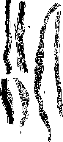
Figure 1. 1 and 2—normal nerve fiber; 3—a, b—Wallerian degeneration thereof, various stages of nerve fiber disintegration.
nerves; almost simultaneously, Türck proved that the fibers of the central nervous system are subject to the same law. The changes occurring in fibers during their aforementioned destruction are termed degenerations and are called secondary, since they develop not independently, but as a result of the separation of fibers from the cell or disease of the cell itself. The degeneration process occurring in the process separated from the cell involves both the axial cylinder and the myelin sheath, whereas the Schwann cells and sheath remain unchanged. Changes occur in the axial cylinder first of all; already on the 2nd day after the disease, the contours of the axial cylinder become uneven, its staining uneven; neurofibrils reveal irregular tortuosity and beaded thickenings. On the 3rd to 4th day, the entire axial cylinder breaks down into separate, variously sized, irregularly twisted segments consisting of a finely granular substance that stains poorly. After 8 to 10 days, small granular lumps remain of the axial cylinder, and then they too disappear. Changes in the myelin sheath begin from the very first days: it loses the regularity of its contours, depressions appear on its outer surface, dividing the sheath into separate segments, which gradually break down into small droplets; changes also occur in the chemical composition of the myelin sheath; it stains differently than a normal one. The disintegration of the sheath occurs throughout the entire length of the fiber, whereas the degree of disintegration is uneven (see Figure 1). The disintegrated myelin is gradually removed from the confines of the fiber; participating in this removal are: 1) Schwann protoplasm with nuclei, dissolving the disintegrated axial cylinders and clumps of myelin located within it, due to which the number of fat droplets and lipoids increases as products of assimilation; 2) surrounding elements of mesodermal origin—wandering cells and lymphocytes. After the disappearance of the myelin sheath, Schwann elements and lymphocytes cease their function. A ribbon-like fiber remains from the medullated fiber, striated in the longitudinal direction, with nuclei, clothed by the Schwann sheath. Owing to the absence of a direct transition of one neuron into another, secondary degeneration
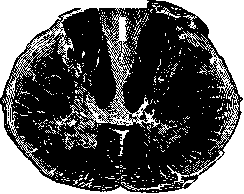
Figure 2. Cross section of the spinal cord;
cervical segment. Ascending degeneration of fibers: in the posterior columns—Goll's tract, in the lateral columns—Gowers' and Flechsig's tracts. fiber propagation extends only to the terminal branching of the neuron, without passing to the next neuron. The central segment of the fiber remaining in connection with the nerve cell also undergoes a change, bearing a different character and called retrograde degeneration. When localizing a pathological process that destroys nerve tissue in the central nervous system, a very complex picture is obtained. Degeneration of nerve fibers spreads in various directions from the site of nerve tissue damage, corresponding to their direction and the location of the cells belonging to them. Thus, when the spinal cord is transected, for example, in the thoracic region, degenerated fibers will be found in both the cervical segments and the lumbosacral ones, depending on the location of the cell giving rise to the fiber (see Figure 2). Fibers whose cells are located below the transection site will degenerate in the ascending direction
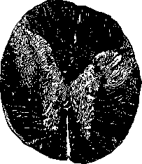
Figure 3. Cross section of the spinal cord; thoracic segment. Descending degeneration of the fibers of the pyramidal tract in the lateral columns.
which is called ascending degeneration; examples of such fibers can be the fibers running in the tracts of Goll, Burdach, Flechsig, Gowers; fibers whose cells are located above the transection site will degenerate (see Figure 3) in the descending direction—descending degeneration (pyramidal fibers, rubrospinal tract, vestibulospinal tract, etc.). Granular balls absorbing the breakdown appear in the places where fibers disappear. Due to the disappearance of nerve elements, neuroglia proliferates at the site of the vanished nerve tissue; cysts form in extensive breakdowns. Secondary degenerations are used to study the course of fibers in the central nervous system, based on the fact that degenerated fibers, in the absence of myelin, do not take up specific stains for myelin, but remain grayish in color, sharply differing from normal tissue. In fresh cases (no more than 3 weeks from the onset of the disease), the Marchi method yields very demonstrative pictures; with this method, small fat droplets stained black with osmic acid are discovered along the course of the degenerated fibers.
Related articles
Mentioned in
Cite this page
“Wallenberg Syndrome and Waller's Degeneration.” Soviet Medical Encyclopedia. English translation of Bolshaya Meditsinskaya Entsiklopediya, 1st ed. (Moscow, 1928–1936), ed. N. A. Semashko. https://sovietmedicalencyclopedia.pages.dev/article/wallenberg-syndrome/