Radial Nerve
Historical document, translated for reference. It reflects medical knowledge of the 1920s–30s and is not medical advice.
Summary
This article from the 1930s Soviet medical encyclopedia details the anatomy, pathology, and clinical management of the radial nerve. It covers the etiology of radial nerve paralysis, including trauma and intoxication, and outlines surgical approaches and treatment methods common to the era.
Encyclopedia article (1928–1936)
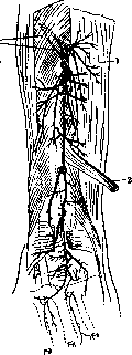
RADIALIS NERVUS, the radial nerve, is one of the main trunks of the brachial plexus. It arises from the posterior cord of the latter, together with the axillary nerve; the segments of the nerve are CVI-CVIII and DI [see Vol. XX (art. 699-700), fig. 22]. By function, the radial nerve is mixed; it innervates the skin, muscles, bones, and joints of the dorsal side of the arm, forearm, and hand [see Vol. XX (art. 749-750), figures 58 and 59]. Typical anatomy. According to the latest research by Sinakevich, the types of branching of the deep branch of the radial nerve are different: in some cases, there is a sequential departure of branches to the corresponding muscles, while in others, the nerve branches divide repeatedly and anastomose, forming plexuses (fig. 1, 2). The muscular branches of the radial trunk can partially run as part of the posterior interosseous nerve of the forearm, separating from the latter only in the lower half of the forearm (figure 2). Comparatively often, the majority of branches to the radius and ulna arise not from the main trunk of the nerve, but from its secondary branches. The degree of branching of the terminal branches of the interosseous nerve, their course, and anastomoses also often vary. In rare cases, an anastomosis with the branches of the superficial branch of the radial nerve is encountered (fig. 1 and 2).
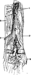
Figure 1. Types of branching of the posterior interosseous nerve (deep branch of the radial nerve): 1-m. supinator; 2- m. extensor pollicis brevis et abductor pollicis longus; 3- ramus superficialis n. radialis; 4-m. extensor pollicis longus; 5-m. extensor digitorum communis.
Pathology of the radial nerve. Of all the nerves of the upper limb, the radial nerve is affected most often. The frequency of lesions is explained, firstly, by its superficial position in the region of the arm under the triceps muscle, and secondly, by its direct proximity to the humerus, on which it lies. Remak, in 242 cases of paralysis of the peripheral nerves of the upper limb, had 105 lesions of the radial nerve. The etiological moment of the disease of the radial nerve can be infection, intoxication, or various traumas. Any infectious disease, both acute and chronic, can cause inflammatory phenomena in the radial nerve (typhus and relapsing fever, malaria, rheumatism, syphilis, leprosy, postpartum processes, etc.). Among intoxications, one should note intoxication with alcohol, lead, arsenic, etc. In the majority of cases, the disease of the radial nerve is of traumatic origin: pressure on the nerve during sleep with the arm placed under the head (most often happens in alcoholics), pressure from crutches, during fractures of the humerus, pressure from bone fragments or the formed callus; strong muscle tension, especially sudden extension of the arm during a rough throwing movement; forcible binding; constricting bandages; injury during subcutaneous injections near the nerve; finally, a puncture of the radial nerve, a blow, gunshot wounds, etc.
Pathoanatomical changes occurring during the disease of the nerve concern both the nerve fibers and the connective tissue that is part of the nerve trunk. In the nerve fiber, under the influence of the process, both the myelin sheath and the axis cylinder suffer (see Neuritis, Polyneuritis, Nerve fibers, Wallerian degeneration). Clinical picture. With paralysis of the radial nerve, the position of the hand is characteristic: the hand is pronated, the fingers are bent at the metacarpophalangeal joints, the hand hangs limply if the forearm is raised to a horizontal position (fig. 3). It is impossible to fully straighten the forearm due to paralysis of the m. triceps; the same paralysis is observed in the extensors of the hand, fingers, in the abductor muscle of the thumb; lateral movements of the hand inward and outward are paralyzed or limited, compression of the hand is somewhat weakened, both supinators suffer, which is expressed in the weakening of supination of the extended forearm (m. supinator brevis) and in the weakening of flexion at the elbow (m. supinator longus). The paralyzed muscles are atrophied and a reaction of degeneration is observed in them. Along with motor disorders, sensory disorders are observed on the dorsal surface of the forearm and hand, on the outer half of the palm, and on the dorsal surface of the fingers. The m. triceps is affected almost exclusively in paralysis from crutches and sometimes only in paralysis from dislocation. In lead poisoning, only the extensors are affected, whereas the mm. triceps and supinator remain unharmed. Sensitivity is subjectively and objectively normal. Often, in lead poisoning, there is a simultaneous lesion of the radial nerve on both arms. Thus, for lead poisoning, the selectivity of the process within the same nerve is characteristic. Persons dealing with lead are subject to the disease: workers manufacturing glaze, painters, typesetters, plumbers, etc. This disease develops slowly and gradually (see Lead).
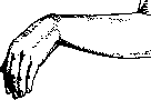
Figure 2. Types of branching: 1-m. extensor digitorum communis; 2- m. extensor pollicis longus; 3-m. supinator. The projections of the bones are indicated by a dotted line. (According to Sinakevich).
Figure 3. Position of the hand in paralysis of the radial nerve. The prognosis in cases of paralysis of the radial nerve is in most cases very favorable. In mild paralysis from pressure, in which electrical excitability does not decrease, recovery occurs in approximately 4-6 weeks. In forms of medium severity, 2-3 months pass before the restoration of function. In case of a breach of the integrity of the nerve, the course of the disease depends on the treatment: when a suture is applied to the nerve, bringing its ends together, the course is very favorable, and complete restoration of function is possible. If the nerve remains unsutured and a reaction of degeneration develops in the muscles innervated by this nerve, the chance for recovery is less favorable; the paralysis may remain for a long time, if not forever. Treatment is the usual one for peripheral nerves: galvanization (cat. 6-8 mA), faradic current and massage can be used. The causes causing the paralysis should be eliminated. Surgical treatment consists of removing tumors, bone calluses compressing the nerve, and suturing the nerve (see Nerve suture, Neurolysis). Wounds. In wartime conditions, gunshot injuries of the radial nerve occupy one of the first places in frequency. In the US Army during the imperialist war, out of 2,390 cases of injuries to peripheral nerves, 51% fell on injuries of the radial nerve (Frazier). In peacetime, mainly injuries are observed, either knife wounds or from bone fragments during fractures. The latter is more often encountered in the middle third of the arm or in the lower part of the forearm. Depending on the place and degree of damage, the conductivity of the nerve is impaired completely or partially. As a result, clinically, there is paralysis or contractures of the corresponding muscle groups. The function of the triceps muscle is rarely impaired, only when the nerve is damaged above the tendon of the m. latissimus dorsi. Injuries of the interosseous nerve or its branches often give a specific clinical picture, the characteristic features of which are edema of the back of the hand, reflex contracture (hand and fingers in a "frozen" semi-bent position), rarefaction of the bones of the hand, and other trophic disorders. Treatment. In traumatic injuries of the nerve (complete anatomical break), its ends are sutured after preliminary excision of the central neuroma and freshening of the peripheral end; in the presence of only scars (without violation of anatomical integrity), neurolysis is performed. Operative approaches to the radial nerve. 1) To the upper part of the nerve (in the axillary cavity) - an incision is made as for access to the axillary artery. In this case, one must spare the branches going to the triceps muscle. It is preferable to use the space between the n. medianus and the axillary vein, pushing them to the sides (Druner). 2) Exposure of the nerve in the canalis spiralis - the incision is made along the posterior edge of the m. deltoideus, in the region of its attachment to the humerus. The nerve is sought between the m. deltoideus and the lateral head of the triceps muscle (fig. 2). 3) Exposure of the nerve in the cubital fossa - the incision is made along the lateral side of the tendon of the biceps muscle. In the depth between the m. brachioradialis and the m. brachialis lies the sought nerve. 4) N. interosseus dorsalis is exposed by an incision made in the middle third of the forearm along the line connecting the head and the processus styloideus of the radius. The nerve lies between the m. abductor pollicis longus and the m. extensor pollicis longus on the interosseous membrane.
A. Vishnevsky, K. Kiselev. RADIAL REFLEX (radiocarpal reflex, reflex from the styloid process of the radius), a periosteal reflex belonging to the category of deep reflexes. It is elicited by a strike of a percussion hammer on the lower edge of the radius (processus styloideus radii). The examiner places the examinee's arm, slightly flexed at the elbow joint, onto their own left hand in such a way that the examinee's forearm is in a position midway between pronation and supination. In response to the strike, a contraction of the m. brachioradialis, m. supinator longus, and m. biceps occurs, along with flexion at the elbow joint, and pronation and flexion of the hand and fingers. The reflex arc of the radial reflex passes through Cv-vi. The radial reflex, like reflexes of the upper limb in general, is normally weakly expressed and may be absent even in healthy subjects; only its increase or absence on one side, while it is clearly expressed on the other, has diagnostic significance. An increase in the reflex is expressed by the fact that a light strike on the periosteum causes sharp flexion of the forearm, hand, and fingers, with not only the m. brachioradialis, m. supinator longus, and m. biceps participating in the movement, but other muscles as well. The disappearance or diminution of the radial reflex indicates a lesion of the gray matter of the spinal cord, or of the anterior or posterior roots in the region of the cervical segments indicated above. An increase in the reflex is observed in lesions of the pyramidal system (see Reflexes).
Related articles
Mentioned in
Cite this page
“Radial Nerve.” Soviet Medical Encyclopedia. English translation of Bolshaya Meditsinskaya Entsiklopediya, 1st ed. (Moscow, 1928–1936), ed. N. A. Semashko. https://sovietmedicalencyclopedia.pages.dev/article/radial-nerve/