Spermatic Cord
Historical document, translated for reference. It reflects medical knowledge of the 1920s–30s and is not medical advice.
Summary
The spermatic cord contains the cremasteric fascia, cremaster muscle, and common vaginal tunic, which houses the blood vessels and nerves of the testis and the vas deferens. It functions to suspend the testis and elevate it through the cremaster muscle, with specific vascular pathways for blood drainage.
Encyclopedia article (1928–1936)
Spermatic cord (funiculus spermaticus) in its composition contains the following structures: fascia cremasterica or fascia Cooperi, m. cremaster, and tunica vaginalis communis, which contains surrounded by loose fatty and connective tissue the blood vessels and nerves of the testis, as well as the vas deferens with its blood vessels and nerves (see figure). In some cases, remnants of the peritoneal appendix that did not obliterate in early childhood may be present in the thickness of the spermatic cord. According to Krymov, an open peritoneal appendix persists in boys up to 1 year of age in 54%, and in adults in 4-5%. The spermatic cord is an organ that suspends the testis and, due to the m. cremaster contained within it, elevates the testis to the inguinal canal. It contains the artery supplying the testis - a. spermatica interna, which originates from the aorta abdominalis. The veins departing from the testis collect into several trunks, with one part of these located in front of the vas deferens (anterior bundle), and another part behind it (2-3 veins of the posterior bundle). The anterior bundle is responsible for carrying blood away from the testis itself. The posterior bundle carries blood away from the epididymis. The venous plexus of the spermatic cord is called v. spermaticae, s. plexus pampiniformis, s. plexus spermaticus int. In the iliac region, plexus pampiniformis merges into one trunk, which under the name v. spermatica int. flows on the right side into v. cava inf., and on the left side into v. renalis. The veins of the posterior bundle mostly flow into v. epigastricae inf. Both bundles have anastomoses with each other, as well as anastomoses
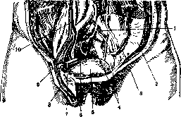
Spermatic cord: 1-recessus intersigmoideus; 2-peritonaeum parietalis; 3-mesocolon descendens; 4-mesocolon sigmoideum; 5-vesica urinaria; 6-intestinum rectum; 7-ductus deferens; 8-funiculus spermaticus; 9-ureter (pars pelvina); 10-ureter (pars abdominalis).
They anastomose with the veins of the tunics of the testis and scrotum. - The lymphatic vessels go along with the veins. - Nerve trunks in the form of delicate plexuses surround the vas deferens, and also accompany the a. spermatica, where they are called the plexus spermaticus, originating from the plexus aorticus. The tunics of the S. k. and m. cremaster are supplied by the n. spermaticus ext. The a. spermatica int., which nourishes the testis, anastomoses with the a. deferentialis (see Seminal duct), so that ligation of the former does not always threaten the death of the testis. Conversely, ligation of the a. deferentialis is much more dangerous, since, according to Tikhomirov, the a. spermatica int. is sometimes absent. The tunics of the S. k. are supplied by the a. spermatica ext., which at the level of the internal inguinal ring anastomoses with the a. deferentialis. The method of medical examination of the S. k. is mainly palpation. Through the thin skin of the scrotum, the structures making up the S. k. can be palpated in detail. In general, the thickness of the S. k. does not exceed that of a little finger. The dilated veins of the S. k. appear as elastic, tense cords or nodularities; the vas deferens is located in the posterior part of the cord as a dense trunk, which in pathological cases can reach the thickness of a pencil or more. In the substance of the S. k., the elements making up a hernia, such as the omentum or a segment of the small intestine, can be easily palpated. In communicating hydroceles of the S. k. with the abdominal cavity, the appearance and disappearance of fluid in the standing and lying positions can be determined both by palpation and visually. When testing reflexes of the m. cremaster, the finger is usually drawn along the inner surface of the thigh and the contraction of the cremaster and the retraction of the testis are observed. The pathology of the S. k. is not extensive. Congenital defects and developmental anomalies include the complete absence of the S. k. in cases of retention of the testis in the abdominal cavity or shortening of the cord when the testis is located in the inguinal canal or at the external inguinal ring. An abnormal position of the S. k. occurs in inguinal hernias: in an indirect inguinal hernia (h. inguinalis obliqua scrotalis), the S. k. is spread apart by the hernia, since the latter lies within its substance; in a direct inguinal hernia, the S. k. is pushed outward. - Tumors of the S. k. are very rare. They originate from the loose connective tissue of the S. k. or its tunics. Most commonly, lipomas, fibromas, and sarcomas can develop here. Lipomas develop between the tunics of the cord, reach various sizes, and on palpation present as soft, lobular formations. Fibromas are more often neoplasms of the tunics themselves; they are more limited and denser than lipomas, and their size varies. Sarcomas of all kinds grow very rapidly, are usually diffuse in relation to adjacent tissues, and quickly involve the testis in their growth or spread into the inguinal canal. In all tumors of the S. k., surgical intervention must be prompt, since benign tumors of the S. k. are prone to degenerate into malignant ones; on the other hand, tumors by their pressure on blood vessels and nerves disrupt the nutrition of the testis and often involve it in the process. When a tumor is limited in development within the spermatic cord, its removal can be easily performed under local anesthesia. Under the influence of injuries and tension of the abdominal press, provided the inguinal canal is wide, the elements of the S. k. are loose, and the absence or elongation of the ligament of Hunter, twisting of the S. k. and along with it the testis may occur. (The painful process is incorrectly called torsion of the testis - torsio testis, whereas the essence of the process lies in the S. k.) Twisting occurs suddenly and is accompanied by severe pains, which sometimes bring the patient to a state of fainting. Along with this, there is significant swelling of the testis, epididymis, and soft parts. The disease is severe and is accompanied by general symptoms - nausea, vomiting, etc. The twisting of the cord can reach 180° or more. In connection with this, there is more or less compression of the blood vessels, and depending on the duration of the process - hemorrhages and swelling of the surrounding tissues, thrombosis of the vessels and gangrene of the testis. - Recognition of twisting of the S. k. is difficult; it can be mistaken for acute orchiepididymitis or a strangulated hernia. Taking into account the fact that with twisting of the S. k. existing for a day, atrophy of the testis occurs, and with a longer duration - its gangrene, it is necessary to be extremely cautious in prognosis and decisive in prescribing surgical intervention. The operation consists in exposing the testis; if gangrenous phenomena have not yet occurred in it, the cord is unwound and fixed with several sutures in the normal position, and the testis is also fixed. The differential diagnosis with a strangulated hernia or acute inflammation of the epididymis and testis is often established only during the operation. If during the operation on the twisted cord gangrene of the testis is found, castration is performed. Under the influence of injuries, hematomas of the S. k. (haematoma extravaginalis funic. spermat.) occur, presenting as a tumor without sharp borders, of varying consistency. - The testis is displaced to the lower pole of the tumor. The tumor is not adherent to the skin. Treatment in the initial stage consists in the application of cold; later, wet or dry heat, high position of the scrotum and pelvis of the patient, etc. When a blood cyst develops from the hematoma, it is incised, evacuated, and either tightly sutured or treated with a tampon. - Acute inflammation of the vaginal tunic of the S. k. (hydrocele acuta funic. spermat.). This disease is possible when the peritoneal process is obliterated not throughout its entire length, but only at the testis and above at the internal opening of the inguinal canal. The immediate etiological factor for the development of the disease is trauma, but the inflammatory process can also spread from neighboring organs: the posterior urethra, prostate, seminal vesicles, epididymis, vas deferens, etc. In all these cases, exudate accumulates in the cavity of the non-obliterated peritoneal process. On palpation of the cord, a sausage-shaped elastic tumor is determined here, extending from the testis and often throughout the entire inguinal canal. In cases where the peritoneal process fuses in several places, the tumor is palpated as separate formations lying one above the other. If the upper segment of the peritoneal process communicates with the abdominal cavity, the tumor disappears in the lying position and forms anew in the standing position. The disease can proceed very acutely with severe pains and symptoms from the intestine and bladder (retention of urine); in such cases, it can be confused with a strangulated hernia. It is always necessary to keep in mind the sharp upper border of the tumor and its translucency when viewed through a stethoscope. However, both of these signs disappear in hydrocele communicans and in bloody or purulent exudate. Treatment consists in the use of anti-inflammatory and analgesic means. - Hydrocele of the S. k. (hydrocele funic. spermat. chronica). For the development of this disease, the same anatomical conditions are necessary as for acute hydrocele of the cord. The etiological factors for hydrocele of the cord are the same as for hydrocele of the testis, but the disease is less common. Recognition of the disease is not difficult, but it is necessary to keep in mind that the palpable tumor can be of very various shapes depending on the above-mentioned variants of obliteration of the peritoneal process. Treatment - surgical excision of the walls of the sac. In diseases of the testis and epididymis, as well as the prostate and seminal vesicles, the inflammatory process can spread to the connective tissue of the spermatic cord (see Funiculitis). For varicose veins of the spermatic cord, see Varicocele.
Related articles
Mentioned in
Cite this page
“Spermatic Cord.” Soviet Medical Encyclopedia. English translation of Bolshaya Meditsinskaya Entsiklopediya, 1st ed. (Moscow, 1928–1936), ed. N. A. Semashko. https://sovietmedicalencyclopedia.pages.dev/article/spermatic-cord/