Appendix
Historical document, translated for reference. It reflects medical knowledge of the 1920s–30s and is not medical advice.
Summary
An anatomical and parasitological overview of the human vermiform appendix from a 1930s Soviet medical perspective, discussing its anatomy, evolutionary status as a vestigial organ, and the role of parasites in causing appendicitis.
Encyclopedia article (1928–1936)
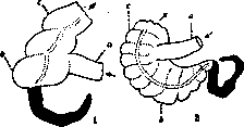
APPENDIX (from Latin appendix - appendage), the vermiform appendix of the cecum (see Figure 1). By its origin, the appendix
Figure 1. Cecum and vermiform appendix (blackened): 1 - human; 2 - chimpanzee; a - small intestine, b - cecum, c - large intestine (arrows indicate the direction of the intestinal canal toward the anus).
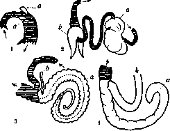
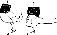
is the underdeveloped end of the cecum. The latter, located at the transition of the small intestine into the large intestine, is found in almost all vertebrates, starting from reptiles (see Figure 2). In some animals, the vermiform appendices of the cecum and their relationships with the latter reach a very high degree of complexity (see Figure 3). The purpose of the cecum is to increase the capacity of the intestine and its digestive surface. Its size depends on the nature of the food usually consumed by a given animal species. Herbivores, feeding on bulky, fiber-rich, and hard-to-digest food, usually have a very large cecum: in the horse, for example, it reaches a length of 80-123 cm, with a volume of 16-68 liters; here the food is retained for up to 72 hours, while the fiber remaining undigested by the overlying sections of the intestine (up to 25% of its total amount) is decomposed here by bacteria and becomes accessible to digestion. In the rabbit, the volume of the appendix exceeds the volume of the stomach by 10 times. In animals feeding on easily digestible and non-bulky animal food, the cecum is weakly developed. In humans and anthropoid apes, its blind distal end strongly narrows, turning into a vermiform appendage, which is lined with a mucous membrane and contains glands and lymph nodes in its wall. In humans, the absolute length of the appendix averages Figure 2. Cecum: 1 - human; 2 - dog; 3 - horse; 4 - bull; 5 - pig (small and large intestines are blackened). 8.5 cm, however, appendices with a length of 2 to 25 cm are encountered. The relative length of the appendix varies greatly with age. In newborns, its length is on average about 3.5 cm, at the age of 10-12 years - 9.5 cm, and in individuals over 60 years it drops to 8.5 cm. At the same time, in newborns, the ratio of the length of the appendix to the length of the large intestine is 1 : 10, and in adults about 1 : 20 (see Figure 4). The cavity of the appendix has a diameter of 3-7 mm, and the opening leading from the cecum into the appendix is about 5 mm. This opening is often closed by a fold. Very frequently, especially in individuals over 50 years old, the lumen of the appendix
Figure 3. Complex types of the cecum. 1 - Myrmecophagus didactylus (anteater): a - two ceca; 2 - Hyrax capensis (rock hyrax): a - unpaired blind appendage of the small intestine, b - paired cecum; 3 - Lagomys pusillus (pika): a - cecum, b - accessory cecum; 4 - Macropus giganteus (kangaroo): a - cecum 42 m long without a distinct appendix (small and large intestines are hatched, blind appendices and appendages are white).
becomes obliterated due to the strong development of connective tissue. At the age of 1 to 17 years, obliteration of the appendix is observed in 4%, from 30 to 40 years - in 25%, from 70 to 80 years - in 58%, while obliteration is observed more frequently the shorter the appendix is. An analogue of the appendix in other animals is the accumulation of glandular (lympho-adenoid) tissue in the blind part of the cecum, for example, in rodents, cats, and others. The appendix is a good example of an underdeveloped (rudimentary) organ that has lost its significance and undergone regression under the influence of the transition from plant food to mixed or purely meat food. The physiological significance of the appendix is negligible, corresponding,
Figure 4. Cecum and appendix: A - human embryo; B - adult human.
perhaps, to one of the numerous glandular accumulations in the small intestines (Peyer's patches); its harm is immeasurably greater because food accumulations getting stuck in it, due to its narrow lumen, contribute to the development of bacterial processes in it, and worms penetrating into it often (whipworm - Trichuris trichiura, pinworm - Oxyuris vermicularis, tapeworms - Taenia solium, etc.), as well as protozoa (for example, the ciliate Balantidium coli) can serve as a source of its diseases (see below).
G. Epstein. The appendix can be inhabited by various parasites. Thus, cases of finding Balantidium coli in it are known, as well as Entamoeba histolytica. In the latter case, we may be talking about amebic appendicitis. Apparently, the appendix much more frequently provides habitation for various parasitic worms, and first of all - pinworms (see Figure 5). According to the statistics of various researchers, in half of all appendices excised during appendectomy, pinworms were found not only in a young state, but even in the form of mature females whose genitalia are overflowing with eggs. According to Russian studies, the whipworm (Trichuris s. Trichocephalus) is found in the appendix much less frequently than pinworms (see Figure 6). Even less frequently, tapeworms (Taenia saginata) and Ascaris (Ascaris lumbricoides) get into the appendix. The presence of parasitic worms in the appendix in many cases does not pass \ Figure 5. Pinworm (in cross section), lying in the depths of a fold of the appendage wall. There is no mucosal epithelium around the pinworm (according to N. I. Solovyov). 6 without a trace for the host. Pinworms are sometimes present in it in tens; secreting irritating substances, they cause marked hyperemia of the mucous membrane of the appendage and its inflammation. In addition, pinworms, sucking onto the walls or getting into the crypts, disrupt the integrity of the mucous membrane and thereby open access inside to secondary infection and, as a consequence, cause the development of appendicitis. A similar action is exerted by the whipworm, which penetrates into the thickness of the mucosa with its thin head end and also disrupts its integrity. Cases are known of a very crude impact of parasites on the appendage, for example, in the form of perforation of it by pinworms or ascarids with the penetration of the latter into the abdominal cavity. If not every case of finding parasites in the appendage entails its inflammation, parasites still play a certain role in the etiology of appendicitis, and perhaps not a small one.
Figure 6. Whipworm (three cross sections) in the submucosa of the appendage, excised during appendicitis. There is no mucous membrane above the whipworm (according to N. I. Solovyov).

Related articles
Mentioned in
Cite this page
“Appendix.” Soviet Medical Encyclopedia. English translation of Bolshaya Meditsinskaya Entsiklopediya, 1st ed. (Moscow, 1928–1936), ed. N. A. Semashko. https://sovietmedicalencyclopedia.pages.dev/article/appendix/