Sarcosporidia
Historical document, translated for reference. It reflects medical knowledge of the 1920s–30s and is not medical advice.
Summary
Sarcosporidia are protozoa of the class Sporozoa that parasitize the muscles of vertebrate animals. They form cyst-like structures called sarcocysts and can cause disease in various animals including humans.
Encyclopedia article (1928–1936)
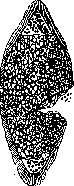
SARCOSPORIDIA, protozoa of the class Sporozoa (or Cnidosporidia according to Hartmann's system), which are parasites of muscles (occasionally also of connective tissue) in vertebrate animals (mammals, birds, reptiles, humans) (fig. 1). First observed by Miescher in mice in 1843, then Rainey discovered them in pigs (1857). Also known as Miescher's or Rainey's pouches. They parasitize within the cells of striated muscle, appearing as pouches (sarcocysts) of oval or round shape, varying in size from several microns to 55 mm (in different animals the size of the parasites varies), whitish-gray or brown in color. They consist of a two-layered shell filled with a granular mass (fig. 2), which under the microscope appears as numerous chambers bounded by partitions and filled with uninucleate protoplasmic spheres (pan sporoblasts, or primary cells), from which sporozoites (spores, or Rainey's bodies) are then formed, sickle-shaped, bean-shaped, rarely spindle-shaped, 3 to 15 μ long, 1 to 5 μ wide (fig. 3); they have finely granular protoplasm with a nucleus and coarse metachromatic grains, and are covered with a shell on the outside. They are non-motile and feed by osmosis. They are sharply stained by ordinary aniline dyes. The biology of the parasite, particularly its developmental cycle, is still insufficiently studied; the routes and methods of infection of the organism with sarcosporidia are unknown. From the parasite, some researchers (Laveran, Mesnil, Teichmann and others) have isolated a toxin (sarcosporidiotoxin, sarcocystin), which in minimal doses is fatal to small experimental animals (mainly to the central nervous system). Among domestic animals, sarcosporidiosis is observed in pigs (Sarcocystis miescheriana; 80% infection and more), sheep (S. tenella), cattle (S. hirsuta), horses (S. Bertrami); also found in dogs, cats, rats, mice, chickens, ducks and other domestic and wild vertebrate animals. Individual cases of sarcosporidia (Sarcocystis Lindemanni) have been found in humans.
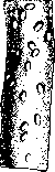
Figure 1. Sarcosporidia
80% infection and more), sheep (S. tenella), cattle (S. hirsuta), horses (S. Bertrami); also found in dogs, cats, rats, mice, chickens, ducks and other domestic and wild vertebrate animals. Individual cases of sarcosporidia (Sarcocystis Lindemanni) have been found in humans. The parasites are located in the body predominantly in certain areas: in sheep, such areas are the abdominal muscles, tongue, and subcutaneous tissue; in cattle, the muscles of the posterior half of the body; in pigs, the abdominal muscles and diaphragm (fig. 4). The infected muscle tissue shows no microscopic changes, and only with extensive lesions do the muscles become flabby and discolored. Old Miescher's pouches, upon dying, become calcified and are easily visible to the naked eye. Upon microscopic examination of muscles (the technique differs in no way from the examination for trichinae), the appearance of the parasites is quite characteristic, and only calcified pouches can be confused with lime concretions, and in pork-with calcified trichinae (unlike the latter, the characteristic absence of changes in the affected muscle fibers, as well as the elongated-oval shape of Miescher's pouches, whose structure is clearly revealed after dissolving the lime in the microscopic preparation with acetic acid). Only in rare cases does the shell of the pouches rupture, and the spores, penetrating into adjacent muscle fibers, cause inflammation and even disintegration of the latter (myositis sarcosporidica). The number of parasites in the infected organism varies: sometimes there are so many of them that the red color of the meat becomes white, like veal. Meat infected with sarcosporidia is considered harmless to humans, which is why it is freely allowed as food. Only with particularly severe infestation, when the meat becomes whitish and watery, the carcasses are rejected, but the fat from them is released from the slaughterhouse without any restriction.
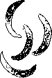
Figure 3. Spores of sarcosporidia.

Figure 4. Miescher's pouches in pork.
Figure 4. Miescher's pouches in pork.
Related articles
Mentioned in
Cite this page
“Sarcosporidia.” Soviet Medical Encyclopedia. English translation of Bolshaya Meditsinskaya Entsiklopediya, 1st ed. (Moscow, 1928–1936), ed. N. A. Semashko. https://sovietmedicalencyclopedia.pages.dev/article/sarcosporidia/