Sarcoma
Historical document, translated for reference. It reflects medical knowledge of the 1920s–30s and is not medical advice.
Summary
Sarcoma refers to malignant tumors of connective tissue origin, though historically it encompassed various malignant tumors resembling fish flesh. The article discusses its classification, occurrence in animals and humans, pathological anatomy, and growth characteristics.
Encyclopedia article (1928–1936)
SARCOMA, sarcoma (from Greek sarcomas--flesh), a term that formerly united malignant tumors of various structures and origins that resembled fish flesh. At present, the term S. is applied only to a group of malignant tumors of connective tissue origin. However, this histogenetic principle is practically difficult to implement completely, as it encounters a number of difficulties associated with the imperfection of the existing histogenetic classification of tumors in general and the impossibility of determining the true histogenesis of tumors from the S. group. Therefore, even at the present time, the definition "sarcoma" derives more from the external structural and clinical features of the tumor than from its histogenesis, uniting malignant tumors originating from the following tissues: 1) connective--fibrous, mucous, cartilaginous, bony, fatty; 2) vascular--blood and lymph vessels; 3) hematopoietic--myeloid and -lymphoid; 4) pigmentary; 5) muscular--smooth and striated; 6) nervous--neuroplastic and glial tissue; 7) in addition, the origin of a number of sarcomas from epithelial tissue through so-called mesenchymoplasia has been established with certainty. S. has wide distribution among animals of various species. They have been found in mollusks. Sarcomas of the ovaries in frogs, sarcomas of the liver in fish have been described; in birds, sarcoma is a fairly common phenomenon; at the same time, sarcoma of Rous (see Chicken sarcomas) receives particular attention from researchers. In mammals, sarcoma occurs frequently (cat, dog, guinea pig, mouse, rat); some of them are used in experimental oncology, where the strain of Ehrlich sarcoma and the strain of Jensen (Jensen) have gained particular fame. Statistics of S. in humans suffer from the lack of precise numerical material. The basis for its compilation, as for other malignant tumors, can only be data from autopsies. The clinical recognizability of sarcoma gives a large percentage of errors, which reaches 10.61% for S. of external organs and 43.23% for S. of internal organs (Lyubarsch). Data based on biopsies could be more accurate in terms of diagnosis, but it is impossible to base oneself on them, since the percentage of patients who underwent biopsies is unknown; therefore, these data do not reflect the general prevalence of S. in the population. The main statistical material for S, as for tumors in general (see), is data from autopsies, all the more so since S. in the vast majority is a disease leading to death, and therefore does not escape the prosector, like other non-fatal diseases. According to Hamburg statistics, for 100 deaths from cancer, there are 4.7 deaths from S. In Merkov's statistics, concerning mortality from malignant tumors in 4 large cities of Ukraine (Kharkov, Kiev, Odessa, Dnepropetrovsk), mortality from cancer and other malignant tumors is given. If we assume that the overwhelming majority of "other" malignant tumors are S, then for 100 deaths from cancer in the years 1923-29 there are "from 8.7 to 11.1 deaths from S. According to these data, the mortality rate from S. fluctuates around 1%. According to sectional data on mortality of the Moscow population for 1923-27 (Davydovsky), mortality from S. reaches 1.52% of all autopsies, and excluding stillbirths--1.75%. According to Lyubarsch's data, mortality from S. among the Berlin population for 1920 reaches 0.96%. As for the susceptibility of S. of individual organs, the main mass of S. according to Moscow statistics and, according to Anschütz's data (Anschtitz), falls on bones. S. affects predominantly young age; the majority of diseases (about 75%) falls on the age up to 30 years; the most frequent of all sarcomas--bone sarcoma--falls on the pubertal age (11-20 years). As for sex, in this respect it is impossible to find any preference. Pathological anatomy. The external appearance of S. differs significantly depending on localization, histo- and cytological structure, course, etc. Common to all forms of S. is the pinkish-white color of the tumors, sometimes more or less pronounced fibrousness, detectable on the surface of the tumor section. S. grows either with single or multiple limited nodes, sometimes reaching a large size (with a fist or a human head), or diffusely infiltrates the organ. Necroses and hemorrhages are often found in tumors. Sometimes S. is a systemic tumor, developing simultaneously in anatomically distant areas of the same physiological system (sarcomatosis of bones, sarcomatosis of the skin). A distinctive histological characteristic of S. is the immaturity of its cellular elements, more or less deviating from the structure of the original maternal tissue. This often makes it impossible to compare S. either with physiologically completed mature forms of the original tissue or with various stages of its development and maturation. In developing and growing normal tissue, its young elements are found in the form of isolated or syncytium-like cellular complexes; in such early periods, a known polymorphism of cellular forms is already observed, depending on the presence of cells in different periods of their maturation. Later, the formation of fibrous, mucous, cartilaginous or basic tissue substance occurs. Thus, between the cellular elements and the basic substance, relationships typical for the functional structure of this tissue are established. A characteristic feature of S. is precisely that its elements do not reach the indicated degrees of development, remaining in their reproduction at a certain stage of maturation of the cells of the original tissue. From granulation tissue, besides cytological features, S. differ in the relationship between cellular elements, basic substance and vessels. In granulation tissue, there is a diversity of cellular forms, depending both on their origin and on their age differences; in S., a greater or lesser uniformity of cellular composition is always observed. Further, granulation tissue is characterized by the systematic distribution of vascular territories, which are centers of tissue neoplasia; in sarcoma, this systematicity is absent, as are, for the most part, territorial grouping and the connection of the proliferating elements of the tumor with vessels. These basic features allow easy distinction of S. from ordinary granulation tissue, while its distinction from specific granulomas is not always easy and gives rise to errors in the diagnosis, classification and terminology of the corresponding granulomas. Such are some processes in the skin, called "sarcoid-ami", among which sarcoids of the type of Boeck and of the type of Darier (Boeck, Darier) are distinguished. Microscopic study allows to detect in S. the stroma and parenchyma. Parenchyma is formed by the cells of S. and the basic substance formed by them. The stroma is formed mainly by vessels and the supporting tissue surrounding them. This supporting tissue is part of the derivative of the tumor, part of the local tissue infiltrated by S. This circumstance extremely complicates, and sometimes makes impossible, the sharp division between specific tumor and non-specific stroma in S. Such a relationship between stroma and parenchyma concerns only S. from proper connective tissue; in S. from myeloid, lymphoid and other tissues (see Lymphosarcoma, Leukosarcomatosis) this relationship is different. The pathogenesis of S. is little studied. In the occurrence of the tumor rudiment, the latter can hardly be distinguished from regenerative neoplasm of connective tissue. The further growth of sarcoma occurs both by intensive multiplication of its elements, which is evidenced by the abundance of mitoses in them, and by the appositional method, at least in the initial phases of tumor development. The possibility of formation of the tumor rudiment on the basis of regeneration is confirmed for many cases by preceding trauma--bruises, fractures of bones with the development of S. in these places after a long time (Fraenkel, Anschütz, Konjetzny, Klemm), the development (rare) of S. in foci of chronic inflammation in lupus. syphilitic lesions. However, one cannot exclude with full certainty the significance of trauma only as a moment of realization in an already existing tumor rudiment, and fractures of bones in some cases have to be considered not the cause of the occurrence of S, but its consequence. The existence of congenital tumor rudiments is confirmed by the development of fetal and congenital sarcomas, heterotopic sarcomatous tumors (mixed), the connection with developmental defects of organs and tissues (S. from birthmarks, from cryptorchid testis, etc.). (The transformation theory of the origin of S.--see Tumors.) For S., as for any malignant tumor, infiltrating growth with invasion and destruction of surrounding tissues is characteristic. The rate of growth of S, the degree of its infiltrating ability are in direct relation to the nature of its cellular elements. In this respect, the greatest malignancy is distinguished by S, consisting of the least mature elements, with negligible or complete absence of development of the basic substance (so-called meristomas - Fischer-Wasels, cytomas-Borst).
Here there is a more or less pronounced parallelism between the malignancy and the immaturity of the cellular elements of the tumor, which determines either the slow growth of the tumor over many years or its rapid development, leading to the death of the patient in a short time. Processes of necrosis and disintegration of tumor tissue with the formation of cavities and ulcers are a frequent phenomenon in sarcomas. The malignancy of sarcomas, in addition to local destructive processes, is determined by their tendency to recur and to spread rapidly throughout the body by the metastatic route. Metastasis occurs predominantly through the bloodstream. Intravascular growth of the tumor is accompanied by thromboses, which are encountered much more frequently than in carcinoma. This infiltration of the vessel walls often manifests in the so-called intimal sarcomatosis, in which the inner surface of the vessel is lined with a layer of sarcomatous cells (Intimasarcomatose); the latter phenomenon, however, may be explained by local hematopoietic processes in the sarcoma and by the formation of blood vessels from elements of the tumor itself. When the tumor grows into a vessel and a thrombus forms, the sarcoma spreads through the blood vessels from small to larger ones. From such thrombi, large particles may break off, giving rise to coarse forms of tumor embolism. In contrast to carcinomas, sarcomas in metastases almost always retain the histological structure and cytological composition of the primary tumor, although phenomena of variability are also known here. General manifestations in sarcoma in the form of cachexia are less pronounced than in carcinoma. Its malignancy is determined by rapid growth with destruction of vital organs and extensive metastasis. The development of sarcoma in hematopoietic organs leads to disruption of blood formation and is often accompanied by anemia. A general clinical prognosis cannot be given for the entire group of tumors grouped under the term sarcoma, since different forms of sarcoma of the same organ, and even more so of different organs, differ in their clinical manifestations of malignancy, and each such form must be considered separately from this point of view. Terminology. Sarcomas from young elements of connective tissue have an epithet characterizing the cytological composition (round-cell sarcoma, giant-cell sarcoma, etc.). Sarcomas from more mature elements are denoted either by a single word characterizing the type of tissue (chondrosarcoma, fibrosarcoma, etc.) or by two words (fibroplastic, osteoplastic sarcoma, etc.). - Individual forms of sarcomas. Sarcomas may consist of both young and mature elements, which accounts for the great variety of sarcomas. - Sarcomas from immature elements. 1) Round-cell sarcoma (sarcoma globocellulare) - an extremely malignant, soft, gray or pinkish-white tumor, growing either in the form of nodules or as diffuse infiltrates with a great tendency to disintegration and hemorrhages. The parenchyma of round-cell sarcoma consists of small or larger round cells resembling lymphoid cells of granulation tissue. Between these cells there is an insignificant amount (sometimes absence) of fibrous stroma or interstitial substance. In sarcomas originating from lymphoid tissue (see Lymphosarcoma), in contrast to ordinary round-cell sarcomas, there is reticular interstitial substance between the tumor cells, sometimes developed to such an extent that each sarcoma cell lies in a loop of this reticular stroma. Round-cell sarcoma grows extremely rapidly, has a great capacity for metastasis via the bloodstream and lymphatic routes. Sites of origin: intermuscular, periosteal, subserous connective tissue, connective tissue of the skin, mucous membranes, meninges, kidney, testis. 2) Epithelioid-cell sarcoma - extremely malignant, externally similar to the previous sarcoma. The parenchyma consists of large cells of epithelioid type, corresponding to the stage of epithelioid cells of granulation tissue. These cells sometimes lie in nests in a well-developed stroma, creating an alveolar structure, similar to alveolar carcinoma, but unlike the latter, between the tumor cells of the sarcoma in the alveoli, the finest fibers can be found, especially after processing by Bel'shovsky's method. This variety is called alveolar sarcoma; it can often be distinguished with difficulty from alveolar carcinoma. Sites of origin of alveolar sarcoma: skin, eye, bone, endothelium of blood vessels (see Endothelioma). 3) Polymorphous-cell sarcoma - a variety of epithelioid sarcoma with extreme polymorphism of cellular elements [see separate table (pp. 711-712), figure 2]. In such a sarcoma, it is sometimes difficult to find two cells resembling each other in size, shape, or nuclear structure. A large number of multinucleated cells and giant mononucleated cells. Also, the sizes of nuclei vary with the most diverse K/P-R (Kern-plasmarelation). Abundance of division figures, direct and indirect, with atypical, monstrous forms, phenomena of degeneration of nuclei and protoplasm, karyorrhexis, pyknosis, etc. - all this gives extraordinary variegation to the cellular composition of such a sarcoma. Sites of origin - the same as for the previous sarcomas. 4) Spindle-cell sarcoma (sarcoma fusocellulare) - a white tumor of somewhat firm consistency, developing in nodules or infiltrates, with a homogeneous or bundle-fibrous surface on section. The parenchyma consists of large or small spindle-shaped cells, irregularly arranged, sometimes forming bundles. In large-cell spindle-cell sarcomas, the cells are richer in protoplasm, the nuclei are oval [see separate table (pp. 711-712), figure 1]. Sites of origin: fasciae, sheaths of blood vessels and nerves, connective tissue of the skin, mucous membranes, intermuscular connective tissue, periosteum. Less malignant than the previous ones; sometimes relatively harmless, having purely local significance and not giving metastases. Giant-cell sarcoma - a tumor growing mushroom-like or in nodules, brownish in color, dense, mostly developing as a periosteal or central tumor of bones (jaw, tubular bones, vertebrae); sometimes originates from joint and tendon sheaths, aponeuroses, especially of the hand and foot. It is characterized by the presence of multinucleated giant cells, scattered in greater or lesser numbers among the main cells of the sarcoma of spindle-shaped or epithelioid type. Sarcomas originating from bones are particularly rich in giant cells. The prototypes of these giant cells are megakaryocytes and osteoclasts. The nuclei in the giant cells are located in the center of the cell, their number sometimes reaching a hundred. The tumor is rich in blood vessels, hemorrhages are usually found in it, accumulations of hemosiderin giving the tumor a brownish color, necrotic areas. Sometimes bone trabeculae are found in the tumor, and it remains unclear whether they are formed by the stroma of the tumor or by the sarcomatous tissue itself. Giant-cell sarcomas are relatively malignant tumors, mostly of local significance; however, malignant metastasizing forms are also known. A special variety of giant-cell sarcomas is the sarcoma of the jaw, known under the name epulis sarcomatosa (features of this tumor - see Epulis). Many pathologists dispute the inclusion of many giant-cell tumors in the category of sarcomas and generally of true tumors, from which they differ in the benignity of their clinical course. At the same time, their connection with fibrous osteitis is proved, in the course of which intense proliferations of fibrous bone marrow sometimes lead to the formation of sarcoma-like tumors containing giant cells (see Fibrous osteitis). Sarcomas from mature elements of connective tissue. 1) Fibrous sarcoma (fibrosarcoma), fibroplastic sarcoma - an intermediate form between fibroma and sarcoma. It forms whitish, relatively dense nodules, with a bundle structure on section. Histologically, it consists of atypically arranged bundles of elongated and spindle-shaped cells, between which a greater or lesser amount of fibrous substance develops. The cells quantitatively predominate over the latter. They are mostly large, richer in protoplasm than in fibroma, and also differ from it by the polymorphism of nuclei. The stroma is poorly developed. The site of origin is the skin, subcutaneous, periosteal, fascial, connective tissue. Relatively benign in course, sometimes encapsulated, similar to fibroma. 2) Myxosarcoma (sarcoma myxomatodes) forms mucoid-like, soft, translucent, grayish-white tumor masses, prone to disintegration and hemorrhages. The cells of such a sarcoma differ significantly in polymorphism; characteristic are spindle-shaped cells with processes and star-shaped cells. Degenerative-necrobiotic changes are frequent in the nuclei. The main characteristic of the tumor is the production of mucoid stroma by the sarcoma cells, which is specially stained by some dyes (Mucicarmin, Mucin) and gives reactions for mucus. Extremely malignant, grows rapidly, metastasizes. Sites of origin: subserous, retroperitoneal, subcutaneous, intermuscular connective tissue; sheaths of nerves, sheaths of the brain, bone marrow. 3) Osteosarcoma, osteoplastic sarcoma.
(osteoma sarcomatodes) is called a sarcoma, the parenchyma of which has a tendency to form bone substance. In such a sarcoma, among round, spindle-shaped, polymorphous, sometimes giant cells, there is bone tissue in the form of delicate beams, plates, or fine cancellous coverings. The site of origin is almost always bone, and two main forms are distinguished: periosteal sarcoma and central, myelogenous sarcoma. The first is characterized by a radial arrangement of the bone trabeculae directed toward the long axis of the bone and a more malignant course. Osteosarcoma usually develops in young subjects at the end of the growth period. Metastases are predominantly in the lungs. 4) Chondrosarcoma, chondroplastic sarcoma often presents as large nodules of neoplasm from dull-white hyaline cartilage tissue, with destructive, sometimes intravascular growth and metastases. It originates mostly from the skeleton, sometimes from soft tissues (pleura). Histologically, it consists of atypically constructed cartilage tissue with the presence of chondroblasts (young cells of cartilage tissue of various shapes). It is rich in blood vessels. Areas of calcification, mucoid degeneration with cyst formation are often found in the tumor. 5) Osteoid sarcoma is characterized by the presence of osteoid tissue, which is normally transitional to ossification (formed, for example, in rickets and osteomalacia). The homogeneous ground substance contains cells in polymorphous cavities that form "bone corpuscles" of sarcomatous osteoid. The osteoid tissue is located either in islands among various sarcoma cells or more diffusely. Depending on the amount of cartilage-like tissue, the designations chondro-osteoid sarcoma, osteoid chondroma are used. It usually develops from the periosteum of large tubular bones. 6) Angiosarcoma is an extremely vague term covering a number of tumors that have both genetic and purely formal connections with the vascular walls, with diverse clinical and morphological features of these tumors. Sometimes the term "angio" is used to describe any type of sarcoma with a richly developed, telangiectatic, cavernous vascular network, sometimes a rare combination of angioma with sarcoma, sometimes a sarcoma in which the parenchymal elements are grouped around blood vessels. The latter form is called perivascular sarcoma, or perithelioma, and, like the first, can be any type of sarcoma in terms of cell composition. Sometimes the perivascular nature of the sarcoma is due to necrosis of large parts of the tumor with preservation of tumor cells in the form of sheaths around blood vessels (pseudoperithelioma). Tumors in which a genetic connection with elements of the vascular wall (endothelium, perithelium) or an angioplastic type of growth is established or assumed are united by broad, also poorly defined terms "endothelioma", "perithelioma", or angioplastic sarcoma, sarcomatous angioma (Borst) (see Perithelioma). Cylindromatous sarcoma, cylindroma is a gelatinous tumor in which hyaline-mucoid masses of club-shaped, spherical, prismatic, cylindrical, sometimes branched shape are found, especially upon fresh examination. It is not an independent type of tumor, as these features are due to the secretion of the mentioned masses by cells of various tumors or degeneration of tumor cells, blood vessels, and tumor stroma (sarcoma, carcinoma, endothelioma, etc.). Such tumors originate from the skin (basalomas), meninges, peritoneum, bones. Most of them belong to the group of endo- and peritheliomas. Tumors of the type of cylindroma of Billroth are those that predominantly develop in the nasal cavity, salivary glands, orbit, on the cheeks and have an epithelial origin. The clinical course of the tumors is determined by the structural features of each in individual cases. Sarcoma pigmentosa, melanoblastoma (see separate table for the article Scarlet fever, fig. 4) is an extremely malignant tumor consisting of cells that produce the pigment melanin. It originates mainly from birthmarks of the skin, from the eye (sclera, conjunctiva, vascular coat), more rarely from the meninges and brain substance, esophagus, rectum, liver, bile duct and bladder, prostatic part of the urethra. Since recent research attributes the ability to form melanin only to epithelial elements, it is now classified as a carcinoma despite the often sarcomatous nature of the tumor cells (see Melanoma). Adenosarcoma is a mixed embryonic malignant tumor developing in the urogenital system (mainly in the kidney), originating from the middle germ layer (or of ectomesodermal origin); histologically it consists of a stroma formed from mature or in places immature (sarcoma-like) connective tissue, in which clusters of indifferent, sarcoma-like cells are embedded. These cells show in places gland-like grouping, in places they form into formed glandular tubes. The sarcoma-like tissue can, in addition, differentiate into mature connective tissue. The degree of malignancy depends on the degree of tissue maturity (see Kidneys). (Clinical course of sarcoma of various organs - see respective organs).
Ya. Rapoport. 4fc * Sarcoma.
°Ь-
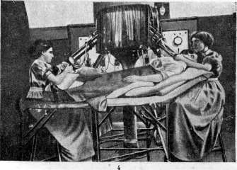
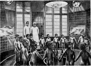
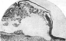
Figure 1. Spindle cell sarcoma. Figure 2. Polymorphous cell sarcoma. Figure 3. Separation of the epidermis by a blister in erysipelas. Figure 4. Finsen's apparatus. Figure 5. Preventive irradiation of children. Idiopathic multiple hemorrhagic sarcoma (sarcoma idiopathicum multiplex haemorrhagicum cutis-Kaposi, syn. acrosarcoma multiplex cutaneum teleangiectodes-Unna, acroendothelioma cutaneum-Radaeli, acroangioma haemorrhagicum-Pospelov), a rare but clinically well-studied skin disease, the nosological position of which and in particular its belonging to the group of Sarcomas is still controversial. Clinically characteristic are: localization on the dorsal surface of the distal parts of the extremities, symmetry, formation of dense, dark blue-brown diffuse spots, swellings and nodules. Since the first description made by Kaposi in 1872, about 200 cases have been published in the literature. Etiology is not clarified. The infectious theory does not have sufficient evidence (auto- and heteroinoculations are fruitless, the causative agent has not been found), although some authors still classify sarcoma idiopathicum multiplex according to hist. structure to chronic inflammatory granulomas. Cases are also known when the development of the disease was preceded by local or general infection. Other authors point to the similarity of idiopathic multiple sarcoma with erythromelalgia (Nikolsky and others) and on the basis of the symmetry of the lesion and the sometimes found infiltration of nerve bundles by leukocytes, pigmentary degeneration of ganglion cells of the intervertebral nodes and degeneration reaction of nerves, speak in favor of a connection with the disease of the nervous system. Sternberg refers idiopathic multiple sarcoma to developmental defects in the sense of hamartomas of Albrecht and explains the development of the disease by the multiplication of detached mesenchymal elements. The variety of histological pictures found in idiopathic multiple sarcoma and their similarity to nevi speak in favor of this theory, but it has no other evidence. Histological changes vary in their picture as in different cases, and often in the same patient on biopsy from different places. Common are: localization of changes in the dermis, neoplasm and significant (sometimes cavernous) expansion of blood and lymph vessels, hemorrhages and deposition of hemosiderin, sometimes also melanin; infiltrate consisting of newly formed spindle-shaped, round cells, among which plasma cells and mast cells may be found. Depending on the predominance of certain elements, the overall picture may correspond to angiocavernoma, to true spindle cell Sarcoma, to granuloma, and finally may be on the border between Sarcoma and granuloma. The latter gave some authors reason to include Sarcoma idiopathicum multiplex in the so-called ?arcoidoids (see).--Patho-anatomical data obtained at autopsies of patients are still very scarce. In some cases, no changes were found in the internal organs. Sometimes nodes, corresponding in structure to skin infiltrates, are found on the mucous membranes, in various internal organs, bones and in the dura mater. The lymphatic system remains unaffected.--Pathogenesis. Undoubtedly, in some cases the development of the first nodes was preceded by local trauma, suppuration or erysipelas. However, wherever the first node appeared, subsequent phenomena have a tendency to localize on the hands and feet. This circumstance speaks in favor of autochthonous, and not metastatic, development of new elements. Development after injuries and infections can be interpreted in favor of Sternberg's theory. It should be noted that idiopathic multiple sarcoma is most often observed in elderly men and relatively often in Jews. Clinic. Sometimes the onset of the disease is preceded by transient edema, paresthesias and pain in the area of the distal parts of the extremities. The skin manifestations, differing in symmetry of localization, consist of local edema, hemorrhages, pigmentation, plaques of infiltrate and individual nodes. The development of the latter is accompanied by sometimes significant pain, and their favorite localization is the hands and the outeri dorsal surfaces of the feet. The skin of the hands and feet swells and pillow-like bulges, and the edema is characterized by considerable density. Against the background of the edematous skin, dark blue-black spots of various sizes develop, which then turn brown-brown. Individual not sharply limited areas are infiltrated and slightly protrude above the level of the skin; against their background or against the background of edema, and sometimes on normal skin, individual nodules of bluish-brown-red color appear, usually very dense, often painful. Their sizes range from a grain to a pea and a nut, and their number can reach several tens and even hundreds. They are located separately or in clusters and can sometimes cover the entire surface of the feet both on the dorsum and the sole in the form of a peculiar papilloma. The nodules are located in the dermis and protrude semispherically above the level of the skin. Sometimes they are on a stalk. The surface of the nodules is smooth and shiny, and on the infiltrates it is sometimes shagreen-like due to the presence of small, closely fitting nodules. Disintegration of the nodules is not mandatory, but possible. Often they disappear spontaneously leaving atrophic or scarred intensely pigmented areas surrounded by a dense red-brown colored roller covered with hard scales, sometimes reaching the degree of keratosis. Eliashev described a pigmented form of idiopathic multiple sarcomatosis, in which extensive pigments and elevated brown infiltrates predominate, while hemorrhages and nodules recede to the background.--After the first changes appear on the feet and hands, the lesions gradually spread along the extremities from the periphery to the center and then appear on the trunk, face and head, genitals and on the mucous membranes. The duration of the course varies and the disease can last 10-20 years or more, without affecting the general condition. In other cases, it ends fatally within 2-3 years with symptoms of exhaustion, fever, sometimes bloody diarrhea and hemoptysis. In such cases, disintegrating nodes are found in the internal organs. When skin nodes disintegrate, secondary infection and development of deep ulcers are possible. Recognition is based on the characteristic features of skin lesions, their localization and symmetry. In doubtful cases, histological research decides.--Prognosis is not absolutely unfavorable, since even without treatment the disease can last for decades.--Of the therapeutic measures, radiotherapy gives the greatest success. Arsenic often remains ineffective. Disintegrated nodes are treated symptomatically.
s. Rosenthal.
Related articles
Mentioned in
Cite this page
“Sarcoma.” Soviet Medical Encyclopedia. English translation of Bolshaya Meditsinskaya Entsiklopediya, 1st ed. (Moscow, 1928–1936), ed. N. A. Semashko. https://sovietmedicalencyclopedia.pages.dev/article/sarcoma/