Cancer
Historical document, translated for reference. It reflects medical knowledge of the 1920s–30s and is not medical advice.
Summary
This article from the 1928–1936 Great Medical Encyclopedia defines cancer as a malignant epithelial tumor, distinguishing it from broader definitions used in other countries. It covers the historical etymology of the term, the pathology of tumor growth, metastasis, the clinical phenomenon of cancer cachexia, and provides statistical data on cancer mortality in the USSR and Europe during the early 20th century.
Encyclopedia article (1928–1936)
CANCER, or in Latin terminology cancer, and in Greek carcinoma, is a concept denoting in our country, the USSR, as well as in Germany and the Baltic states, a malignant epithelial tumor. In contrast to this, in some other countries, for example in France, the word "cancer" encompasses various forms of malignant neoplasms (for example, carcinomas, sarcomas, chorioepitheliomas, endotheliomas, etc.). The designation "cancer" originates from very ancient times, when concepts regarding the essence of this pathological process were most primitive and when the primary attention was drawn to the external form of this tumor. The definition of cancer is connected with a very distant resemblance of the external appearance of some neglected cases of breast cancer in women to a river crayfish. With sufficient imagination, in the central focus of the neoplasm, an analogy was seen with the body of a river crayfish, and in the dilated, tortuous veins of the skin surrounding the painful focus, they wanted to see a resemblance to the claws or tentacles of sea creatures. Representing a tumor originating from epithelium, cancer can develop almost everywhere where epithelial elements are present. Thus, we know of cancer of the skin, mucous membranes, parenchymal organs, etc. In view of the destructive character of the growth of this neoplasm, the development of the tumor in a living organism is in most cases connected with the constant and systematic destruction by the neoplasm of the maternal soil and even the entire organism. The destructive character of the growth of cancer, which destroys fascia, bones, etc., as it spreads in width and depth, is also manifested by the property of cancer to infiltrate the surrounding environment by way of the ingrowth of cancerous strands into the underlying tissues. Cancer cells easily undergo necrobiosis, the vessels of the neoplasm are often thrombosed, as a result of which necrosis of entire sections of the tumor appears with the inevitable consequence in the form of the formation of a so-called "cancerous ulcer," distinguished by thickened, everted edges and sometimes ichorous discharge. Local cancerization of normal epithelium, being, as we shall see subsequently, the result of very deep disturbances of nutrition and physical-chemical changes in the structure of cell colloids, once rooted in the organism, continues to develop, often despite very energetic therapeutic measures. This ability of a cancerous tumor to return to the site of a primarily (incompletely) removed focus is termed the ability of cancer to recur. The latter can sometimes immediately follow the removal of the primary focus or appear after certain intervals of time. The most characteristic feature of cancer is its ability to form metastases, i.e., the transfer and implantation of cancer cells, following lymphatic [see separate table (art. 255-256), fig. 5] and blood pathways, into regional lymph nodes or into distant organs [see separate table (art. 255-256), fig. 6]. A classic example of lymph node involvement in cancer is the involvement of the axillary lymphatic system in breast cancer. Thus, for example, Küster, in 117 operated cases of cancer mammae, did not find cancer cells in the axillary nodes in only three cases. It is interesting that in metastases, cancer cells can retain their physiological function, e.g., liver cancers retain the function of bile formation [see separate table (art. 247-248), fig. 7]. The ability of cancer cells for active amoeboid motility explains to us their energetic advancement along lymphatic pathways to regional nodes. A significant majority of cancer cells perish, undergoing phagocytosis and dissolution. Although viable cancer cells can be histologically proven even in circulating blood, the majority of them still perish here, just as a considerable number perish in lung tissue (demonstrated on histological preparations by Goldman, Schmidt, and others). The most unfavorable living conditions for cancer cells are apparently encountered in the sinuses of the spleen, where metastases of cancer are extremely rare. As the cancerous neoplasm develops, the function of the organ on which the cancer is primarily localized or in the territory of which it metastasizes is disturbed; concurrently with the ulceration or decay of the neoplasm, a severe reaction of the organism occurs, manifesting as a state of cancerous poisoning or cancerous cachexia. The causes of cancerous cachexia lie, firstly, in the disturbance of the physiological functions of the diseased organs (e.g., cancer of the esophagus, stomach, urinary bladder), whereby nutrition, excretion, etc., suffer, or in the disturbance of the chemistry in the juices and tissues of the organism. There are grounds to explain cachexia not by a specific cancerous poison (Seiderhelm, Lampe), but rather to think about glycolytic, proteolytic, and hemolytic enzymes produced by certain cancers and acting destructively on surrounding tissues. As a result of the action of these enzymes, there occurs, apparently, an atypical cleavage of protein particles, leading, on the one hand, to anemia, and on the other, to increased and pathological carbohydrate and protein breakdown, as a result of which unoxidized reaction products poison the organism. To reflect the frequency of cancerous diseases, statistical information from various countries can be used, compiled, however, not always according to the same principle. The most reliable turn out to be pathological-anatomical autopsy data, which, according to Regaud, reveal the presence of cancer almost twice as often as is established by simple medical death certificates issued. The quantity of cancers diagnosed during life is barely 10% not confirmed by autopsies. Statistical material collected according to data on cancer morbidity, compiled by attending physicians, is fraught with diagnostic errors, although it covers broader strata of the population. The method of accounting most corresponding to our capabilities consists in the registration of certificates issued by physicians regarding the causes of death of patients. In the latter case, a significantly greater accuracy of diagnosis is possible, since it usually concerns far-advanced cases. Systematic works on the frequency of cancers in civilized countries belong to Hoffmann. They are compiled according to death certificates. Usually, it is a matter of calculating the annual mortality from cancer per 100,000 inhabitants. Mortality figures from cancer per 100,000 people for various countries for 1881-1925 are given in Table 1 (according to Hoffmann). We have approximately analogous data, although for a later period, for the USSR as well. According to data from Mamonov and Novoselsky, reported in 1914 at the 1st All-Russian Congress on the Fight Against Cancerous Diseases, as well as according to materials collected and processed by S. A. Kholdin, the average mortality from cancer per 100,000 inhabitants of the RSFSR is currently 80-90. Comparative data on mortality from cancer in cities of the RSFSR (1913, 1925, and 1926) are as follows: Table 2. Cities Years Mortality per 100,000. In 121 cities with a total population of 4,775,000 without Leningrad, Moscow, and Odessa.... In cities of the RSFSR with a population above 50,000... 1913 / 1915 \ 1928, 86. Considering the population of the RSFSR by 1930 to be approximately 108,942,000 and assuming that the rural population suffers from cancer approximately as often as the urban, it must be assumed with a significant degree of probability that about 95,000 people die from cancer annually in the RSFSR. Our information on mortality from cancer in the major centers of the Union is more complete (Tables 3 and 4). The general coefficients of mortality from cancer per 100,000 inhabitants in Moscow and Leningrad are higher than in other cities of the Union. For Moscow, they are equal to 94.5, and for Leningrad 114.3, while for other cities of the RSFSR this coefficient, at least for 1926, is equal to 90. The increased coefficient of cancer mortality for large centers has its special explanations and is not proof of an alarming increase in the cancer danger or an actual increase in the incidence of cancer. According to the conclusions of the well-known authority Hoffmann, who familiarized himself with Russian cancer statistics in Leningrad, it must be admitted that the frequency of cancer in the USSR does not exceed that in other civilized countries of Europe and America. That an alarming increase in the cancer danger is not observed can also be judged by the constant percentage of cancer autopsies to the total number of autopsies performed, for example, in Leningrad. Thus, N. G. Pozoeva (Oncological Institute in Leningrad), based on autopsy material from 5 of the city's largest hospitals (1920-30), established that the ratio of the total number of autopsies to the number of cancer autopsies remains approximately constant over 10 years. According to these... Table 3. Mortality from cancer in Moscow per 100,000 inhabitants.
Years Mortality Years Mortality 1922 1923 1924 1925 1926 99.3 103.3 102.5 111.2 118.0 1927 1928 1929 1930 122.9 120.3 123.7 119.3. According to studies, out of 28,464 autopsies, there were 4,203 cancer patients, which constitutes an average of 14.8%, i.e., a figure approximately constantly established for the frequency of cancer autopsies. 14.8%. The figures presented by Pozoyeva for Leningrad coincide, for example, with the frequency of cancer autopsies in Berlin hospitals (Redlich), where we have: in 1895-1900 13.24%, in 1900-1905 13.00%, in 1908-1918 12.35%, and in the hospitals of Paris—in 1802-1816 (out of 2,556 autopsies)—14% (Charité), and 100 years later—in 1902-1916—10-12% (Tenon). According to data (1923-27) from Moscow prosectoriums (I. V. Davydovsky), the percentage of cancer autopsies fluctuates between 12.2-14.2, averaging 12.9 (out of a total number of 21,383 autopsies, counting from the age of 16). The significance of age for the frequency of cancer diseases is an irrefutable fact. The cancer risk, as a rule, increases with the aging of the organism, which, however, does not exclude the possibility of cases of cancer in childhood and even in infancy. According to Arkhangelsky, out of 100,000 living people of each age, the number of deaths from cancer in Brussels in 1911 was (Table 5): Table 5. Age Mortality Age Mortality 0-10 years 10-20 » 20-30 » 30-40 » 40-50 » 8.5 13.0 22.5 92.0 286.5 50-60 years 60-70 » 70-80 » 80-90 » 369.5 820.5 828.5 499.5. According to data announced by Kholdin at the 1st Oncological Congress in Kharkov in 1931, cancer mortality by age is as follows (Table 6): Table 6. Cancer mortality for 1930. From the tables provided, it is evident beyond doubt that the cancer risk for humans increases sharply in the period from 40 to 60 years. In women, in particular, the risk of contracting cancer, due to the earlier aging of the organism, begins at age 30. In deep old age, this threat gradually decreases. Despite claims found in literary sources about the increase of cancer in young people, there is still no reliable statistic confirming the "rejuvenation of cancer" (McConnell). The significance of sex for cancer morbidity is currently sufficiently clarified. It has the greatest significance for the localization of cancer and only secondarily for its general frequency. According to older statistics, women contract cancer significantly more often than men. According to newer statistics, the difference in the general frequency between the cancer morbidity of men and women is smoothing out more and more; for example, according to Toichkin, in Moscow in 1878, for every 100 men who died of cancer, there were 213 women. Subsequently, this noticeable difference gradually smoothed out, and by 1912, for every 100 men, there were 129 women. According to the latest information collected by the economic-statistical sector of the Leningrad Regional Planning Commission, the ratios are as follows: in 1922, out of 953 registered deaths from cancer, there were 452 men and 501 women; in 1924—615 men and 636 women; in 1928—1,016 men and 1,029 women; in 1930—1,201 men and 1,234 women. The reason for these changes in statistics by year is connected with the significant improvement in the diagnosis of cancer of internal organs, while the recognition of cancer diseases of areas of the human organism easily accessible to inspection and palpation has not improved as strikingly over the last decades. The significant frequency of cancer morbidity in women in old years was attributed mainly to cancer of the uterus, mammary gland, and other organs easily accessible for examination. The equalization of the morbidity figures for men and women in recent years is connected with an increase in the number of cancer diseases mainly of the digestive and respiratory tracts in men, i.e., forms that remained often unrecognized in old years. Not without interest are the data collected by Lautenborn (1905-07) on the statistics of comparative mortality of men and women from cancer depending on age groups in Germany. The author's conclusions coincide with the conclusions of Berencisy and Wolff for Budapest and Arkhangelsky for Moscow and St. Petersburg. They state that women, who generally age earlier than men, are affected by cancer more often in relatively early years of life, while subsequently, this difference gradually smooths out. One way or another, it can be concluded that cancer morbidity in women is somewhat higher. As for mortality (and lethality), it is the same, and perhaps even somewhat higher in men. The reason for this is the greater operability of cancer in women. The frequency of cancer morbidity by organ can be judged by the corresponding statistics provided by Hoffman in 1915. The annual average mortality from cancer in England per 100,000 inhabitants from 1901 to 1910 by organ is as follows (Table 7): Table 7. Organs Men Women Uterus 16.53 22.41 Mammary gland 9.57 17.27 Stomach 7.84 14.6 Liver and gallbladder 5.09 13.61 Rectum 4.29 6.13 Esophagus 6.4 1.59 Tongue 2.4 0.48 Intestine 1.78 8.13 Urinary bladder and urethra 1.21 0.96 Skin 2.21 1.16 Lower lip 1.52 0.1 Jaws 0.76 Larynx 0.47. The latest statistics for Leningrad, collected by Kholdin (1931), also indicate that the vast majority of deaths from cancer are due to diseases of the digestive tract (data for 1922-30). Thus, among all those who died from cancer, cancer of the digestive organs was 61.4% (67.2 in men, 48.3 in women), cancer of the uterus—23.7%, cancer of the intestine—7.6%, cancer of the lungs—7.9%. For Moscow (1923-1927), similar data from sections, covering 2,765 cancer cases, are expressed in the following figures (Davydovsky) (Table 8): Table 8. Organs In percent Stomach 34.87 Bronchi 7.5 Esophagus 7.3 Uterus 6.98 Intestine 6.3 Pancreas 4.34 Upper respiratory tract 3.47 Ovaries 2.89 Bile ducts 2.8 Breast 2.27 Liver 2.2 Kidney 1.12 Skin 0.97 Prostate 0.89 Testicles 0.53 Thyroid gland 0.39 Pharynx 0.36 Tongue 0.3 Others (larynx, penis, jaw, pituitary, branchiogenic formations, etc.). In general, a comparison of morbidity figures and mortality figures yields significantly differing results for cancer, which is understandable in view of the significant operability of many cancers, e.g., skin, gynecological. On the other hand, mortality figures obtained through mass sections will differ greatly from the same figures obtained by methods of ordinary (non-sectional) registration of causes of death, especially in relation to difficult-to-diagnose cancers, for example, cancer of the bronchi. Finally, it is natural that in cities, as more cultural centers, mortality with the same morbidity, e.g., in relation to cancer of the uterus, will be much lower than in the provinces. One way or another, statistics on morbidity, mortality, and lethality of cancer can yield significantly differing figures depending on a whole series of factors, such as: the age contingent of the population, its cultural level, the organization of sectional work, surgical aid, etc. The general mortality coefficients from cancer per 100,000 inhabitants in Moscow mentioned by us above are 94.5% and in Leningrad 114.3%. These figures continue to rise, from which it by no means follows that there is a catastrophic increase in the so-called "cancer risk." However, one cannot deny a certain increase in cancer cases, although, according to the conclusion of Wolff, this threat has increased again due to cancer of the digestive tract, the diagnosis of which continues to improve. For large centers, the increase in cancer risk from year to year depends on several reasons: firstly, on the opening in some of them of special anti-cancer centers, institutes, etc. Secondly, the explanation for this fact is also justified by the well-known paradox of Wells, who says that "the better the sanitary condition of a given country or city, the greater the cancer risk." This assertion is apparently fully justified for the following reasons.
Thanks to better medical care, the life expectancy of patients is increasing, mortality from childhood infections is decreasing, and thus the prerequisites are created for an increase in cancer due to older people. According to Mayo's data, cited by Regaud, the average life expectancy in the USA was 40 years in 1810, 45 years in 1875, and 50 years in 1927. In connection with the lengthening of life expectancy, the number of diseases characteristic of older age naturally increases; for example, not only does the frequency of cancer increase (by 86%), but also other diseases, e.g., heart disease (by 71.6%), nephritis (by 68%), etc. For the USSR, the influx of the adult population to large industrial centers, which concentrate specialists aged 30 to 50, also provides material for similar conclusions about the apparent increase in cancer; thus, according to information from the Leningrad Economic-Statistical Sector (Novoselsky, 1931), data on the age composition of the population for Leningrad, non-capital centers, and rural areas are presented as follows. The percentage of the population from 30 to 60 years old in Leningrad is 37, in other cities 31, and in rural areas 28. The curious paradox of Greene, who speaks of an increase in cancer in the USA from 1910 to 1924 in the same proportion as the increase in the number of doctors, is illustrated to a certain extent by our data for Leningrad and Moscow regarding the increase in mortality from tuberculosis. With the fall in mortality from tuberculosis, mortality from cancer increases. The latter, of course, is not a true increase in the cancer danger, but only an inverse reflection of the fall in tuberculosis morbidity. The increase in life expectancy thereby entails a more prominent manifestation of a stable percentage of mortality from cancer among people reaching old age. Comparative data on mortality in Moscow per 10,000 inhabitants from tuberculosis and cancer are as follows (Table 9): Table 9. Years Total mortality from tuberculosis from cancer 1925........ , 232 363 131 130 25.0 39.7 15.7 16.0 9.3 8.3 9.2 10.3 The fall in the percentage of total mortality in 1930 in relation to mortality in 1914 was 30%, and the fall in mortality from tuberculosis was even 33%, while at the same time in relation to cancer...
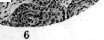
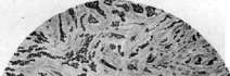
Figure 1. Cancer planus; anaplasia of the epithelium. Figure 2. Cancer (of the lung) small-cell; ingrowth into a vein with thrombosis of the latter. Figure 3. Colloid cancer. Figure 4. Cancer (adenocarcinoma), growing into the wall of the stomach. Figure 5. Basal cell cancer (basalioma). Figure 6. Cancer (cancer keratodes) with formation
pearls. The rise in mortality is calculated at 40%. However, as has been mentioned and as the majority of authors assert (e.g., Willcox, Wolff, and others), the "cancer danger" is actually increasing very slowly and in reality turns out to be much lower than the apparent rise. Cancer is not an exclusive privilege of the human race. In the plant kingdom, we know of so-called "frost cancers" on roses in the form of knotty, bizarre growths; we see approximately the same tumors on cabbage, the so-called "cabbage cancer," etc. However, at the present time, these plant tumors are regarded as infectious granulomas rather than true neoplasms. The difference between them and true tumors lies, among other things, in the fact that while the transplantation of cancer in animals succeeds only within the limits of the same species, Smith's bacteria cause tumors in various species of plants. Furthermore, the absence of a corresponding cytological tissue reaction in plants does not allow for the identification of plant "cancers" with the cancer of animals and humans. While cancer in invertebrates has been studied almost not at all, cancer in vertebrates is well known on the basis of accidental observations and experimental data. Thus, for example, in fish, especially in ponds, cancer is known in salmonids and cyprinids. According to Joest, among birds, particularly chickens, 20 cases of cancer have been registered. The maximum number of cancerous tumors, not counting humans, is observed in mammals, especially in white mice. Thus, for example, according to Bashford, there is on average 1 case of spontaneous cancer per 3,500 mice. Nevertheless, independent (not artificially transplanted) adenocarcinomas of the mammary gland in mice have not been observed frequently in Russia recently; in mice, they have been noted by Krontovsky, Vereshchinsky, Krotkina, and others. According to Schütz and Sticker, in Berlin, up to 50 cases of cancer were observed per 10,000 dogs, which is a fairly high percentage of morbidity among this type of mammal. Unlike the so-called cancer in plants, cancer in animals appears to be formations that are completely identical to cancer in humans, especially those standing on lower rungs of cultural development; therefore, the previously prevailing opinion about cancer as a privilege of only civilized peoples does not seem to correspond to the truth (cf. Hoffmann). Wells believes that the rarity of cancer in countries with primitive culture is far from proven, especially because the average life expectancy there is lower than in civilized countries. Hence, the apparently unproven position of Niceforo and Pittard regarding the increase in cancer from homo mediterraneus to homo nordicus. The fact is that while in southern countries, for example in India, the average human life expectancy is 36 years, and in Bengal only 17.8% of inhabitants live to the age of 40, in England over 27% of the population reaches the age of 40. Since cancer is the lot of older age, it follows that, regardless of the geographical location of the state, the frequency of cancer must increase with the life expectancy of the inhabitants of a given country. According to Rogers, the percentage of mortality from cancer in people over 40 years of age in Calcutta and highly civilized London is approximately the same. Consequently, the degree of civilization does not have decisive significance for the number of cancer cases. This position, however, requires some corrections in relation to the specific habits and living conditions of individual countries. Thus, for example, in Kashmir, cancer of the skin of the abdomen is very common, i.e., forms of cancer usually very rarely encountered in general clinical practice. This unusually frequent localization is explained by the habit of the natives of warming themselves with pots of hot coals, which they carry under their clothing on their abdomen. As a result of chronic burns, skin cancers develop. The chewing of betel plant nuts, which is very common in the Sunda Islands, leads to a specific frequency of cancer of the oral cavity (betel chewing carcinoma), so that, for example, in Travancore, this form accounts for up to 70% of all registered cancers in the population. The pathological anatomical structure of cancer, both from the macroscopic side and from the side of the finest structure of cellular elements, has given rise to a huge literature. The external appearance of a cancerous tumor is extremely diverse depending on the affected organ and tissues and the nature of the neoplasm: barely noticeable skin thickenings of the hyperkeratosis type, papillary growths [see separate table (pp. 255-256), Fig. 3], mushroom-like forms, large and nodular nodes penetrating deep into the underlying tissue and deeper layers, etc. The integrity of the most superficial layers of the epithelium, as the process develops, gives way to ulcerative forms with deep, undermined, and dense edges. While in the majority of cases primary cancerous foci are single, in rarer cases we have multiple forms, which is quite often encountered in skin cancers and less often on mucous membranes. On the latter, in particular especially along the gastrointestinal tract (in stomach cancer), one encounters either single or multiple polypoid cancers, sitting like mushrooms on a broad base (c. fungosus). Forms are known that grow in the form of nodular thickenings, either with extensive necrotic areas on the periphery or covered by a more or less intact mucous membrane. Flat, skillet-shaped, and saucer-shaped forms with wide ulcerations and crater-like depressions are noted alongside so-called "planar, infiltrating" varieties, in which it is impossible to determine the actual boundaries of the tumor either by eye or by touch. Along with shrinking, dense, fibrous forms, so-called "scirrhus," we have soft or encephaloid cancers and cancers of semi-liquid consistency, so-called "colloid" forms. Especially often on the cervix of the uterus, we see so-called "excrescent," "exophytic" forms of cancerous tumors with extensive growths of the neoplasm, which acquires an external resemblance to cauliflower. If we are talking about glandular organs, for example, breast cancer, then the macroscopic form of the neoplasms also varies widely here, starting from the superficial, so-called "cancerous eczema" (Paget's form), developing in the nipple area, and reaching deep, often immobile nodes and infiltrates, sometimes capturing the entire gland, which sometimes reach such extensive dimensions that the entire human chest cavity turns out to be enclosed in a cancerous infiltrate, as if in a shell. By histological structure, cancer represents a typical "organoid" tumor, i.e., built according to the scheme of an organ and consisting of connective tissue stroma and epithelial parenchyma. The participation of the connective tissue stroma from the morphological side occupies such a prominent place in a cancerous tumor that some classify cancer as "fibro-epithelial" tumors. It is customary to distinguish a whole series of individual forms of cancerous tumors, which are encountered either in their typical and classical varieties or are combined with various amounts of connective tissue, a larger amount of vascular elements, sometimes with a sarcomatous character of the stroma (sarco-carcinomas), etc. Under all circumstances, however, the parenchyma of a cancerous tumor continues to retain its epithelial nature, although it is also characterized by extreme atypia. The epithelium anaplasizes [see separate table (pp. 239-240), Fig. 1], loses the physiological isolation and systematic nature of its arrangement; it penetrates into the underlying tissues in strands and is not held within the physiological boundaries defined by the limiting membrane. Epithelial elements in cancer lose their "complexity" (i.e., they are not arranged in continuous strands but are interrupted, for example, by connective tissue fibers) and also lose their "polarity," i.e., the regularity of the arrangement of the two cellular poles [cells, as is known, have a nutritive pole directed toward the underlying tissue and a laborative one directed toward the free surface (bipolarity)]. The leading significance in cancer in terms of its growth rate, metastasis, and other nuances of behavior belongs to its epithelial parenchyma. Therefore, the type of cancer epithelium and the nature of its arrangement are taken as the basis for the classification of cancer. Histologically, one distinguishes, firstly, squamous cell cancers, predominantly observed on skin integuments and mucous membranes covered with squamous epithelium. In these latter, so-called "keratinizing" forms of squamous cell cancer, often called spinocellular cancers, are especially common. So-called "cancer pearls" in these varieties represent islets of complete keratinization of the epithelium, the scales of which are layered on top of each other in the form of an onion; the pearls, in the form of whitish shiny balls, are squeezed out of the cancer specimen together with the so-called "cancer juice." Keratinizing squamous cell cancers [see separate table (pp. 239-240), Fig. 6], as highly differentiated, are sometimes also called cancroids. Cancers originating from the deepest layer of Malpighian cells are usually called "basocellular cancers" or "basaliomas" [see separate table (pp. 239-240), Fig. 5]; they are more often of the non-keratinizing type.
On mucous membranes covered with cylindrical epithelium, and in glandular organs, one encounters cylindrical-cell glandular cancers, in which the alveoli either retain a glandular appearance with a lumen (adenocarcinomas) [see separate table (pp. 239-240), fig. 4] or turn out to be completely filled with cells that have either not lost (c. cylindrocellulare) or are losing their cylindrical shape and have the appearance of small polygonal elements (simple, or solid cancer). In some cancers, the epithelial cells retain the secretory function characteristic of the epithelium that served as the source of the cancerous growth. This may include the aforementioned production of keratin (keratinization) in squamous cell cancers, the production of mucus by adenocarcinoma cells, which gives rise to mucous (c. myxomatodes) and colloid cancers (c. colloides, s. gelatinosum) [see separate table (pp. 239-240), fig. 3], the secretion of bile in some liver cancers, the secretion of colloid in thyroid cancer, the production of melanin pigment by cancers originating from pigment epithelium (pigmented cancers, melanocarcinomas), etc. Some cancers are classified as "lymphoepithelial tumors" (of the Schmincke type), where, in addition to epithelial cells, one finds a proliferation of lymphadenoid tissue (tumors of this type are encountered predominantly in the tonsils). The stroma in a cancerous tumor, as in other types of neoplasms, has only a subsidiary, though still far from clarified, significance. Varying amounts of denser or looser fibrous connective tissue are supplied with varying amounts of vascular, nervous, and other elements. Strong proliferation of the stroma and its apparent predominance over the parenchyma results in dense, fibrous cancers, called scirrhus [see separate table (pp. 255-256), fig. 4]; conversely, with a sparse stroma, the tumor has a soft consistency and is designated as encephaloid cancer, or medullary cancer. In the case of papillary, villous growths of the stroma (and epithelium), one speaks of c. papillaris, c. villosum [see separate table (pp. 255-256), fig. 3]. Very often, cancer cells morphologically completely lose their epithelial properties, becoming lymphoid, small [see separate table (pp. 239-240), fig. 2] or, conversely, giant, polymorphonuclear (small-cell and giant-cell cancers). Sometimes the morphological boundaries between the proliferating epithelium and the stroma are lost, and the cancer cells take on elongated forms, resembling fibroblasts. There is, however, an opinion regarding the actual transition here of epithelial elements into connective tissue elements (Koritsky, see Desmoplasia) - c. desmoplasticus. The center of morphological interest undoubtedly focuses on the cancerous parenchyma and its cellular elements, to the study of which an extensive literature is devoted (Hansemann, Galeotti, Krompecher, Trambusti, Boveri, Lipschütz, Sokolov, Shirokogorov, and others). Research by Harrison, Weil, Macklin, Goldschmidt and Fischer, Lockwood, and others (Harrison, Weil, Macklin, Goldschmidt u. Fischer, Lockwood) is specifically devoted to the structure of cellular elements of cancerous tissue in in vitro cultures. According to the data of review works by Lewin and Klossner, we usually distinguish three sizes of cancer cells: 1) elements of "normal size" or reduced size, 2) large cells, so-called "semi-giant cells," 3) truly giant forms (giant cells). As for cellular poles and the character of cell arrangement, one speaks of bipolar elements and non-polarized ones (i.e., those that retain or do not retain polarity). The former, i.e., bipolar ones, are encountered, for example, in fibroadenomas, cancers of the mammary gland (usually of the adenocarcinoma type), and the latter—almost as a rule—in solid forms of cancer (the so-called carcinoma solidum). As for the nucleus and nucleoli of the cancer cell, which are inconstant in their number, it must be said that many authors have attempted to attach symptomatic significance to their dimensions when determining the malignancy of cells. The nucleus of a cancer cell, which is often distinguished by indefinite, uneven contours, indentations, and notches, reaches a diameter of 10.5x5.4 μ according to Klossner's measurements, with a corresponding nucleolus size of 2.8x1.8 μ, while, for example, cells of benign forms of tumors, such as fibromas of the mammary gland, have maximum nuclear dimensions of 8.4x6.7 μ, and nucleoli of 1.7x1.6 μ. In encephaloid cancers, where nuclei often acquire the most fantastic sizes and unusual outlines, their diameters can reach colossal dimensions, e.g., 52.7x26.2 μ. Some authors have tried to link the increase in the size of the cancer cell nucleus with the interpretation of the essence of malignant tumors. An increase in the size of the cell nucleus in tissues in their "precancerous" state is viewed by some as a symptomatic striving of cells toward the path of cancerous degeneration, although the cells do not yet show a tendency toward infiltrative growth at this time. As for the reticular apparatus of the cells (cytoreticulum), in contrast to the delicate and fairly regular structure of this formation in cells of benign tumors that retain their bipolarity (e.g., adenomas), the picture changes in cancer cells, especially in dense carcinomas. In non-polarized elements, the threads of the reticulum thicken, shift to one of the poles of the cell, lose the uniformity of their arrangement, or collect into clumps. According to the majority of authors, the more atypical the cancer cell is and the more polymorphic it is, the more sharply defined the cellular network is within it. An exception to this rule can be considered the small cells of scirrhous forms, in which the cytoreticulum is usually weakly expressed. Particular attention in the study of the cancer cell has been attracted by the location and shape of the microcenter. In cells of benign tumors, e.g., in fibroadenomas of the mammary gland, i.e., bipolar glandular elements, the microcenters are located at the apical pole of the cell near the free edge. In cancerous degeneration, in truly cancerous cells, the displacement of the central apparatus is more pronounced the more sharply the cancerous nature of the cellular element is expressed. In dense forms of cancer, the microcenter is located extremely irregularly; it can be seen in any part of the cytoplasm, sometimes in its center, sometimes in a depression in the outline of the nucleus. In thyroid cancer, for example, according to research by Masson, microcenters can even shift to the other pole of the cell and be located, for example, between the nucleus and the basement membrane. Changes can also concern the number of centrioles, where their number, for example according to Leydenius, sometimes increases significantly. As for the shape and mutual arrangement of the centrioles, here too there are deviations from normal ratios in cancer cells. The centrioles can be unequal: one of them larger, the other very small, one round, the other elongated, etc. Besides the characteristic reticularity of the protoplasm of the cancer cell, which is altered compared to the norm, we also see its different character in different zones. Thus, for example, in elements that retain their bipolarity, we see a light field surrounding the microcenters; this cleared section of protoplasm is usually limited quite sharply. It is otherwise in a cancer cell that has lost its complexity: the centrioles lie in a darker field and are often found to be displaced. From this morphological observation, attempts have been made to derive a regularity, consisting supposedly in the fact that the less the microcenter and cytoplasm differ from the normal type, the lighter the field surrounding the centrioles itself, and conversely, the closer the cell stands to the formation of a new malignant "race," the darker and more blurred its centrosphere. Mention should also be made of some silver-impregnating cellular structures of cancerous elements (impregnation by the Golgi method). In cancer cells, two forms of these formations are distinguished: some of them approximately coincide with the concept of "apparato reticolare interno" of nerve cells and consist of thick threads intertwining back and forth among themselves; other forms consist of small granules, droplets, and spots; such formations have been described by Tello, Veratti, and others. According to Klossner, the arrangement of the mentioned elements is also less regular in localization and structure the higher the degree of malignancy of the cells of a given tumor variety (cf., for example, transitions in fibromas, fibroadenomas, adenocarcinomas, dense cancers). Despite the technical difficulty of silver impregnation of such, until now, mysterious cellular formations, the persistence of their appearance still rivets the attention of researchers. W. Jacobs (1927) insists that some, albeit unclarified, important significance should be attributed to the Golgi apparatus in the vital activity of cancer cells. Chondriosomes in cancer cells are observed in the form of threads, rod-like formations, etc., with, however, inconstancy in their mutual arrangement. Chondriosomes in the form of droplets and globules have been described in cancer cells by Regaud, Favre, Sokolov, and others.
The arrangement of chondriosomes, unlike their uniform distribution, for example, in fibroadenomas or in fibrous mastitis of the mammary gland, is distinguished in breast cancer by extreme inconstancy and irregularity; cancer cells in some fields of view are completely studded with them, while in some cells they are not visible at all (Saltzmann, 1931). Although Saltzmann found a large number of chondriosomes everywhere in cancer cells, he nevertheless expressed the opinion that their number is smaller the more pronounced the anaplasia of the cell. A large number of chondriosomes specifically in cancer cells were seen by Martinelli, Favre, and Klosner. Shirokogorov believes that the number of chondriosomes in cancer cells is very inconstant. It is symptomatic, allegedly, according to some authors (Porcelli, Titone), that in non-polarized cells, for example in cells of squamous cell cancer, chondriosomes are distributed extremely unevenly in the protoplasm, which we do not see in cells of more benign formations, for example in adenomas, which retain the bipolarity of their elements. Klosner considers it possible to use the number and character of the distribution of chondriosomes, as well as the character and location of microcenters, for the pathological-anatomical diagnosis of the malignancy of tumor elements. Questions of karyokinesis of cancer cells have long been sought to be linked to the "problem" of cancer. Cell division and the number of chromosomes in all the minute details of this issue have been at the center of attention of many pathologists over the last 40 years. It is undoubted that division figures in cancer elements often deviate significantly from the normal prototype of this process. The mentioned 3 types of small, medium, and giant cancer cells are also linked (Lewin) to the number of chromosomes—to their doubled, quadrupled, or so-called "polyploid" number. So-called "diploid cells" usually contain a number of chromosomes equal to the somatic number of the given variety of animal. Small cancer cells with the usual number of chromosomes during mitotic division usually form bipolar forms of division figures; tripolar or quadripolar figures, as is known, are also encountered not infrequently; they have been described by Krompecher, Trumbusti, and others. Cells with a double number of nuclei usually indicate an imperfection of the division process, but do not argue, as some believed, for the fusion of cell bodies. All studies of chromosomes after the division of the above-described types of cells point to changes in the normal (diploid) number of chromosomal elements. Cells with a so-called "subdiploid and hyperdiploid" number of chromosomes are encountered not infrequently. A part of the chromosomes as a result of splitting is pushed out of the nucleus into the cytoplasm, where the chromosomes dissolve. So-called "semi-giant" cells of cancerous tissue usually have a doubled or quadrupled number of chromosomes. In external structure, semi-giant elements differ from ordinary varieties only in size; their nuclei divide mitotically; the division of the cell body does not always accompany the division of the nucleus. Tripolar and multipolar division figures are not rare here. In giant cancer cells, single- or multi-nucleated, lobed nuclei are often seen, but these forms of them are not a sign of amitosis, as some authors assumed. The number of chromosomes in giant cells can be increased many times over compared to their normal number (up to 300 and more). Division figures are often very abnormal, sometimes four-poled, and usually are not accompanied by the constriction of the protoplasm. The splitting of chromosomes in giant cancer cells occurs in most cases independently of spindle-shaped division figures and often has a self-sufficient character. The final fate of repeatedly dividing chromosomes is not always clear; a part of them undoubtedly dissolves and is pushed out by the cytoplasm; perhaps the products of this dissolution contribute to the further division of cancer cells. Hansemann (1890) counted 40 chromosomes in human cancer cells. Subsequently, these counts were checked by Galeotti, Walter, and others. The determination of the number of chromosomes in cancer cells acquired interest as the number of chromosomes in normal human tissue cells was accurately counted. The somatic number of chromosomes in humans is 47-48 (Winiwarter) and in haploid varieties 23-24. These counts were confirmed by Penter (1923), Evans, and Swezy (1929). Some prominent morphologists assert that the cells of an organism acquire the properties of malignancy in connection with abnormalities of the splitting and distribution of chromosomes in the nucleus. This opinion was expressed by Hansemann, Galeotti, Boveri (1914), and others. Winge (1927), among others, holds the same opinion in relation to plant neoplasms (cabbage cancer); Heiberg and Kemp (1929) specifically engaged in counting chromosomes in human cancer cells; they used biopsy material. The authors cite figures of 94-96 chromosomes, and some cells according to their counts had over 100 (Table 10). Further studies in this direction belong to Lockwood, Goldschmidt, and Fischer. The latter researchers, however, believe that deviations in the number of chromosomes can hardly be linked to the problem of tumor origin, since it is quite possible that they are changes that arise sequentially in cancerous tissue.
The long series of morphological features of cancer cells presented speaks in sum for a completely definite pathological-anatomical face of these elements. Special studies devoted to the study of the finest structure of cells of a whole range of forms and pathological processes, at one end of which stand normal tissues, and at the other—a sharply pronounced cancerous process, speak more than anything against the view of a critical, immediately occurring change of a cell upon its transition into the category of malignant ones. In particular, the studies of Klosner, Wahlgren, and others illustrated this position with detailed histological studies of a whole range of inflammatory diseases, benign tumors, and malignant neoplasms in relation to the mammary gland. In this sense, Orth expressed himself, speaking of the fact "that an epithelial cell does not turn into a cancer cell immediately, as this phenomenon of transformation turns out to be the result of a more or less prolonged development of the cell in a certain direction." If physical-chemical anaplasia precedes morphological, then it can be assumed on the basis of the collected factual material that in the doubled variety of polyploid number of chromosomes 48 14-18 40 40 42 18 94-100 26-- 32-36 23 80, - 80
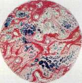

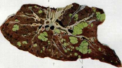
Fig. 14. Remains of round-cell infiltration. Fig. 2. Siderosis (iron deposition) in the... Fig. 3. Nodule of aponeurosis in rheumatism. Transformation of fibrous tissue into... Fig. 4. Nodule of aponeurosis in rheumatism. Concentration of cells (fibrocytes) around the focus (same field of view as in Fig. 3). Fig. 5. Disordered proliferation of cartilage (blue) among osteoid (pink) tissue in cancer. Fig. 6. Rachitic rosary. Disordered masses of cartilage among osteoid tissue with hyperemic vessels of the bone marrow. Fig. 7. Cancer of the liver (d) | f - vessels. In the epithelial elements, in the karyoplasm and cytoplasm of cancer cells, specific changes in structure occur even before the cells acquire the properties of infiltrative growth and all the signs characterizing the cancerous process. Experimental studies have contributed no less than pathological-anatomical investigations and clinical observations to the deepening of our knowledge about the cancerous process. Artificial reproduction of cancer can be very diverse. The transplantation of ready-made tumors to animals of the same species (in most cases from mouse to mouse) belongs in its original form to Novinsky, who developed this method of transplantation in 1885 in the laboratory of Professor Rudnev (St. Petersburg, Military Medical Academy); Jensen (Jensen, 1903) worked very energetically in this same direction in Copenhagen. Strictly speaking, the inoculation of crushed cancer tissue within animals of the same species is not an experimental reproduction of a new cancer. It is rather an artificial metastasis in a new animal, since the growth of the tumor has its origin in the surviving cells of the transplanted neoplasm. From the field of experiment, an interesting observation relates to salmon fish placed in ponds where so-called epidemics of thyroid cancer rage. Healthy fish released there contract cancer of the thyroid gland (P. gl. thyreoideae); however, these same diseased specimens, when transferred to new containers with healthy fish, do not infect them, since the cause of this experimental cancer lies apparently not in microorganisms, but in the special properties of the mud of the ponds in which the fish suffering from thyroid cancer live. Not only the laboratory, but life itself creates 'clinical experiments' that are of great interest for the reproduction of cancer in animals under the influence of various external irritants. At the present time, the so-called 'occupational cancers' of radiologists, appearing under the influence of many years of skin irritation by X-rays, can be attributed to the field of clinically 'experimental' cancer. For example, Lazarus (P. Lazarus) collected from the literature (1914) 100 cases of skin cancer that developed after 4–14 years of application of X-rays. From the field of pure experiment, exclusive attention has recently been paid to the reproduction of cancer in animals (mice, rabbits) by the method of prolonged smearing of the skin with coal tar. In 1915–18, Yamagiwa and Ichikawa published classic works on this topic. The regularity of the appearance of cancer in white mice under the influence of tar smearings lasting for months is no longer something accidental, but a strictly verified observation. Thus, according to Jordan, mice that withstand smearing for over 4 months develop cancer in 100% of cases at the site of tar application to the skin. It is remarkable that the smearing of experimental animals with tar to obtain cancer does not at all have to continue until the appearance of cancer as such. From a certain moment, for example, from the appearance of reactive papillomas on the skin, the cessation of smearing in some cases leads to the regression of hyperkeratoses, and in others, on the contrary, to their even more rapid growth and the appearance of true cancer. The moment of the onset of an apparently irreversible reaction, consisting in the cancerization of the cell, cannot always be strictly determined. By the method of chronic irritation of normal tissues, cancerous degeneration of the cell can be achieved in various ways. The conditions of chronic irritation of the epithelial cover can be created both on the skin and on mucous membranes, not only by tar smearings, but also by other irritative factors, such as parasites, bacteria, etc. The works of Fibiger, who worked on the experimental reproduction of cancer in the stomach of rats using the worm Spiroptera neoplastica (see Tumors), are classic in this respect. From the above brief review of various experimental methods of reproducing cancer, it is evident that the process of tissue malignization can be achieved experimentally in different ways. Since the essence of the cancerous process remains the same in all cases, it is apparently possible to achieve a disturbance of the biological equilibrium of the cell in the sense of malignization in various ways, leading, however, to one invariable effect, consisting apparently in the disturbance of the physical-chemical state of the colloids of the nucleus and protoplasm. The histogenesis of cancer and the gradual morphological picture of its development were especially demonstratively illuminated by experimental material after the famous experiments of Yamagiwa and Ichikawa, who developed the methodology for the artificial production of cancer in mice as a result of prolonged skin smearing with tar. It turned out that the hyperkeratoses, fissures, and warty outgrowths of the skin occurring as a result of these smearings entail a change in the appearance and relationships of the epithelial cells; the constant regularity of the growth of cellular elements changes, the tendency of epithelial strands to submerge into the underlying dermis increases, and active proliferation of the epithelium begins, reaching the so-called pre-cancerous state (praecancer). The potential possibility of cancer formation precedes morphological phenomena in the cell that are detectable by a microscope. In connection with the onset of cancerization, the cells enlarge, swell, and become lighter; they acquire a resemblance to embryonic cellular elements; both these and cancer colloids turn out to be highly hydrated; the high hydration of cancer colloids is accompanied by their increased dispersity. Changes in the underlying connective tissue, according to Bierich, are divided into two phases: the phase of swelling of the collagen fibers of the connective tissue and the phase of destruction, coinciding with the beginning of malignant growth. According to Waterman, the connective tissue surrounding the future cancerous focus becomes depleted of calcium salts and thereby supposedly lowers its resistance to the active growth of the epithelium. The mentioned physical-chemical transformations of the normal epithelial cell under the influence of various etiological factors can also occur depending on the most diverse biological processes associated with intrauterine life or later periods of development. At the same time, one cannot deny the significance of certain dystopically displaced groups of cells, which, under suitable conditions, are a source for the further cancerous process (Cohnheim). The trigger for the manifestation of the latent growth potential contained in the displaced embryonic elements can be the most diverse factors: injuries, irritations, etc. Chronic irritations, especially multiple and even non-specific ones, are capable of modifying tissues in the direction of their malignant degeneration (Menetrier). To this area belong the so-called 'pre-cancerous states of tissues', inflammatory hyperplasias, erosions, hyperkeratoses, benign tumors with a tendency to cancerous degeneration, etc. The root cause of the appearance of cancer in a certain area is obviously rooted not in some mysterious properties of the given region of cells, but in a significant number of cases depends on the persistence and systematic nature of the applied irritation. To illustrate this view, it is sufficient to point to the following observations. Cancer of a spontaneous nature in rabbits is known in the literature as a rare exception and has been described in approximately 40 cases; among these forms, there is almost no skin cancer. At the same time, the same skin cancer in a rabbit on the ears can be experimentally induced by tar smearing of the rabbit's ears in a significant percentage of experimental series cases. The histogenesis of cancer in individual epithelial organs (cancer of the skin, stomach, etc.) was studied mainly by the so-called 'marginal' areas, where older researchers considered it possible to understand the mechanism of the cancerogenic process more easily. The authors, investigating 'marginal growth' [Waldeyer for skin cancer, Hauser for cancer of the mucous membrane], spoke of the 'cancerous morbidity' of normal cells adjacent to the cancerous focus, as a result of which new 'cellular races' were created. These cells, therefore, underwent a fundamental change in their cytological properties, the essence of which process was interpreted differently by morphologists. Hansemann spoke of anaplasia, Lubarsch viewed the changes not as the acquisition of 'new properties' by the cell, but as a change in the degree of its individual properties.
Ribbert initially allowed for some unexpected 'emancipation' of the cell; Marchand and others spoke of a reverse return to embryonic stages of normal cells through intermediate generations. As a result of prolonged tissue regeneration, cells with special growth energy are created, a point insisted upon by Hauser, Lubarsch, Verse, and others ('...herangezuchtet werden...'). Borst speaks of an endogenous congenital factor, of a pathological predisposition of the cell, of elusive variations in the biological essence of cellular elements. The origin of cancer occurs once in solitary nodes, although even in multiple cancerous foci, in each growth focus, however, it is only 'for itself'; if the initial cancerous foci are multiple (e.g., in multiple skin cancers), then in each focus this origin also occurs only 'for itself' and the cancer grows 'from itself' (see also Tumors). Whether this origin occurs from special cells isolated during embryogenesis or from ordinary elements that have lost their normal physiological and morphological structure is difficult to say with certainty. Regarding the histogenesis of cancer of mucous membranes, Bormann, like others, believes that the growth focus originates from the smallest cell groups, which we will hardly ever be able to see under a microscope. The entire subsequent growth of cancer occurs 'from itself' and from within these cell clusters. Thus, consequently, appositional growth, in his opinion, is ruled out. Hansemann says that the true, initial beginning of the histogenesis of cancer cannot be investigated; Borst asserts: 'we are usually faced with a fully developed cancer; the processes of its origin are usually long finished for us, and we are occupied with questions of cancerous growth.' One way or another, the questions of the origin of cancer and its histogenesis remain largely unclear. The question of the contagiousness of cancer must at present be categorically resolved in the negative. The non-contagiousness of cancer can be proven both experimentally and clinically. Tens of thousands of mice in the American laboratory of Slye (M. Slye) were studied over many years from the point of view of the contagiousness and heredity of cancer. Healthy specimens of mice, specially placed in cages with animals that had ulcerated cancer, never contracted cancer. Placing healthy mice into cages that had long contained cancerous specimens also did not lead to the appearance of cancer in the healthy animals. Even more conclusive is the fact that surgeons who operate on an innumerable number of cancer patients and who, even if rarely, inevitably sustain wounds to their hands during operations, have never been infected with definite cancer. The only case of Lecene and Lacassagne, published in 1926, where it concerned a student who wounded his hand and 'contracted' sarcoma while bandaging a cancer patient, is apparently explained more by trauma than by direct infection. Valuable observations in this regard were published in 1928 by Regaud, who studied the contagiousness of cancer through long-term observations of cancer shelters in France. Care for severe ulcerated cancer patients in these shelters is entrusted to middle-aged women who have dedicated themselves specifically to charity, have no special medical education, and thus do not take systematic protective measures against possible infection. Nevertheless, cases of direct illness among any of the indicated personnel have not been noted to date. So-called 'cancer quarters' or 'cancer houses' in cities should also apparently be relegated to legends. Juillerat in Paris examined cancer mortality by residence for 1906-09 and found that 9,953 deceased cancer patients lived in 8,675 different houses. If cases of increased mortality from cancer are observed in certain areas or houses, this can be explained more by special predisposing external moments of living conditions, special life habits, heredity, age selection, etc. The question of the heredity of cancer is of no less interest than its contagiousness. That there exist so-called 'cancer families,' where not only several members of the same family suffer from cancer, but where a familial predisposition to this disease is also evident, cannot be disputed. However, it is a long way from this to the assertion of any dominant significance of heredity in the question of the etiology of cancer in humans. At the same time, there is no shortage of authors who have tried to prove a fairly high percentage of cancer heredity in humans; for example, Williams determined the hereditary factor in 9.3% of 235 cases of breast and uterine cancer. At the same time, Menetrier, although quite rightly pointing out the small number of such observations, still believes that cancer heredity can be admitted in 13% of human cancer cases. Heredity in the incidence of cancer does not at all concern the direct transmission of a cancerous tumor to descendants, but only a predisposition to cancer, transmitted by inheritance. This predisposition often has an organ-specific character. Thus, if there is a predisposition to uterine cancer in a family, then it is most likely that in the case of cancerous disease in female offspring, the cancer will be located precisely in the uterus, and not in the stomach, intestine, etc. Examples of this type have recently been collected by Auvray, Bauer, and others. The question of heredity appears more clear in the light of experiment. Recognizing a priori the great importance of heredity for cancer incidence, Slye was nevertheless able, over many years and on a huge amount of material, to prove that only artificially created selection of individuals who spontaneously developed cancer (for example, mice) leads to generations clearly prone to hereditary cancerous processes. Crossbred specimens of white mice that had already had a cancerous tumor over many generations can turn into such 'cancer families' in which the frequency of cancer can be arbitrarily brought up to 100, 50, etc., percent. And conversely, the selection of crossbred individuals taken from families not burdened by cancer heredity can produce offspring free of any tumor for many years. Localization or so-called organ-specificity of the disease can itself become an object of experiment. By specifically selecting individuals who have breast cancer, one can create a generation of mice that have cancer mammae in 100% of cases. This same observation applies to other rarer localizations of cancer, e.g., thyroid cancer in mice (cf. works of Slye). Observations in this direction have been carried even further and expanded by data on organ heredity in mice under the influence of specifically localized trauma. For example, mice taken from families predisposed to the appearance of cancer from injury to the hind limb do not get it if the trauma is inflicted on the head, back, etc. What the essence of this artificially created cancerous predisposition consists of, in which heredity essentially manifests itself, remains unclear to this day. Whether this is a kind of 'biological inferiority' of certain organs or tissues—so-called dysembryoplasia (Letulle), i.e., a state predisposing to the appearance of cancer under certain external conditions, or something else, remains open. The brief data presented on the heredity of cancer are sufficient to assign only secondary importance to this factor in humans at the present time, in view of the fact that we do not encounter artificial selection of cancer representatives among people in life, especially over long years and many generations. Thus, the extreme judgments of some French authors who spoke out against marriages of persons burdened with cancer heredity must be recognized as poorly founded, as, incidentally, Delbet has proven. The prevention of cancerous diseases differs significantly from the prevention of other pathological forms, for example, infectious diseases. Data on the etiology and pathogenesis of cancer outline some milestones on the path of preventive measures in relation to cancer. From a preventive point of view, all moments leading to systematic irritation of tissue elements must also be systematically eliminated to avoid the accumulation of a maximum of biological irritation of the cell, as a result of which, as we have seen, its malignant transformation occurs. Broad social prevention of cancer has not yet been sufficiently developed and has not been placed at the proper level. It belongs to the tasks of the near future. For now, the current state of the question of cancer prevention follows two paths: the sanitary-public path and the path of individual hygiene. Practically, attention should be paid to eradicating the significance attributed to cancer heredity, which is clearly exaggerated among the masses. The false idea of cancer as an incurable disease must also be eradicated by sanitary-educational measures. Among general measures related to everything stated above about the essence and pathology of cancer, attention should be paid to the prevention of early aging of the organism as one of the most prominent factors contributing to the appearance of this neoplasm; here, of course, we can speak of physical culture, general body hygiene, etc.
Measures must be taken to eliminate chronic tissue inflammations and to eradicate diseases predisposing to cancer, such as syphilis, tuberculosis, etc. Among general hygienic measures, abstinence from the ingestion of substances that irritate tissues is recommended: alcohol, tobacco, strong spices, etc.; the latter should be forbidden especially to persons suffering from chronic stomatitis, tonsillitis, chronic catarrhs of the gastrointestinal tract, etc. To avoid congestive phenomena, particularly in the intestine, regular functioning of the gastrointestinal tract should be a subject of attention for the purpose of preventing cancer of the rectum; for these same reasons, long-unhealing fissures in the anal region should be eliminated in a timely manner. Sanitation of the oral cavity, removal of carious teeth, and elimination of prostheses that irritate the gums also belong to the most important local preventive measures in relation to cancer of the oral cavity. Improvement of the female reproductive apparatus, hygiene of childbirth, breastfeeding, and timely elimination of erosions on the cervix uteri belong to the most important preventive tasks. Finally, the regulation of the general routine of life, the eradication of alcoholism, etc., currently belong to the field of general anti-cancer prophylaxis. Occupational anti-cancer prophylaxis currently attracts medical attention in the USSR more than anywhere else. The significance of the profession, at least for some forms of cancer, cannot be disputed. The established fact of the connection of malignant tumors with the profession touches upon questions: a) about the significance of occupational factors for understanding the etiology and pathogenesis of cancer, b) about the practical role of occupational factors in the occurrence of cancer, and finally c) about the practical use of occupational organizations for the implementation of cancer prophylaxis. The study of the question of the significance of the profession for the etiology of cancer has provided extensive material [chimney sweep cancer, discovered in England (Pott), cancer of radiologists, coal and other cancers (Table III)] and for the correct understanding of the pathogenesis of the malignant transformation of normal cells of the organism into cancer, sarcoma, etc. As a result of comparing occupational and experimental cancers, it was possible, among other things, to establish a fairly unshakable position that the cessation of the action of a harmful agent after its long influence does not always prevent or stop the development of malignant growth (ablata causa non cessat effectus). The practical role of occupational hazards in the occurrence of cancer, however, undoubtedly cannot occupy a large place. Since the vast majority of cancer forms relate to the digestive tract, it often proves impossible to link them without strain to the conditions of occupational hazards; thus, for undoubtedly occupational cancer, a fairly modest place must be assigned in statistics. For example, in England, where mortality from cancer is calculated at approximately 40,000 people per year and where the occupational cancer of chimney sweeps ("chimney sweep's cancer of the scrotum") was repeatedly described, it still turns out that this variety of cancer affects no more than one in 1,000 chimney sweeps (Leitch). According to Kennaway, an average of 4 chimney sweeps die annually from cancer of the scrotum in all of England. The occupational cancer of radiologists was already mentioned above. The cancer of spinners attracts great attention, usually observed on the hands, face, nape, and other places in clear dependence on friction by clothing soaked with mineral oils, which takes place in connection with the constant lubrication of various parts of spinning machines with these oils. Also relatively frequent is cancer among workers of briquette factories from pitch that sprays during processing (Teutschlander); it is in this profession that occupational figures are particularly demonstrative. It turns out that among briquette workers working for over 5 years in their profession (Baden enterprises - Teutschlander), up to 40% fall ill with cancer; out of 13 workers, 12 usually have "pitch dermatitis," warty growths, etc., i.e., are in a precancerous state of tissues. The use of liquid pitch for the manufacture of briquettes is a method of prevention of this so-called "briquette cancer." Cancer is also not uncommon among workers engaged in the distillation of coal tar. According to statistics in England (1920-23), 133 cases of tar cancer were discovered. The well-known aniline cancer of the bladder (discovered by Rehn in 1895) is a relatively rare form of cancer: such observations are currently registered in the literature in only about 60 cases. According to Nassauer, cancer of the bladder is observed in approximately 27% of cases among workers in aniline production; Curschmann, however, found only 177 cases of bladder cancer per 100,000 workers in aniline production. Some authors consider aromatic substances (benzene, toluene) to be the causes of "aniline cancer," while others see the evil in the impurity of arsenic in aniline. Occupational cancers must also include malignant tumors of the lungs encountered among miners of the Schneeberg mines in Saxony. This disease is linked to chronic poisoning with speiss cobalt containing arsenic. According to Kolsch, half of the workers employed in the Schneeberg production allegedly die from lung cancer, although according to Lehmann, this figure is actually significantly lower. Analogous examples of occupational cancers, albeit in a small percentage of cases, are observed among those working with creosote, fuel oil, asphalt, hot glass, etc. At the present time, it can be said with certainty that even all reliable occupational cancers taken together, the number of which is negligible in comparison with the number of non-occupational forms, do not affect the general cancer mortality of the country. Even the famous occupational "chimney sweep's cancer," the mortality from cancer among whom in England is higher for men than in any other profession, does not at all fall on the cancer of the scrotum typical for chimney sweeps, but on cancer of the digestive and respiratory tracts. In general, the frequency of occupational cancer apparently cannot yet be reliably established, since the cited statistics suffer from great contradiction (Wolff). For professions of female labor, some authors mention skin cancers from needle pricks in seamstresses, stomach cancer from constant tasting of hot food by cooks, etc. These indications, however, are not very competent. In particular, the latter observation can rather be explained by secondary domestic hazards associated with the abnormal conduct of the postpartum period in these professions (Petrov). Apparently, one has to think that it is precisely secondary occupational domestic hazards that play a particularly prominent role in the mass pathogenesis of cancer. In quantitative terms, they undoubtedly have greater significance than individual specific irritants inseparably linked to relatively few harmful professions. From this position, the principles of occupational anti-cancer prophylaxis also follow. Where the connection between cancer and occupational hazard is beyond any doubt (and such cases are in the minority), all harmful moments and products must be removed from processing, or protective measures must be implemented that paralyze the harmful action of the agent. That is, here one could speak of replacing the spraying pitch in briquette production with liquid; in aniline production, where the urinary tract is threatened during long-term work, it would be necessary (in addition to the necessary constant control of urine) to speak of transferring workers to other workshops after certain periods, etc. Since for the mass of industries of the leading industry, the specific influences that malignize tissues are not yet known, among these workers, general hygienic provisions should be implemented more widely as occupational-preventive measures, the improvement of living conditions should be introduced more deeply, and thereby a reduction in the number of chronic catarrhs should be created, the hygiene of childbirth, the postpartum period, etc., should be improved (Table 11).
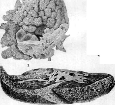
Measures of preventive struggle also include the timely treatment of precancerous diseases, such as, for example, the removal of benign tumors, extirpation of cysts of the mammary gland, treatment of leukoplakia of the tongue, ruptures of the cervix uteri, radical treatment of stomach ulcers, etc. If at the present time we are still very far from asserting that we have mastered
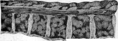
Figure 1. Psammoma (fibroma psammosum). Figure 2. Radix: a-infiltrate, b-root. Figure 3. Papillary tuft of ovarian cancer. Figure 4. Scirrhus of the mammary gland. Figure 5. Carcinoma of the pleura. Figure 6. Metastases of cancer in the spine. Table 11. Occupational cancer. Profession: Chimney sweeps, Coal miners, Briquette makers, Asphalt workers, Creosote workers, Paraffin workers, Oil refinery workers, Arsenic production workers, Cotton mill weavers, Locksmiths, X-ray workers, Glassblowers, Dye factory workers, Schneeberg mine workers. Cancer-causing agent: Soot, coal tar; Soot, coal tar; Tar, coal, tar; Tar, coal, tar; Creosote, tar; Paraffin; Fuel oil; Arsenic; Mineral oils; Radiant and ordinary heat; X-rays, rays; Thermal irritants, rosin, pine tar; Aniline dyes; Cobalt, arsenic. Patient age: 40-50, 40-50, 40-50, 40-50, 55-60, 37-79, 35-50, 40-50, 55-60, 45-60, 40-55, 51 years, 40-50, 50-60. Prof. experience: 15-20, 15-20, 15-20, 25-30, 25-30, 15-25, 20-26, 25-60, 15-45, 9-25, 15-25, 15-20. Localization: Scrotum, outer surface of thigh, buttocks, forearms, hands; Scrotum, outer surface of thigh, buttocks, forearms, hands; Skin of face, forearms, hands; Skin of face, forearms, hands; Forearms, hands; Scrotum, lower leg, forearms, hands; Scrotum, forearms, hands; Hands, forearms; Scrotum, thighs; Face, hands; Hands, fingers; Skin of face, hands; Urinary bladder; Lungs, bronchi, pleura. If we talk about cancer prevention, then the observance of many of the above-mentioned guidelines should practically lead to a noticeable decrease in cancer incidence in the USSR. The treatment of cancerous tumors represents only one side of the entire edifice of the anti-cancer struggle. At the present time, it should be said that combined methods of cancer treatment appear to be the most reliable. The main stage of treatment is undoubtedly surgical aid, based on the radical removal of the primary focus within absolutely healthy tissues. Exclusively operative aid finds application in the majority of cancers of the intra-abdominal organs (for example, cancer of the stomach, cecum). In view of the dangers associated with the bloody method, predisposing to so-called implantation metastases as a result of introducing viable cancer cells onto the surface of a fresh wound, one can recommend, in addition to general antiblastic measures (for example, swabbing the wound with iodine, washing hands and the wound surface with alcohol, etc.), the use of an electrodiathermic knife instead of a scalpel. In such cases, either electroexcision or electrocoagulation of the cancerous focus is performed. Actinotherapeutic methods come to the aid of surgery in the fight against cancer: X-ray therapy and curietherapy. The direct introduction of radium into the operative wound as the final act of the intervention is the most demonstrative method of combined therapy. In cases where operative and actinotherapeutic aid cannot be provided with sufficient hope for success, palliative measures remain in reserve. Here, one can speak of vaccine therapy and toxin therapy for cancer. Treatment with Coley's bacterial toxins is based on observations of the favorable influence of spontaneously arising erysipelas on the cancerous process. Serotherapy, e.g., Deutschmann's Typho-rocidin or the serum of animals immunized against Micrococcus neoformans (Doyen), does not give any reliable results. Vaccine therapy, also called histiotherapy and extractotherapy according to Fichera, gives somewhat more than serum treatment. This method has been studied mainly in experiments from the point of view of the prophylaxis of animals in relation to transplanted cancer. Inoculations of tumor extracts of autolysates, as a therapeutic method primarily on animals having cancer, gave a positive result according to Blumenthal, Jensen, and others. Not small hopes were placed until recently on various types of chemotherapy for cancer, e.g., with colloidal lead solutions. Despite individual striking successes even in incurable cases, the method of intravenous injection of colloidal lead proved to be so dangerous that it has not yet received wide distribution. Thus, one can hardly doubt that the key to the success of cancer therapy lies in the early seeking of medical help by patients; only early diagnosis gives in most cases maximum hope for subsequent lasting cure. Hardly the most striking illustration of this position is the material from the Mayo Clinic, published by Sistrunk. Thus, for example, out of 86 cases of breast cancer operated on radically during the period when axillary glands were not yet palpable, 49 patients (57%) were alive after five years; out of 132 patients operated on when the glands were already affected, only 22 were alive after the same period, i.e., 17%. The same can be said about other clinical forms. Cancer of the lower lip, treated in the period of the absence of palpable glands, can be cured in almost 92% of cases, and in the presence of even mobile glands on the neck, only in 34% (Berven). König, having reviewed the cancer material of several surgical clinics in Germany over 19 years, could conclude that cancers of various localizations, operated on without involvement of lymph glands, yielded a cure, followed up for 5 years, in almost 100% of cases (with enlarged glands, a 5-year cure is given in 39%, and a 10-year cure in only 10%). Neither the three-year nor the 5-year period of postoperative observation for treated cancer yet fully resolves the question of a lasting cure, although each of these periods successively reduces the probability of cancer recurrence. The vast majority of recurrences are observed in the first year after treatment. It must be said, however, that so-called late recurrences (after 10-15 years or more) belong, as the duration of the cure increases, to increasingly greater rarities and are considered by many no longer as true recurrences, but as a new disease of cancer (which, however, in most cases is incorrect). It is almost impossible to give a general clinical picture for all types of cancer in a summary form, since the main signs of the disease depend to a significant extent on the localization of the tumor and the affected organ. While in young people cancer often takes a lightning-fast course and carries off patients in a few months, cancer in old people, for example, so-called creeping skin cancers of the face, scirrhus of the mammary gland in elderly women, sometimes drag on for years. On the other hand, if the average life expectancy of a cancer patient is calculated at 1 1/2-3 years, then often, even without achieving a radical cure by palliative methods, one can restrain the progressive development of the disease, periodically lowering the viability of cancer cells by X-ray therapy, curietherapy, etc. ("keeping the tumor in obedience" - "tenir en respect"). In all forms and localizations of cancer, as the tumor develops further, the general cachectic state of the organism is increasingly felt, against the background of which secondary complications in the form of pneumonia, sepsis, hemorrhages, etc., easily join. The prognosis depends, of course, on the entire sum of clinical signs of the given form of cancer, on the involvement of vital organs, on the histological structure of the cancer, etc. The richness of the tumor in cells, the degree of differentiation of the cellular elements of the cancer influence the prognosis; the more sharply expressed the "anaplasia", the less mature the cells, and the higher the karyokinetic index, the higher the malignancy and, consequently, the worse the prognosis. Individual clinical forms. Skin cancer, in most cases observed in men at a more advanced age, has the appearance of superficial excoriations or turns into deep ulcers with the invasion of underlying tissues and sometimes a tendency to unilateral scarring from one edge (ulcus rodens). Involvement of the lymphatic apparatus, mainly when the skin cancer is located on the face, is not a rarity. Among other forms of cancer, skin cancer ranks 5th in frequency. By virtue of its good operability, it occupies 14th place in general cancer mortality (cf. Davydovsky). According to materials from the Oncological Institute in Leningrad (175 cases, cf. Shanin), skin cancer among other forms is encountered in 4.9%; metastases in it are noted in 3.4% to 17.6%. Serious therapeutic advantages in any sharply expressed forms are possessed by radiation methods of therapy. Thus, recurrences after actinotherapeutic methods are encountered in 4.8% to 15.5%, and after operative removal - in more than 24%. At the same time, operative removal of non-neglected forms of skin cancer according to Swedish collected statistics (Nyström) gives long-term recoveries up to 5 years in 76%. More numerous are the observations of individual authors on the long-term results of treating skin cancer with radium: according to Regaud there are 58.5% of recoveries, according to Deland - 62%, according to Forssell - 86% for superficial and 51% for deep forms. Directly adjacent to skin cancer is lip cancer, which clinically manifests itself in the form of ulcers and infiltrates of various sizes, usually developing from cracks, leukoplakias, non-healing scars, irritations from many years of pipe smoking, etc. Carcinomatous involvement of the lymphatic apparatus in lip cancer, especially of the lower lip, turns out to be a frequent complication of the disease (in 60% of all cases at the start of treatment according to the material of Lacassagne, Paris).
Unfortunately, even now many patients with lip cancer seek medical help too late. For instance, out of 173 patients who visited the outpatient clinic of the Oncological Institute (Leningrad) with lip cancer, 57 had to be immediately recognized as advanced and incurable. In men, cancer of the lower lip occurs in approximately 93% of all cases, and in women in 7%. Treatment of lip cancer can be surgical only, radiological only, or combined. For non-infiltrating, limited tumors, wedge excision, especially with simultaneous surgical removal of the cellular tissue and lymph nodes in the neck, yields good results. Berven in 1923 reviewed material on 637 cases of lip cancer. It turned out that in initial forms of the process (without nodes), there was a 66% 5-year cure rate; for the same superficial forms, also treated surgically but with simultaneous surgical removal of non-palpable cervical nodes, there was a 92% recovery rate, while with palpable nodes after the same 5 years, only 34%. Similar data were published by Nyström in 1928—60% 5-year recoveries. Currently, the widely used curietherapy for lip cancer also yields excellent results, especially in more difficult cases. Quick, in 202 cases, had an 80% 5-year cure rate for superficial forms after radium therapy, and 41% for deep forms. Forssell, who also possesses a large amount of material, had 68% absolute recoveries for a 5-year period for all cases of lip cancer treated with curietherapy. Among the severe forms of cancer that must still be recognized as almost incurable are esophageal cancer, which occupies third place in general cancer mortality (Davydovsky, Moscow, 1923–27) (see Esophagus). One of the most frequent clinical forms of cancer is undoubtedly stomach cancer. Its frequency among other cancer forms reaches 40%. In general cancer mortality, stomach cancer (Davydovsky, Moscow, 1923–27) occupies first place, far leaving behind other cancers (of the uterus, esophagus, bronchi). Intestinal cancers occupy 5th place in general cancer mortality (Moscow, 1923–27). Rectal cancer. Rectal cancer proves to be a practically important form. The regrettable confusion of this form with hemorrhoids, the unsuccessful treatment of which is sometimes carried out by doctors for months or even years, creates a large pool of inoperable cases. It must be mandatory for a doctor to examine all those patients who complain of any ailments related to the rectum, especially in cases of bleeding from it, constipation, and general weight loss (see Rectum, cancer). Breast cancer is the third most frequent cancer in women. Among all other forms of cancer, it occurs in approximately 8%. In our Union, over 10,000 women die annually from this form. In general cancer mortality (Moscow, 1923–27), breast cancer occupies 10th place. The most frequent cancer in women is uterine cancer. In the USSR, over 35,000 women die annually from this disease. In general cancer mortality, uterine cancer occupies 4th place (Moscow, 1923–27). All old erosions, cervical tears, untreated gonorrhea, prolonged catarrh of the mucous membranes, as well as a number of other chronic irritations are among the frequent etiological causes of this form (see Uterus, cancer). Jaw cancer occurs in approximately 3% of all cancers of the human body. In the majority of cases, these are squamous cell keratinizing forms originating from the mucosa of the maxillary sinus or the alveolar process (see Jaws). A. Vereshchinsky. Cancer tests. The overwhelming majority of numerous tests for cancer proposed by various authors are based on the premise of the entry into the blood, excretion in urine, or secretion into the stomach cavity (in stomach cancer) of various products of tumor vital activity, which are detected by the corresponding test. The expansion of the field of study of chemical and biochemical anaplasia and the metabolic characteristics of cancerous tumors expands the circle of proposed reactions. Nevertheless, most of them are based on scientifically poorly grounded empiricism or on attempts, unsuccessful to date, to find a specific 'Krebs-gift' (see Oncology). Verification of all cancer tests without exception has shown that they are not strictly specific, yield a high percentage of errors in cancer, can be positive in non-cancerous subjects, or are non-specific reactions associated with cachexia. Therefore, the diagnostic value of the overwhelming majority of tests is negligible, and they have found no application in clinical practice; a significantly smaller number of them have only relative significance among other symptoms of a tumor as indirect evidence of its presence or absence. The first group includes the reaction for colloidal nitrogen in urine by Salkowski, the color reaction with urine by Davis, the Gluzinski test for hydrochloric acid in gastric juice, the test for peptolytic enzymes (tryptophan test, proposed by Neubauer and Fischer), the Kelling test for the heterolytic ability of the serum of cancer patients in relation to the erythrocytes of various animals, complement fixation reactions (Dungern), the antitryptic reaction, and many others. To the second group, one could assign the following tests: 1) The test for protein or nitrogen in gastric contents in stomach cancer (Salomon's method); a positive result is given only by an ulcerated tumor, and therefore a negative result is not proof of the absence of a tumor at all. The presence of bile or blood in the gastric contents makes the reaction unsuitable, as do cases with significant amounts of free HCl (i.e., and pepsin). Thus, this reaction can be used only as a differential diagnosis between achylia of non-cancerous and cancerous origin. Technique: the day before, the patient is given only liquid food, and from the evening, the stomach is thoroughly washed clean; in the morning, it is washed once more with 400 cm3 of physiological saline, pouring it in and out twice. 100 cm3 is taken from the lavage fluid, and the protein is determined in it according to Esbach or nitrogen according to Kjeldahl. In the absence of a decaying tumor, the Esbach reagent does not give a flocculent precipitate; the amount of nitrogen according to Kjeldahl should not exceed 20–30 mg in normal conditions. 2) The precipitation reaction of Freund and Kaminer (Trübungsreaction) is based on the assumed presence in the blood of cancer patients of antibodies supposedly specific to the cancer antigen. Verification studies have shown the non-specificity of this reaction; furthermore, it is technically difficult. Therefore, despite its good practical results, it has found little application. Technique: an emulsion of cancer cells is prepared from crushed tumor in 1% Natr. biphosphoric; the emulsion is pressed through gauze, the cells are allowed to settle, and they are diluted with 1% NaCl. The emulsion is unstable, and instead of it, a tumor extract can be prepared: to 100 cm3 of emulsion, 5 cm3 of 5% acetic acid is added, heated on a water bath to 80° for 1/2 hour, filtered, and after cooling, neutralized with 10% Natr. carbonic. to litmus, heated again to 80°, and filtered. To 3 cm3 of emulsion or extract, 10 drops of the patient's blood serum are added. Cancerous sera give precipitates in the emulsion and turbidity in the extract; non-cancerous sera leave the extract transparent and clarify the emulsion due to the dissolution of cancer cells. 3) The meiostagmin reaction is based on the lowering of the surface tension of immune serum when it combines with an antigen, which is determined by a decrease in volume and, accordingly, an increase in the number of drops in a certain amount of serum, measured by a stalagmometer. The reaction is non-specific, occurring in chloroform anesthesia and in pregnant women, but nevertheless has relative diagnostic value. Instead of a specific cancer antigen, non-specific ones from lecithin, etc., can be used. 4) The Botelho test, a turbidity and flocculation reaction (Flockung-, Trübungsreaction), is based on the qualitative change in blood proteins and lipoids in cancer patients. It is non-specific but yields a high percentage of positive results in cancer patients and disappears after the removal of the tumor. Technique: to 2 cm3 of a 5% citric acid solution and a 1% formalin solution, 0.5 cm3 of the patient's blood serum, diluted by half with 0.75% NaCl, is added. To the mixture, 0.7 cm3 of I + KI (1.0 I + 2.0 KI in 100 cm3 of water) is added. A precipitate forms, which dissolves if the serum is taken from a non-cancerous patient; in the latter case, the precipitate appears only when another 0.3 cm3 (i.e., a total of 1 cm3) of the I + KI solution is added. 5) The Abderhalden reaction—see Abderhalden reaction.
Related articles
Mentioned in
Cite this page
“Cancer.” Soviet Medical Encyclopedia. English translation of Bolshaya Meditsinskaya Entsiklopediya, 1st ed. (Moscow, 1928–1936), ed. N. A. Semashko. https://sovietmedicalencyclopedia.pages.dev/article/cancer/