Anus
Historical document, translated for reference. It reflects medical knowledge of the 1920s–30s and is not medical advice.
Summary
The anus is the external opening of the rectum, surrounded by sphincter muscles that control defecation. The article describes its anatomical structure, muscular layers, and surgical procedures for creating artificial anus openings.
Encyclopedia article (1928–1936)
ANUS, the Latin anatomical term for the external opening of the rectum; physicians by the term A. usually mean the anal canal (pars analis recti, canalis analis), which represents a narrow passage connecting the lower part of the rectum (ampulla recti) with the external environment. Its average length, with an empty rectum, is about 3 cm; when the rectum is distended, its length becomes somewhat smaller. Its anteroposterior dimension is 1-1.5 cm. The longitudinal axis of the A., running from front to back and from above downward, is inclined to the axis of the rectum at almost a right angle. The anal canal, thus, represents the extrapelvic part of the rectum, well-fixed, surrounded by two sphincters (m. sphincter ani externus and m. sphincter ani internus) and fibers of the muscle that elevates the anus (m. levator ani). The walls of the canal are kept in contact by the sphincters. In front of the anal canal in men is the bulbus urethrae (cavernous bulb of the urethra), in women-the lower part of the posterior vaginal wall. The wall of the anal canal consists of layers-muscular, submucous, and mucous. The muscular layer, consisting of smooth muscle fibers, divides into two parts-external and internal; in the external part the muscle fibers are arranged longitudinally, in the internal part-circularly. Upward the longitudinal fibers of the anal canal pass directly into the longitudinal fibers of the rectum;
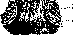
The anal segment of the rectum (opened from behind and unfolded): 2-sphincter internus; г-columnae rectales (Morgagni); 3-sphincter externus (according to Braus).
in the area of attachment to the canal wall of the muscle that elevates the anus, its fibers mix with the longitudinal fibers of the canal, descend with them downward, pass between the external and internal sphincters, and finally attach to the skin surrounding the anal opening. The circular part of the muscular wall of the rectum at the very upper end of the anal canal begins to thicken sharply, and this thickening also ends abruptly, not reaching, approximately, 1 cm to the anal opening. This thickened area of smooth circular fibers is called the internal sphincter (sphincter ani internus). The mucous membrane covering the upper 2/3 of the anal canal forms 6-8 longitudinal folds (columnae Morgagni), the wide bases of which arc into one another, as a result of which depressions (sinus Morgagni) are formed in this place; swallowed foreign bodies (needles, bone fragments, etc.) can be retained in these depressions, and then the removal of these foreign bodies requires artificial assistance. The mucous membrane of the upper 2/3 of the anal canal is covered with cylindrical epithelium, which below the Morgagnian depressions gradually transitions into multilayered: at the edge of the anal opening the multilayered epithelium imperceptibly transitions into the epidermis of the skin. From under the submucous tissue of the anal opening, thin bundles of smooth fibers radiate, which attach to the skin surrounding this opening (the muscle that corrugates the skin of the anal opening-m. corrugator cutis ani). This muscle turns the mucous membrane of the lower end of the anal canal inward after this membrane has been everted during the act of defecation. The voluntary muscles surrounding the anal canal are the external sphincter (sph. ani externus) and the pubo-anal portions of the muscle that elevates the anus. Together with the internal sphincter they form a muscular mass, the main purpose of which is to close the anal opening. They can also help in the evacuation of feces, worm-like contracting from top to bottom.-Pathology of A.-see Rectum. A. Deshev. ANUS PRAETERHATURALIS, artificial anus; a fistula created surgically on the large intestine with the calculation that all intestinal contents exit through it outward. It has to be created, as a preliminary act, in the excision of the rectum for malignant neoplasms, in the resection of the large intestine for the same reason, in inoperable cancers and some inflammatory processes of the rectum, and in obstruction due to various causes. In some cases, after eliminating the disease and restoring natural relations, A. p. can be closed (temporary A. p.), in others-it has to be left forever (permanent A. p.). Two basic requirements are made to A. p.: 1) all intestinal contents must exit outward through it and must not enter the lower part of the intestine, 2) A. p. must retain feces and, if possible, gases, i.e., it must possess the function of a natural anus. The striving to fulfill these requirements created many ingenious methods of creating A. p., from which the entire history of A. p. was formed. The first thought about A. p. was expressed by Littré in 1710 in connection with a case of atresia of the rectum. He pointed out that in such cases it is possible to sew the intestine lying above the stricture into the abdominal wall and open it. This operation was first performed by Pyllore in 1776. The operation method according to Littré consists of the following: in the left iliac region 2-2.5 cm above the anterior superior spine with an incision of 6-7 cm, parallel to the inguinal ligament, the abdominal cavity is opened, the S-shaped intestine is grasped and brought out into the wound; its seromuscular membrane is sutured to the peritoneum and skin, the abdominal wound is closed. The intestine is opened in 2-3 days (if necessary-immediately). Such A. p. did not meet either of the above requirements. In 1881, Sebinzinger proposed to create A. p. by cutting the S-shaped intestine, at which the lower end is sewn up tightly and lowered into the abdominal cavity, while the upper is sewn into the skin wound. The same was proposed by Madelung, why the method is called the Sebinzinger-Madelung method. With this A. p. all intestinal contents are evacuated outward, but it is doomed to incontinence, and in inoperable cancers of the rectum in the lower segment above the site of stricture a closed sac with septic contents may form. Therefore Kni as early as 1888 recommended to sew into the abdominal wall also the lower segment of the intestine. In the same year Maydl proposed to ensure the exit of feces from the upper segment and their non-entry into the lower to bring out the loop of the S-shaped intestine so as to form a spur. Witzel, in 1894, when creating a spur, passed the intestinal loop through the rectus muscle, hoping to create a sphincter. However, with this method complete closure of A. p. is not achieved, which led to a series of new methods aimed at compressing the end of the intestine with the help of spring devices. For a time the methods of Hacker and Kappis gained greater popularity (see fig. 1-3); the principle comes down to creating a skin tube for the exit of feces, which can be easily compressed by a spring clamp. Kleinschmidt, applying Hacker's method 12 times, remained satisfied with the results. The advantages of this method: 1) it can be applied to an existing A. p., as long as the latter is applied deeply enough on the S-shaped intestine, 2) the intestine is opened away from the place of formation of the valve,
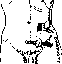
Figure 1. Formation of a skin flap according to Kappis (above). The lower segment sewn into the wall (below).
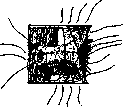
Figure 2. The anterior wall of the rectus muscle has been removed; formation of a skin tube and application of sutures for its subsequent closure.
is closed. The intestine is opened in 2-3 days (if necessary-immediately). Such A. p. did not meet either of the above requirements. In 1881, Sebinzinger proposed to create A. p. by cutting the S-shaped intestine, at which the lower end is sewn up tightly and lowered into the abdominal cavity, while the upper is sewn into the skin wound. The same was proposed by Madelung, why the method is called the Sebinzinger-Madelung method. With this A. p. all intestinal contents are evacuated outward, but it is doomed to incontinence, and in inoperable cancers of the rectum in the lower segment above the site of stricture a closed sac with septic contents may form. Therefore Kni as early as 1888 recommended to sew into the abdominal wall also the lower segment of the intestine. In the same year Maydl proposed to ensure the exit of feces from the upper segment and their non-entry into the lower to bring out the loop of the S-shaped intestine so as to form a spur. Witzel, in 1894, when creating a spur, passed the intestinal loop through the rectus muscle, hoping to create a sphincter. However, with this method complete closure of A. p. is not achieved, which led to a series of new methods aimed at compressing the end of the intestine with the help of spring devices. For a time the methods of Hacker and Kappis gained greater popularity (see fig. 1-3); the principle comes down to creating a skin tube for the exit of feces, which can be easily compressed by a spring clamp. Kleinschmidt, applying Hacker's method 12 times, remained satisfied with the results. The advantages of this method: 1) it can be applied to an existing A. p., as long as the latter is applied deeply enough on the S-shaped intestine, 2) the intestine is opened away from the place of formation of the valve,
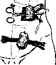
Figure 3. Formation of a skin tube from a flap (above). The lower segment sewn into the wall (below).
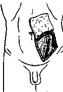
Figure 4. Formation of a skin flap according to Lambret.
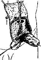
Figure 5. Method of Lambret.
The segment of intestine is grasped with a clamp and brought through the upper wound. Consequently, this field is protected from contamination, 3) The artificial anus retains feces well. Among other methods, the one most accepted at present is that of Lambret, who proposed creating an artificial anus in the form of a proboscis covered with skin (see fig. 4-8). Lambret first cuts out a quadrangular flap with the base upward and to the middle or with the base downward and to the side. If the flap is taken with the base upward, the course of the operation is as follows: the abdominal cavity is opened at the lower edge of the incision, with the muscles being separated along the course of the fibers. An S-shaped loop of intestine is extracted, and the place where it should be divided is determined. The mesentery is incised in a radial direction. It is important that the upper end, brought out externally, be of sufficient length (up to 12 cm). At the selected place, after crushing, the intestine is divided, and the ends are sutured with a purse-string suture. The lower end is sutured especially carefully and is lowered into the abdominal cavity. If necessary, it can be sutured into the wound. When the removal of the rectum is contemplated, the lower end to be lowered should be detached downward, which is better achieved through a preliminary median incision. On the left index finger introduced into the abdominal cavity at the base of the skin flap, a sufficiently wide passage is made through the muscles and peritoneum. With the help of forceps, the upper end of the intestine is pulled through here and sutured. The lower abdominal wound is sutured. The protruding end of the intestine is covered with the skin flap. After 36-48 hours, the end of the intestine is cut off somewhat deeper than the edge of the surrounding skin, so that when the edges of the intestine and skin are sutured, the latter is turned inward. Retention of feces is achieved by either the opening of the artificial anus being closed with a strip of adhesive tape, or the protruding proboscis being pressed with a pessary. Among complications, the author himself observed one case of necrosis of the entire skin flap, - it was necessary to create a conventional artificial anus; in several cases there was partial necrosis, which, however, did not affect the function. Korganova-Müller and Kruglov introduced some changes into the method. All these improvements have application when creating a permanent artificial anus. When it is necessary to create a temporary artificial anus, it is better to use the method of Maydl or similar methods, since an artificial anus created by these methods can be closed. The closure of the artificial anus itself is not without difficulties. If a spur is present, one can, following the example of Dieffenbach and Mikulicz, crush it in several sessions, and after the restoration of wide communication between the afferent and efferent ends, suture the opening of the artificial anus. This method has major drawbacks: 1) crushing of the spur is performed blindly, 2) nerves of the mesentery may be subjected to crushing, which can cause shock phenomena, 3) bleeding may occur in case of damage to blood vessels. Based on these principles, Hohlbaum recommends closing the anus praeternaturalis by resection. In the clinic of Payr, 42 patients were operated on by the resection method, and only in one case did failure occur due to insufficient surgical technique.
When it is necessary to create a temporary artificial anus, it is better to use the method of Maydl or similar methods, since an artificial anus created by these methods can be closed. The closure of the artificial anus itself is not without difficulties. If a spur is present, one can, following the example of Dieffenbach and Mikulicz, crush it in several sessions, and after the restoration of wide communication between the afferent and efferent ends, suture the opening of the artificial anus. This method has major drawbacks: 1) crushing of the spur is performed blindly, 2) nerves of the mesentery may be subjected to crushing, which can cause shock phenomena, 3) bleeding may occur in case of damage to blood vessels. Based on these principles, Hohlbaum recommends closing the anus praeternaturalis by resection. In the clinic of Payr, 42 patients were operated on by the resection method, and only in one case did failure occur due to insufficient surgical technique.
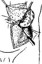
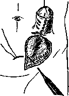
Based on these principles, Hohlbaum recommends closing the anus praeternaturalis by resection. In the clinic of Payr, 42 patients were operated on by the resection method, and only in one case did failure occur due to insufficient surgical technique.
Fig. 7. Lambret's method. Closure of the skin wound.
V. Gricep.
Related articles
Mentioned in
Cite this page
“Anus.” Soviet Medical Encyclopedia. English translation of Bolshaya Meditsinskaya Entsiklopediya, 1st ed. (Moscow, 1928–1936), ed. N. A. Semashko. https://sovietmedicalencyclopedia.pages.dev/article/anus/