Balantidium Coli
Historical document, translated for reference. It reflects medical knowledge of the 1920s–30s and is not medical advice.
Summary
This article from the 1928–1936 Soviet Great Medical Encyclopedia details the biology, pathology, and epidemiology of Balantidium coli, a parasitic protozoan that causes balantidiasis in humans.
Encyclopedia article (1928–1936)
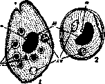
BALANTIDIUM COLI (Claparede et Lachmann, 1858), a ciliate genus of the order Heterotrichida. More than 10 species are known, parasitic in all classes of vertebrates, among mammals (in humans, monkeys, pigs, horses, sheep, guinea pigs) and among invertebrates (coelenterates, mollusks, and insects). Morphologically typical and the only pathogenic representative of this genus is B. c. (Malmsten, 1857), parasitic in humans and causing ulcerative lesions of the intestine. B. c. (see Figure 1) has an oval (or pear-shaped) body measuring, on average, 50–80 x 30–60 µm. At the anterior end of the ventral surface is a peristome opening outward with a wide aperture and ending in the body with a pharynx. At the posterior end of the body is a perforated anal opening (cytopyge). The body contains two contractile vacuoles. The nucleus consists of a sausage-shaped or spherical macronucleus and a small micronucleus. The body is covered by longitudinal spiral rows of cilia, 4–6 µm long. The oral aperture is surrounded by long cilia 8–12 µm long, which form the so-called adoral spiral, continuing deep into the pharynx. Reproduction of B. c. occurs by division. Conjugation has also been described (Brumpt, 1909, 1913). B. c. form irregular, slightly elongated, spherical protective cysts, about 50–60 µm in diameter, covered by a double membrane. Experimental studies with inoculation of Balantidium coli into other animals showed that monkeys (Macacus cynomolgus) developed balantidiasis upon infection with B. c. obtained both from humans and from pigs (Brumpt), whereas pigs in natural conditions apparently do not suffer from balantidiasis, although they are carriers of B. c. Experiments on rabbits and dogs gave negative results. Cultures of B. c. were obtained on a weakly alkaline medium consisting of 1 part inactivated human serum and 16 parts of a 0.5% NaCl solution. The medium is poured into test tubes in 8 ml portions and seeded with 0.1 ml of feces per tube. The best growth is obtained at 37°C after 48–72 hours. Transplants are made after two days; under such conditions cultures lasted up to 32 days (Barrett, Jarbrough, 1921). Balantidium coli can be a pathogenic parasite for humans and cause the picture of balantidiasis.
E. Epstein. Balantidiasis, an acute or chronic lesion of the human large intestine caused by Balantidium coli. - Pathological anatomy. A series of deep ulcers with gangrenous, decomposing bases is formed in the large intestine. Lesions of the mucous membrane of the cecum and small intestine have been described. B. c. penetrate into the lumen of the glands of the large intestine, where, from their presence, desquamation of the epithelium occurs and thus paths for the penetration of B. coli into the interglandular tissue are created, where they multiply. Microscopic examination of the ulcers, besides numerous B. c., reveals coagulative necrosis, round-cell infiltration, and sometimes local eosinophilia (see Figure 2 and 3). - Pathogenesis. For a long time it was considered that B. c. are not pathogenic for humans until in 1901 N. S. Solovyov in the Kurlov clinic (Tomsk), and then others, discovered B. c. in the thickness of the intestinal wall. In general, B. c. are found only in persons suffering from diarrhea, perhaps only because they do not examine healthy people. Some think that a person can be a long-term carrier of balantidia, which only under the onset of special conditions, for example, upon intensification of putrefactive processes in the intestine, penetrate deep into the tissues (Rzhannitsyn, 1925). Walker on the Philippine Islands found that out of 57 local patients with balantidiasis, only 11 had enteritis symptoms. Mortality ranges from 29% (on 111 cases of Strong, 1904) to 7% (on 57 cases of Walker in 1913). - Clinical picture. Balantidiasis, in general, gives the same clinical symptoms as amebiasis: liquid stool, often with mucus, blood, and pus; tenesmus, colic, and tenderness of the large intestine upon palpation; loss of appetite, thirst, and general malaise. Diarrhea may be continuous or alternate with remission until it becomes chronic. The temperature falls to 40%, edema appears on the face and legs and ascites, which leads quickly to a fatal outcome. Mild forms have also been described. - Diagnosis is based on the presence in microscopic examination of fresh feces of characteristic, motile B. c. (or their cysts). In the absence of intestinal disorders, a saline laxative (Natr. or Magn. sulfur., 25–30 g per dose) must be given and wet smears prepared from fresh feces, fixed with Schaudinn fluid and subsequently stained with Heidenhain iron hematoxylin or hemalaun (Mayer). - Prevention. It is not clear how cysts of B. c. get to humans; Strong in 1904 showed that about 25% of patients with balantidiasis have a connection with pig farming; for these persons caution is recommended, namely: drink only boiled water, observe cleanliness of hands (especially before eating) and under-nail spaces. It is important that shepherds, workers on pig farms and slaughterhouses, as well as workers on small sausage factories, who have to inflate intestines with their mouths, be protected from the ingress of cysts (per os) into them. Cysts can also be carried by flies. In England B. c. in pigs is encountered frequently, and balantidiasis of local origin in humans has not yet been found (Dobell and O'Connor, 1921). In Leningrad 25% of pigs in slaughterhouses are infected with B. c. (Yakimov). - Geographical distribution of balantidiasis is wide: Europe, Asia, Africa (Wenyon), America, Philippine Islands. In the USSR several cases of balantidiasis were discovered in Moscow, Siberia, Turkestan, Georgia, and Armenia. - Epidemiology. Brumpt infected monkeys from each other with fecal injections of motile B. c.; he succeeded in infecting a young pig from a monkey and vice versa. Walker (1913) infected 12 out of 13 monkeys per os with cysts of B. c. from a pig, as well as two out of four monkeys per rectum with vegetative B. c. from humans; experimentally infecting humans with cysts from pigs has not yet been successful (Grassi and Calandruccio, 1888), nor from monkeys (Ziermann, 1925). MacDonald (MacDonald, 1922), studying B. c. of pigs in the U.S.A., came to the conclusion that there are two species of B. c. in pigs: one identical with those encountered in humans, and the other not encountered in humans. - Statistics of balantidiasis in all countries is still very incomplete, and up to 1927 only about 172 cases of finding B. c. in humans have been described in world literature.
E. Epstein.
Figures 2 and 3 are described in the text as showing changes in the epithelium of the gland orifice upon contact with a beginning to invade balantidium (Fig. 2) and changes in the gland upon retention of Balantidium coli in its lumen (Fig. 3), according to F. A. Rzhannitsyn.

E. Epstein.
Related articles
Mentioned in
Cite this page
“Balantidium Coli.” Soviet Medical Encyclopedia. English translation of Bolshaya Meditsinskaya Entsiklopediya, 1st ed. (Moscow, 1928–1936), ed. N. A. Semashko. https://sovietmedicalencyclopedia.pages.dev/article/balantidium-coli/