Opisthorchiasis
Historical document, translated for reference. It reflects medical knowledge of the 1920s–30s and is not medical advice.
Summary
Opisthorchiasis is a liver disease caused by the trematode Opisthorchis felineus, first discovered in humans in Tomsk. The parasite is widespread in certain river basins of the USSR, particularly in Siberia, and can cause significant pathology in the liver and pancreas.
Encyclopedia article (1928–1936)
Opisthorchiasis, opisthorchiasis, a liver disease caused by the trematode Opisthorchis felineus (Rivolta; 1885) (synonym: Distomum sibiricum Winogradoff, 1892). First discovered in humans by Winogradoff in Tomsk. Opisthorchis felineus is
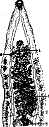
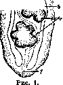
a member of the family Opisthorchidae, has a leaf-shaped body, tapering more in its anterior part, 8-13 mm long and 1.2-2 mm wide. The ventral sucker. (fig. 1) is somewhat smaller than the oral sucker, which reaches 0.25 mm in diameter. Characteristic is the location in the posterior part of the body of two lobed testes (6), between which the excretory canal (7) is bent in the shape of a Greek letter gamma, opening in the posterior part of the body. In front of the testes lies the seminal receptacle (4) and then the ovary (3); even further forward, winding between blindly ending intestinal loops (5), are the loops of the uterus (2), occupying the middle part of the parasite. At the same level, but outside the intestinal loops, are the grape-like vitellaria (2). The genital openings (male and female) lie in front of the ventral sucker, **1 f4^?^r; and to the male is connected a powerful, winding vas deferens (6). The eggs are very delicate, pale-yellow, provided with a lid, and on the opposite pole - a small conical thickening of the shell (fig. 2). Dimensions: 0.026-0.030 mm x 0.010-0.015 mm. Development of the parasite occurs with two intermediate hosts, of which the first is not precisely established; mollusks of the genus Dreissena and Limnaea stagnalis are suspected; the second intermediate host is various species of freshwater fish: roach, dace, bream, rudd. Infection of the definitive host occurs when eating raw fish or lightly boiled and fried salted or smoked fish infected with Opisth. felineus metacercariae. Definitive hosts besides humans are cats, dogs, pigs and some other mammals. Opisthorchis felineus is distributed in certain river basins of both the European and Asian parts of the USSR. This parasite is especially frequent in Siberia, where the Ob River basin represents a large focus of spread of O. among people and animals. Thus, according to data from the 70th Union Helminthological Expedition (K. I. Skryabin and V. P. Podyapolskaya), Voguls on the North Sosva River are infected on average in 89%, and adults even in 96%; similarly intensely infected in the area of Obdorsk and other small northern peoples (Khanty, Zyryans). Workers (Russians) of the Tobolsk and Obsk (Obdorsk area) canneries are infected in 86%. All the mentioned population groups become infected from eating raw fish. In these same areas
domestic carnivores are also very widely and intensely infected. In the upper reaches of the Ob River on the Tom River (Tomsk) and in the Omsk area, O. is also widespread in humans, but to a lesser extent. From other areas of the USSR, opisthorchiasis has been recorded in the basin of the North Dvina, in the North Caucasus region, in the lower Volga, in the Kazakh SSR, in Kharkov. Besides the USSR, this disease also occurs in Romania, northern Germany, Holland, France, on the Scandinavian Peninsula, Hungary, Italy, Japan. Localization of the parasite - the bile ducts of the liver, the gallbladder and less frequently - the pancreas. Occurs in numbers from a few specimens to several thousand. According to Askanazy, who worked extensively in this field, pathoanatomically cholangitis is noted; dilated bile ducts are filled with parasites and their eggs, surrounded by mucus. The histological picture consists of glandular proliferation of the walls of the bile ducts, cellular infiltration, partially with eosinophils. Similar phenomena occur in the pancreatic ducts, where hemorrhages and necroses are also noted. Later, cirrhotic processes develop, connective tissue proliferations in the liver, in some cases atypical and finally cancerous neoplasms from the side of the epithelium. Winogradoff noted in the gallbladder loose concretions with eggs of the trematode in the center, on which crystals of cholesterol and bile pigments were deposited.
* The clinical picture of O. is still poorly studied. With insignificant invasion, clinical manifestations may apparently be completely absent. With more intense invasion, constant dull pains in the liver and stomach are noted, which sometimes turn into severe pains in the right hypochondrium; usually there is a decrease or absence of appetite (less often its increase), sometimes disturbances in stool; yellowing of the sclera is noted or less often a more pronounced jaundice; sometimes ascites. In some cases, O. can proceed under the picture of chronic recurrent cholecystitis. - Diagnosis of O. can be established accurately only by finding Opisthorchis felineus eggs in the feces or in the bile, obtained from the duodenum by probing with a thin probe. In weaker invasions, the latter method is the most reliable.--Therapy of O. Has not been developed, and there is as yet no means that surely kills Opisthorchis felineus. The best of the tried means in O. is emetine. With its help, it is possible to achieve both a decrease in the number of eggs in the bile and sometimes a significant improvement in the general condition of the patient. Sometimes for a fairly long period after treatment, it is not possible to find Opisth. felineus (fig. 2) eggs either in the feces or in the bile. However, neither a persistent disappearance of eggs nor a complete disappearance of all clinical manifestations is usually achieved. Dosage of emetine: 3 cm3 of a 2% solution of hydrochloride of emeteine under the skin for 3 days in a row. After three-day intervals, repeat again. Treatment is long.-Preventive measures should consist of broad sanitary helminthological education and protection of water bodies from contamination by feces of humans, dogs, cats, pigs. The above animals should not be fed raw fish. For personal prevention, consumption of fish should be done exclusively after thorough thermal processing.

Related articles
Mentioned in
Cite this page
“Opisthorchiasis.” Soviet Medical Encyclopedia. English translation of Bolshaya Meditsinskaya Entsiklopediya, 1st ed. (Moscow, 1928–1936), ed. N. A. Semashko. https://sovietmedicalencyclopedia.pages.dev/article/opisthorchiasis/