Paragonimiasis
Historical document, translated for reference. It reflects medical knowledge of the 1920s–30s and is not medical advice.
Summary
Paragonimiasis is a disease primarily affecting the lungs, caused by the trematode Paragonimus Ringeri. The parasite has a complex life cycle involving two intermediate hosts, and while mainly found in East Asia, cases have been documented in the Soviet Union.
Encyclopedia entry (1928–1936)
PARAGONIMIASIS (paragonimosis), a disease mainly of the lungs, caused by the trematode Paragonimus Ringeri (Cobbold, 1880) (syn. Paragonimus Westermanni), belonging to the family Troglotrematidae Odhner, 1914. The parasite is 7.5 to 13 mm long with a rather thick, egg-shaped body, with an expanded posterior and somewhat narrowed anterior end. The diameter of the oral sucker is 0.75 mm, and that of the ventral sucker, located 4.3 mm from it, is 0.8 mm. The size of the pharynx is 0.4x0.3 mm, and the length of the esophagus is 0.3 mm. The genital openings are located behind the ventral sucker. Symmetrically arranged lobate testes are located in the posterior quarter of the body (Fig. 1).
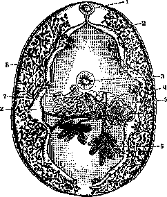
Fig. 1. Paragonimus Ringeri: 1- oral sucker; 2- intestine; 3- ventral sucker; 4- Mehlis' gland; 5- uterus; 6- testes; 7- ovary; 8- vitelline glands. The strongly lobate ovary lies in front of the testes. The vitelline glands occupy the lateral parts of the body. Bursa cirri is absent. The eggs are 0.0754-0.0884 x 0.0442-0.0546 mm, light brown with rounded poles, with an operculum on one of them (Fig. 2). Definitive hosts besides humans: pigs, dogs, cats, and some wild mammals. The normal localization is the lungs, where the parasite is located more often in pairs, rarely singly, in cysts the size of a walnut; these cysts are in direct connection with the small bronchi. More rarely, the parasite can be localized in the brain, liver, diaphragm, and other tissues. The main area of distribution of the parasite is East Asia, especially Japan, Formosa, the Philippine Islands, and Korea. On the territory of the USSR, several imported cases of paragonimiasis have been recorded. In 1928, the 60th Union Helminthological Expedition proved the existence of a local Far Eastern paragonimiasis, discovering Paragonimus Ringeri in the lungs of two cats (out of 46 examined) in the lower reaches of the Amur River (Nikolaevka-on-Amur) and finding eggs of this parasite in one Gilyak on Liangr Island (south of the Sea of Okhotsk). Fig. 2. Paragonimus Ringeri develops with the help of two intermediate hosts, of which the first is numerous species of mollusks of the genus Melania, and the second intermediate host is crabs of the genera Eriocheir, Potamon, and the crayfish Astacus japonicus and others. Within the Far East of the USSR, the existence of Eriocheir japonicus has been proven. The eggs of Paragonimus Ringeri, excreted into the external environment with sputum or when swallowed with feces, do not yet contain an embryo; for their maturation, they must enter a freshwater environment, in which over several weeks (from 16 to 60 days depending on the time of year, under the climatic conditions of Korea), a miracidium develops in the egg, opening the egg operculum and floating for some time in fresh water. Then, having penetrated into a mollusk, the miracidium sheds its ciliated cover, turning into a sporocyst, which usually forms one redia. The latter passes from the subcutaneous layer of the mollusk into the liver, where, by parthenogenetic reproduction, it gives rise to a generation of cercariae; the latter exit from the mollusk's body, again enter fresh water, actively bore into crabs, and, encysting in the muscle tissue of the abdomen, the muscles of the limbs, and sometimes in the liver, turn into metacercariae. When eaten raw or insufficiently cooked by the definitive host with a crab, the metacercariae enter the latter's intestinal tract. The digestive juices of this host dissolve the cyst and release the small parasite, which, in addition to suckers and digestive organs, is equipped with a special boring stylet, with the help of which it bores into the thickness of the intestine and penetrates into the abdominal cavity. By boring through the diaphragm, the young parasites pass from the abdominal cavity into the thoracic cavity, and then through the pulmonary pleura into the lung tissue and bronchioles. The maturation of metacercariae in the crab's body (i.e., the period of time from the moment of penetration of the cercaria into the crab until the metacercarium is able to continue development in the definitive host) occurs within 42-54 days. The transformation of the parasite in the human lungs into the sexually mature stage occurs 90 days after infection with the metacercarium. Clinical picture. In P., phenomena from the respiratory organs are mainly observed: cough with sputum, sometimes stained with blood; there can be independent pulmonary hemorrhages. The disease is characterized by a chronic, often multi-year course. Most often, P. is confused in clinical symptoms with tuberculosis of the lungs. Numerous cases of finding tumors or foci of softening in the brain with Paragonimus Ringeri and its eggs in Jacksonian epilepsy have been described. - Diagnosis of pulmonary P. is made mainly on the basis of finding parasite eggs in sputum (as well as in feces). Radiodiagnosis is also of great importance, as with the help of X-rays it is possible to prove the existence of small scattered foci of shadowing in the lung tissue (cysts with parasites). - The prognosis for pulmonary paragonimiasis is more or less favorable. Therapy. No specific remedy is available. Some authors point to emetine as a good therapeutic agent, however most consider it to be of little effectiveness. Prevention consists in consuming crabs and crayfish only in a thermally processed form.
Related articles
Cite this page
“Paragonimiasis.” Soviet Medical Encyclopedia. English translation of Bolshaya Meditsinskaya Entsiklopediya, 1st ed. (Moscow, 1928–1936), ed. N. A. Semashko. https://sovietmedicalencyclopedia.pages.dev/article/paragonimiasis/