Fascioliasis
Historical document, translated for reference. It reflects medical knowledge of the 1920s–30s and is not medical advice.
Summary
Fascioliasis is a helminthiasis of humans and other mammals caused by trematodes of the genus Fasciola, including Fasciola hepatica and Fasciola gigantica. The article provides detailed descriptions of these parasites, their characteristics, synonyms, and includes a table of fasciculi with their anatomical locations and functions.
Encyclopedia article (1928–1936)
FASCIOLIASIS, helminthiasis of humans and other mammals, caused by trematodes of the genus Fasciola L., 1758 and family Fasciolidae Railliet, 1895: 1) Fasciola hepatica L., 1758 and 2) Fasciola gigantica Cobbold, 1856. 1. Fasciola hepatica (figs. 1 and 2). Synonyms: Distomum hepaticum Retzius, 1786; Fasciola humana Gmelin, 1789; Distomum caviae Sonsino, 1890; Cladocoelium hepaticum Stossich, 1892 and others. Brief characteristics. Large trematodes up to 30 mm in length and up to 12 mm in width. The cuticle is armed with spines. The diameter of the oral sucker is 1.0 mm, the ventral one is 1.6 mm. The eggs are large, 0.111-0.151 (most often 0.126-0.140) mm in length and 0.063-0.078 (most often 0.066-0.072) mm in width. Table of individual fasciculi, s. tractus, s. fibrae. 0< Name Where it begins Where it passes Where it ends Functional value In which article is it mentioned Fasciculus anterior proprius (Flechsig) In the gray matter of the spinal cord In the anterior columns of the spinal cord In the gray matter of the spinal cord, in segments lying above and below Association tract of the spinal cord Spinal cord Tractus antero-lateralis superficialis (Gowersi) See fasciculus Gowersi Fibrae arcuatae externae anteriores, s. ventrales In the arciform nucleus, partially formed by Flechsig's bundle On the anterolateral surface of the medulla oblongata They form part of the restiformis and together with it end in the cerebellum (vermis) Projection fibers, efferent system of the cerebellum Medulla oblongata, Cerebellum Fibrae arcuatae internae In the nuclei gracilis and cuneatus of the medulla oblongata In the medulla oblongata, around the central canal After the decussation, they pass into the medial lemniscus Projection fibers, second sensory neuron Sensitivity, Medulla oblongata Fibrae arcuatae posteriores, s. dorsales In the nuclei gracilis and cuneatus On the posterior surface of the medulla oblongata They form part of the restiformis and together with it end in the cerebellum (vermis) Projection fibers, afferent system of the cerebellum Medulla oblongata, Cerebellum Fasciculus arcuatus (Fo-Tille) * See fasciculus obliquus pontis ? Fasciculus arcuatus, s. fasciculus longitudinalis superior In the cells of the frontal lobe cortex Parallel to the upper edge of Reil's insula In the cortex of the parietal and temporal lobes Association tract Association fibers 8 9 Fasciculus Arnoldi See fasciculus temporo-thalamicus Tractus bulbo-cerebellaris lateralis In the cells of the lateral column nucleus of the medulla oblongata In the medulla oblongata They form part of the restiformis Projection tract, afferent system of the cerebellum to Tractus bulbo-cerebellaris posterior In the cells of the external cuneate nucleus Tracto bulbo-thalamicus, s. lemniscus medialis In the cells of the gracilis nucleus and cuneate nucleus in the medulla oblongata After the decussation, the fibers pass in the trunk and in the cerebral peduncle In the external nuclei of the thalamus opticus Projection tract, second sensory neuron Sensitivity, Thalamus opticus Fasciculus Burdachi In the cells of the intervertebral ganglia In the posterior columns of the spinal cord It passes into the fasciculus cuneatus of the medulla oblongata Projection tract. Conducts deep sensitivity ; Burdach's bundle, Medulla oblongata № Name Where it begins Where it passes Where it ends Functional value In which article is it mentioned Fibrae caudo-thalamicae In the cells of the caudate nucleus Through the internal capsule In the cells of the anterior parts of the thalamus opticus Projection fibers Thalamus opticus 15' Fasciculus cerebello-bulbaris, s. fastigio-bulbaris, s. tr. uncinatus, hook-shaped bundle From the cells of the cerebellar nuclei - nucleus fastigii and possibly nucleus globosus The fibers pass over the brachium conjunctivum and enter the inner part of the inferior cerebellar peduncle In the nucleus Deiters, partially in the nuclei of motor cranial nerves Projection tract, efferent system of the cerebellum Cerebellum Fasciculus cerebello-spinalis, s. spino-cerebellaris See fasciculus Flechsigi Fasciculus cerebello-tegmentalis mesencephali In the cells of the dentate nucleus and nucleus fastigii As part of the brachium conjunctivum In the red nucleus Projection fibers, efferent system of the cerebellum Cerebellum Fibrae cerebello-vestibulares In the cells of the fastigial nucleus of the cerebellum Through the inner part of the inferior cerebellar peduncle In the vestibular nuclei Projection fibers, efferent system of the cerebellum Cerebellum Fasciculus cerebro-spinalis anterior See fasciculus cortico-spinalis anterior Fasciculus cerebro-spinalis lateralis See fasciculus cortico-spinalis lateralis Fasciculus cervico-lumbaris dorsalis In the cells of the gray matter of the cervical part of the spinal cord In the posterior columns of the spinal cord In the gray matter of the spinal cord in the sacral region Association descending tract Spinal cord Fasciculus cortico-bulbaris See fasciculus cortico-nuclearis Tractus cortico-habenularis From the pyramidal cells of Ammon's horn and from the cells of the fascia dentata As part of the fornix, and then in the striae medullares In the habenular ganglion Projection-association fibers Fornix Fibrae cortico-luysianae (existence is disputed) In the cells of the cerebral cortex Through the internal capsule In the corpus Luysi Projection fibers, transmitting the influence of the cerebral cortex Corpus Luysi Tractus cortico-mamillaris From the pyramidal cells of Ammon's horn and from the cells of the fascia dentata As part of the fornix The majority of fibers in the corp. mamillare in turn, a smaller part in the opposite Projection-association fibers; belongs to the olfactory system Fornix J* Name Where it begins Where it passes Where it ends Functional value V men-tionedinthear-ticle 25 26 Fibrae cortico-nigrae In the cells of the anterior central gyrus Through the internal capsule and cerebral peduncle In the ventral parts of the substantia nigra Projection fibers, transmitting the influence of the cerebral cortex Substantia nigra Fasciculus cortico-nuclearis, s. fasc. geniculatus In the cells of the anterior central gyrus, in its lower third Through the genu of the internal capsule, pes pedunculi In the motor cranial nerve nuclei located in the brainstem Projection tract, transmits the motor impulse from the cortex to the nuclei of the cranial nerves Pyramidal system Fasciculus cortico-pontinus In the cells of the frontal, temporal and occipital lobes Through the internal capsule, pes pedunculi (where it occupies the inner and lateral parts) In the nuclei of the pons, where the middle cerebellar peduncles originate Projection fibers, transmit the impulse from the brain to the cerebellum through the pontine nuclei Pons, Cerebellum Tractus cortico-spinalis anterior, s. cerebro-spinalis ant., s. pyramidalis anterior In the cells of the anterior central gyrus Through the internal capsule, brainstem, in the anterior column of the spinal cord In the anterior horns of the spinal cord Projection tract, transmits the motor impulse to the cells of the anterior horns Pyramidal system 30 31 Tractus cortico-spinalis lateralis, s. cerebro-spinalis lateralis, s. pyramidalis lateralis Through the internal capsule, brainstem, after the decussation in the lateral column of the spinal cord In the anterior horns of the spinal cord Fibrae cortico-striales (existence is disputed) In the cells of the cerebral cortex In the internal capsule In the globus pallidus according to some authors, in the putamen according to others Projection fibers, transmitting the influence of the cerebral cortex Basal ganglia Tractus cortico-tectalis In the cells of the occipital and other lobes of the brain Through different parts of the internal capsule and through the brachium of the quadrigeminal body In the cells of the anterior quadrigeminal body Projection fibers, transmit the impulse from the cortex to the quadrigeminal body Corpora quadrigemina Tractus cortico-tegmentalis In the cells of the cortex 1) occipital lobe, 2) temporal and 3) frontal 1) In the retrolenticular part of the internal capsule, 2) in its sublenticular part, 3) in its anterior crus 1) In the anterior quadrigeminal body, 2) in the posterior and 3) in the red nucleus Projection fibers Visual pathways, Auditory pathways, Frontal lobe 33 34 Tractus cortico-thalamicus In the cells of the cortex of various areas of the brain In the peduncles of the thalamus opticus In the cells of various parts of the thalamus opticus Projection fibers, transmit to the thalamus opticus the influence of the cortex Thalamus opticus, Brain Fasciculus cuneatus It is a continuation of the fasciculus Burdachi (see) In the posterior columns of the medulla oblongata In the cuneate nucleus, which appears among its fibers Projection tract, conducts deep sensitivity Medulla oblongata Fasciculus fastigio-bulbaris See fasciculus cerebello-bulbarts № ;
Name Where it begins Where it passes Where it ends Functional value In which article is mentioned Fasciculus Flechsigi, s. epino-cerebellaris dorsalis In the cells of Clark's column, located in the posterior horns of the spinal cord In the lateral columns of the spinal cord and medulla oblongata, and then enters corpus restiforme In the cortex of vermis on its own and opposite sides Projection bundle, transmits irritation from the periphery to the cerebellum Flexig's bundle Fasciculus Foreli See fasciculus lenticularis and fasciculus thalamicus Fasciculus Foville'n See fasciculus obliquus pontis Fasciculus fronto-centralis In the cells of the cortex of the frontal lobe of the brain In centrum semiovale In the cortex of the central gyri Association bundle 40 41 Fasciculus fronto-occipitalis At the outer edge7 of the lateral ventricle In the cortex of the occipital lobe, partially in nucl. caudatus Association bundle Association fibers Fasciculus geniculatus See fasciculus cortico-nuclearis Fasciculus Golli In the cells of the spinal ganglia In the posterior columns of the spinal cord Passes into fasc. gracilis Projection bundle; first neuron conducting deep sensitivity Golli's bundle Fasciculus Gowersi In the cells of the posterior horn of the spinal cord In the lateral column of the spinal cord and trunk on the opposite side In the cortex of vermis Projection bundle, afferent system of the cerebellum, transmits irritations from the periphery to the cerebellum Gowers' bundle Fasciculus gracilis Is a continuation of fasc. Golli In the posterior columns of the medulla oblongata In nucl. gracilis Projection bundle, first sensory neuron Medulla oblongata Fasciculus Gudden'a See fasciculus mamillo-tegmentalis Fasciculus Ш See fasciculus thalamicus Fasciculus Ha See fasciculus lenticularis Tractus habenulo-peduncularis See fasciculus retroflexus Meynerti Fasciculus Held'a In nucleus ventralis n. cochleae In the most posterior parts of corp. trapezoid. in the pons Varoli Enters the composition of lemnisci lateralis, together with it ends in primary auditory centers Projection bundle, enters the composition of the 2nd auditory neuron Auditory conducting pathways Name Where it begins Where it passes Where it ends Functional value In which article is mentioned Fasciculus Helweg, s. fasc. olivo-spinalis, s. fasc spino-olivaris, s. fasc. periolivaris; paraolivaris; synonym triangular bundle Not clearly established in which direction it goes - descending or ascending; begins either in oliva inferior or in the gray matter of the spinal cord At the border of the anterior-lateral column of the spinal cord peripherally, well expressed in the cervical region In the gray matter of the spinal cord, or conversely, in oliva inferior Projection bundle; apparently has relation to the statics of the head Spinal cord, Medulla oblongata, Helweg's bundle Fasciculus Hoche See fasciculus septo-marginalis Fasciculus Inferior pontis In the cells of the nuclei of the pons Transverse fibers in the lower part of pons Varoli In the cortex of the cerebellar hemispheres Projection fibers; afferent system of the cerebellum Fibrae interluysianae Fibers connect both corpora Luysi, pass in the middle parts of commissurae subopticae poster. Foreli Commissural fibers Corpus Luysi Fibrae interpedunculares, In the cells of substantiae nigrae, nucl. ruber, nucleus of Darkshevich, nucl. interstitialis In the composition of commissurae poster. near aquaeductus Sylvii In thalamus, in hypothalamus and in the corresponding nuclei Fibrae interpulvinares Fibers connect both pulvinar, pass in the posterior parts of commissurae poster. Commissural fibers Fasciculus lateralis plexus brachialis, s. fasc. superior Formed by the anterior branches of the first two primary trunks of the plexus In the axillary fossa Continues partially in n. medianus and n: musculo-cutaneus Projection peripheral bundle Brachial plexus Fasciculus lenticularis (Foreli), s. fasc. H2 In the cells of nuclei lenticularis (globus pallidus) In the intermediate brain, through commis. Foreli passes to the opposite side In nucl. ruber, corp. Luysi, zona incerta, nucl. periventricularis of its own and opposite sides Projection bundle, transmitting impulse from the striatal system to a number of autonomic centers Regio subthalamica, Extrapyramidal system Fasciculus longitudinalis inferior In the cortex of the temporal lobe Along the outer wall of the inferior and posterior horns of the lateral ventricle In the cortex of the occipital lobe Association bundle Association fibers 59 60 61 Fasciculus longitudinalis posterior, s. medialis, s. dorsalis (Schulz) From various cellular groups of the brain stem In the brain stem along the floor of the IV ventricle and in the anterior columns of the spinal cord In the anterior horns of the spinal cord Projection bundle, efferent pathway of labyrinthine reflexes and other posture reflexes Fasciculus longitudinalis posterior Fasciculus longitudinalis praedorsalis See fasciculus tecto-spinalis Fasciculus longitudinalis superior See fasciculus arcuatus Name Where it begins Where it passes Where it ends Functional value In which article is mentioned Fasciculus LOwenthali, a. vestibulo-spinalis; synonym anterior marginal bundle In the cells of nucl. Deitersl In the anterior parts of the lateral column of the medulla oblongata; peripherally in the anterior column of the spinal cord between the exit of the anterior roots In the anterior horns of the spinal cord Projection bundle; efferent bundle of the extrapyramidal system Lenthal's bundle, Spinal cord, Cerebellum Tractus mamillo-tegmentalis, s. mamillaris princeps, s. fasc. tegmenti-(Gudden) In the cells of nucl. interni corp. mamill. Through the cerebral peduncle In the pons Varoli in nuclei tegmenti dorsalis et ventralis (Gudden) Projection fibers, have relation to the olfactory system Regio subthalamica, Corpora mamillaria Fasciculus mamillo-thalamicus, s. fasc. Vicq d'Azyr'a In the cells of nucl. interni corp. mamill. In regio subthalamica In nucl. anterior thalami optici Projection bundle, has relation to the olfactory system Corpora mamillaria, Regio Subthalamica Fasciculus Marie et Leri; synonym residual bundle, angular bundle Moeli In gangl. opticum basale, s. nucl. supraopticus In tractus opticus, along its outer edge In the retina Projection bundle Regio subthalamica Fasciculus medialis plexus brachialis Is a continuation of the anterior branch of the third primary trunk of the brachial plexus In the axillary fossa Continues partially in n. medianus, in nn. ulnaris, cutaneus brachii medialis, cutaneus antibrachii medialis Projection peripheral bundle Brachial plexus Fasciculus medius pontis See fasciculus obliquus pontis Fasciculus Meynerti See fasciculus retroflexus Fasciculus Monakowi, s. fasc. rubro-spinalis In the large cells of nucleus rubri forms a cross Forel'H In the lateral columns of the pons Varoli, medulla oblongata and spinal cord In the cells of the anterior horns of the spinal cord Projection bundle, efferent bundle of the extrapyramidal system Spinal cord, Cerebellum, Nucleus ruber Fasciculus Miinzer'a See fasciculus, s. tractus tecto-bulbaris et tecto-pontinus Fasciculus nuclei caudati In the cells of the gyri lying above corpus callosum Under the corpus callosum, under the ependyma of the ventricle In nucl. caudatus Projection bundle Basal ganglia Fibrae nuclei caudati In the cells of nucleus caudati Through capsula interna In nucl. lenticularis, thalamus opticus, regio subthalamica Projection fibers Fasciculus obliquus pontis, s. medius pontis, s. fasc. arcuatus Foville'a In the cells of nuclei located at the base of the pons Varoli Transverse bundle of the pons Varoli, goes in an oblique direction Ends in the cortex of the cerebellar hemispheres Projection bundle, afferent bundle of the cerebellum Pons Varoli, Cerebellum Ju Name Where it begins Where it passes Where it ends Functional value In which article is mentioned Fasciculus occipitalis transversus cunei In the cells of the cortex of the upper lip of fis. calcarinae In the white matter of the tem-poral lobe In the cortex of the gyri on the outer surface of the occipital lobe Association bundle Association fibers Fasciculus occipitalis transversus lingualis In the cells of the cortex of the lower lip of fis. calcarinae Fasciculus occipitalis verticalis In the cells of the cortex of occipital and temporal gyri on the lower surface In the white matter of the occipital lobe in a vertical direction In the cortex of occipital gyri on the outer surface and in the lower parietal gyri Association bundle Occipital lobe Tractus olfacto-ammonicus Zuckerkandli In the cells of substantia perforatae anter. Goes in an ascending direction as part of fornix, partially crosses In the cortex of cornu Ammonis Projection-association bundle, relating to the olfactory system Olfaction Tractus olfacto-habenularis In the cells of bulbi olfactorii In tractus olfactorius, and then in striae medullares In the cells of gangl. habenulae.
Tractus olfacto-mesence-phalicus (Wallenberg's) From the plexuses of primary olfactory centers Forms the olfaction radiation In tuber clnereum, corpus mamillare, substantia reticul. tegmenti Projection bundle, basal olfactory bundle Tractus olfactorius In the cells of the olfactory bulb On the lower surface of the brain along the olfactory fissure Passes into the trigonum olfactorium Projection bundle, second olfactory bundle Tractus olivo-cerebellaris In the cells of the inferior olive of the medulla oblongata In the medulla oblongata partially crosses and enters the corpus restiforme of its own and opposite side In vermis cerebelli Projection bundle; enters the afferent system of the cerebellum Cerebellum, Medulla oblongata Fasciculus opticus, s. tractus opticus Continuation of the optic nerve after the chiasm At the base of the brain In the primary visual centers Projection bundle, first visual neuron Visual centers and fibers Tractus pallido-infundibularis In the cells of the globus pallidus Passes through the diencephalon between ansa lenticularis and tractus opticus In the lower part of nuclei mamillo-infundibularis and nucl. pallido-infundibularis Projection bundle, belonging to the vegetative system Regio subthalamica, Vegetative nervous system Fasciculus pallidus In the cells of the globus pallidus Through the internal capsule In thalamus opticus, regio * subthalamica Projection bundle, connecting gl. pallidus with thalamus and with vegetative centers Regio subthalamica Fasciculus paraolivaris See fasciculus Helweg's № Name Where it starts Where it passes Where it ends Functional value In which article is mentioned 86 87 Fasciculus parieto-centralis In the cells of the parietal lobe cortex In the white matter of the brain In the cortex of the central sulci Association bundle Fasciculus parieto-occipitalis pedunculi In the cells of the occipital and parietal lobe cortex Through capsula interna and the outer part of the pedunculus In the nuclei of the oculomotor nerves and in the nuclei of the pons Projection bundle Brain-peduncular fibers Tractus peduncularis transversus In the cells of the retina of the eye Along the lateral surface of the cerebral crus Outside and behind corpus mamillare Projection bundle - Fibrae perforantes corporis callosi Exit from tr. olfacto-am-monicus Perforate the corpus callosum Enter into the composition of striae longitudinalis and end in the Ammon's horn Projection bundle Corpus callosum Fasciculus periolivaris See fasciculus Helweg's Fibrae pontis, s. taenia pontis In the cells of sulci mesencephali lateralis Extends along the anterior edge of the Varolius bridge In sulci nervi oculomotorii Projection fibers, the most anterior bundles of the cerebellar peduncles to the pons Varolius bridge, Cerebellum Tractus ponto-cerebellaris In the nuclei of the base of the Varolius bridge Cross and enter into the composition of the middle cerebellar peduncles In the cortex of the cerebellar hemispheres Projection bundle, afferent system of the cerebellum Cerebellum Tractus posterior plexus brachialis From the posterior branches of three primary trunks In the axillary region Continues in the p. radians Projection peripheral bundle Brachial plexus 94 95 98 97 Fasciculus praedorsalis See fasciculus, s. tractus tecto-spinalis Fasciculus praepyramidalis Thomas In the cells of substantia reticularis of the medulla oblongata In the lateral column of the spinal cord, in front of the pyramidal tract, parallel to the anterior horn In the cells of the anterior horn of cervical segments Association bundle between the respiratory centers of the spinal cord and medulla oblongata Spinal cord, Medulla oblongata Fasciculi primarli Form from the roots CV-CVIII and DT In the subclavian fossa Divides into three secondary trunks Projection peripheral bundle Brachial plexus Fibrae propriae gyrorum (Meynerti), s. librae, arcuatae Arnoldi In the cells of the cortex on the walls of the cerebral convolutions On the floor of the grooves dividing the convolutions In the cortex of the neighboring convolution, on its wall Association fibers Association fibers Fasciculus proprius dorsalis; syn. main bundle of the posterior column In the cells of the posterior horn of the spinal cord In the anterior parts of the posterior column In the posterior horn Association bundle Spinal cord № Name Where it starts Where it passes Where it ends Functional value In which article is mentioned 101 102 103 Fasciculus proprius lateralis; syn. main bundle of the lateral column In the cells of the posterior, lateral and anterior horns of the spinal cord In the internal parts of the lateral column In the posterior, lateral and anterior horns Association bundle Spinal cord Fasciculus proprius ventralis; syn. main bundle of the anterior column In the cells of the anterior horns of the spinal cord Immediately near the anterior horn In the anterior horn Fasciculus pyramidalis anterior See fasciculus cortico-spinalis anterior Fasciculus pyramidalis lateralis See fasciculus cortico-spinalis lateralis Fasciculus reticulo-spinalis lateralis In the cells of substantia reticularis of the brain stem of the opposite side By scattered fibers go in the lateral column of the spinal cord In the anterior horns of the spinal cord Projection bundle, efferent bundle of the extra-pyramidal system Spinal cord Fasciculus reticulo-spinalis ventralis In the cells of substantia reticularis of the brain stem By scattered fibers in the anterior column of the spinal cord Fasciculus retrollexus Meynerti In the cells of gangl. habenulae In the midbrain along the inner edge of nucl. rubri, then bends back After the chiasm, in the cells of nucl. interpeduncularis and substantiae perforatae poster. Projection bundle, efferent system of both-avoiding reflexes Regio subthalamica, Rhinencephalon 106 107 Fasciculus rubro-bulbaris In the upper parts of nucl. rubri Goes with fasc. Monakowi In the nucleus of the lateral column of the medulla oblongata Projection bundle, afferent bundle of the extra-pyramidal system Extrapyramidal system Fasciculus rubro-reticularis In the small cells of nucleus ruber In brachium conjunctivum, participating in its chiasm In substantla reticularis pons Varoli, its continuation is served by fasc. reticulo-spinalis Fasciculus rubro-spinalis See fasciculus Monakow's Fasciculus septo-marginalis, s. fasc. Hoche In the cells of the spinal cord 1 In the cervical and thoracic seg- 1 In the gray matter of the spin- 1 Association bundle of the spinal cord ments of the posterior columns cord of the brain Spinal cord Fasciculus septo-thalamicus In the cells of septi pellucidi Passes under coramissura [ To the anterior parts tha-anterior | of thalami optici, where it enters | taenia thalami Projection bundle, afferent bundle of thalami optici - 111 112 113 Name : Where it starts Where it passes Where it ends Functional value In which article is mentioned Tractus solitarius In the ganglia of the vagus nerve and glossopharyngeus nerve In the medulla oblongata, near the floor of the IV ventricle In nucl. terminalis, surrounding fasc. solitarius Projection bundle; relates to the taste system Taste, Vagus nervus, Glosso-pharyngeus nervus Tractus spinalis n. V. In the cells of Gasser's ganglion In the lateral column of the trunk and spinal cord (cervical part) In the cells of substantia gelatinosa Rolandi Projection bundle, relates to the sensory system Trigeminus nervus Fasciculus spino-bulbaris dorsalis, s. posterior In the cells of the gray matter of the spinal cord Goes in the posterior column of the spinal cord, partially crosses In the nuclei of the medulla oblongata Projection bundle, partial conduction of tactile sense is assumed Spinal cord, Medulla oblongata 115 Fasciculus spino-bulbaris ventralis In the cells of the gray matter of the spinal cord Goes in the anterior column of the spinal cord together with fasc.sulco-marginalis ascen-dens In the nuclei of the medulla oblongata Projection bundle, connecting the SPINAL CORD with the lower part of the medulla oblongata Spinal cord, Medulla oblongata Fasciculus spino-cerebellaris dorsalis See fasciculus Flechsigi Fasciculus spino-cerebellaris ventralis See fasciculus Gowersi Fasciculus spino-olivaris See fasciculus Helweg's 118 120 Fasciculus spino-tectalis In the cells of the posterior horns of the spinal cord After the chiasm goes in the composition of fasc. Gowersi and lemnisci lateralis In the posterior quadrigemina, and partly in the anterior Projection bundle, afferent bundle of the sensory-reflex subcortical center of Gowers Gowers' bundle, Corpora quadrigemina Fasciculus spino-thalamicus In the cells of the posterior horn of the spinal cord After the chiasm goes in the composition of fasc. Gowersi, and then in lemniscus lateralis In the latero-ventral nucleus of thalami optici Projection bundle, second sensory neuron Sensitivity, Spinal cord, Gowers' bundle Fibrae strio-hypothalami-cae In the cells of nucl. lenticula-ris-globus pallidus Through capsula interna In regio subthalamica Projection fibers, efferent system of the striatum Regio subthalamica Fibrae strio-luysianae In the cells of globus pallidi Through capsula interna In corp.
Luysi in its outer parts Corpus Luysi Fibrae strio-nigrae In the cells of the apex and lower side of the globus pallidus In substantia nigra Substantia nigra. № Name Where it begins Where it passes Where it ends Functional value In which article is it mentioned Fibrae strio-rubrae In the cells of the apex and lower side of the globus pallidus Through the internal capsule In the red nucleus Projection fibers, efferent system of the striatum Nucleus ruber Fibrae strio-thalamicae In the cells of the lenticular nucleus-globus pallidus Through the internal capsule In the ventral part of the external nucleus of the thalamus Projection fibers Basal ganglia 125 126 Fasciculus subcallosus, s. stratum subcallosum In the cells of the cortex of the gyri lying above the corpus callosum, as well as formed by collaterals from the fasciculus of the caudate nucleus Under the corpus callosum in the outer angle of the ventricle In the cortex of the gyri lying under the corpus callosum, partially passes into the tapetum Association-projection bundle Brain Fasciculus sulco-marginalis In the adhesive cells of the spinal cord After the decussation, it lies along the anterior sulcus of the spinal cord and along the periphery of the anterior column In the optic thalamus, partially in the medulla oblongata (fasciculus spino-bulbaris ventralis) Projection bundle, efferent system of the thalamus optici Spinal cord Fibrae superiores pontis In the pontine nucleus In the upper parts of the Varolian pons along the anterior surface They enter the composition of the middle cerebellar peduncle and together with it ends in the cerebellum Transmission of impulses from the cerebral cortex to the cerebellum - Tractus tecto-bulbaris Miinzeri In the cells of the superior colliculi of the quadrigeminal body, partially in the inferior Together with the tractus tecto-spinalis through the stem part of the brain In the nuclei of the medulla oblongata Projection bundle, optical-acoustic reflex pathway 129 130 Tractus tecto-cerebellaris In the cells of the quadrigeminal body The path is not precisely clarified In the cortex of the cerebellum Projection bundle j Tractus tecto-pontinus (Miinzer), s. tecto-protrudentialis Goes along the periphery of the tegmentum of the cerebral peduncle In the nuclei of the base of the pons near the pyramidal bundle Corpora quadrigemina Tractus tecto-reticularis In the cells of the quadrigeminal body, mainly the anterior Goes together with the previous one along the periphery of the tegmentum of the cerebral peduncle In the reticular substance of the Varolian pons and medulla oblongata - Corpora quadrigemina 13! Tractus tecto-spinalis, s. praedorsalis, g. longitudinalis praedorsalis In the cells of the quadrigeminal body, mainly the anterior, forms Meynert's decussation Passes through the trunk, through the anterior lateral funiculus of the spinal cord In the cells of the anterior horns of the spinal cord Projection bundle Spinal cord, Corpora quadrigemina 3 Tractus tegmento-mamillaris, s. pedunculus mamillaris In the cells of the tegmentum of the Varolian pons Together with the fibers of the main loop passes through the midbrain in front of the decussation of the brachium conjunctivum In the nuclei of the corpora mammillaria on the contrary, opposite side; part of the fibers ends in the tuber cinereum Association bundle Corpora mammillara Jfi Name Where it begins Where it passes Fasciculus temporo-pontinus, я. Тазе. Turdci Fasciculi temporo-thalamici Arnoldi ! JS1 14! Tractus thalamo-corticalis In the cells of the cortex of the temporal gyri In the cells of the cortex of the temporal gyri In the cells of various parts of the thalamus optici Tractus thalamo-habenularis Tractus thalamo-mammillaris Through the sublenticular part of the internal capsule and through the outer part of the cerebral peduncle Passes in the inferior peduncle of the thalamus opticus Where it ends Functional value In the nuclei of the base of the pons In the cells of the pulvinar part Through the peduncles of the thalamus optici, forming the corona radiata thalami In the cells of the zona thalami optici In the composition of the stria medullaris In the cortex of the cerebral hemispheres In the cells of the habenular nucleus Projection fibers, connecting the cerebral cortex with the cerebellum through the nuclei of the pons Projection bundle. Transmits regulatory and inhibitory influence of the temporal lobe on the pulvinar Projection bundle. Transmits to the cerebral cortex the irritations it has received In which article is mentioned Cortico-pontine fibers Thalamus opticus Thalamus optici Association bundle See fasciculus, s. tractus mammillo-thalamicus Tractus thalamo-olivaris In the cells of the internal nucleus of the thalamus optici Traelus thalamo-reticularis | In the cells of the internal nucleus of the thalamus optici Fasciculus thalamicus (Foreli), s. fasc. H1 In the periventricular nuclei of the diencephalon Enters the composition of the fasciculus centralis tegmenti Through the reticular substance of the midbrain Passes in the diencephalon above the incerta Ends in the cells of the inferior olive of the medulla oblongata In the reticular nucleus and in the central nuclei of the tegmentum of the pons In the lenticular nucleus, partially in the thalamus opticus Projection bundle, efferent extrapyramidal system Projection bundle, efferent extrapyramidal system, its continuation is the fasciculus reticulo-spinalis Projection bundle Thalamus opticus, Medulla oblongata Thalamus opticus Regio subthalamica Л"9 142 143 Name Where it begins Where it passes Where it ends Functional value In which article is mentioned Fasciculus Thomas See fasciculus praepyramidalis Fasciculus tuberis cinerei In the gray matter of the tuber cinereum Directed upward and backward and crosses with the bundle of the opposite side In the central gray matter of the aquaeductus Sylvii, internal nucleus of the thalamus optici and in the infundibulum Association bundle Regio subthalamica, Vegetative nervous system Fasciculus Tiircki See fasciculus temporo-pontinus Fasciculus uncinafus; syn. hook-shaped bundle In the cortex of the lower part of the frontal lobe At the level of the insula Reili threshold In the cortex of the temporal pole Association long bundle Association fibers i 147 14S 149 Fasciculus verticalis See fasciculus occipito-verticalis Fibrae vestibulo-cerebellares In the cells of the vestibular nucleus Through the inner part of the inferior cerebellar peduncle In the vermis of the cerebellum Projection fibers, afferent system of the cerebellum Cerebellum Fibrae vestibulo-oculomotoriae In the cells of the vestibular nucleus Through the fasciculus longitudinalis posterior To the nuclei of the oculomotorius, abducentis, trochlearis nerves Projection fibers Oculomotorius nervus Fasciculus vestibulo-spinalis, s. fasc. Luwenthali; syn. bundle of Luenthal See fasciculus Lowentnali Fasciculus Vicq d'Azyr'a, s. fasc. mammillo-thalamicus See fasciculus mammillo-thalamicus Fasciculus Wallenberg'a See fasciculus olfacto-mesencephalicus £81 width, yellow or yellow-brown in color, with a thin smooth shell, a lid on one pole and a flat tubercle on the other (fig. 3). Definitive hosts are most often herbivorous animals (sheep, goat, cattle, camel, horse, reindeer), rodents (hare, rabbit, squirrel, nutria, guinea pig, beaver, porcupine) and man. Localization-biliary ducts of the liver. Intermediate hosts: mollusks, mainly of the genus Limnaea-L. truncatula and others (fig. 4). Distributed in all parts of the world. 2. Fasciola gigantica (fig. 1 a) differs from F. hepatica in size (up to 75 mm in length) and body shape-the lateral edges of the body are almost parallel. Eggs 0.150-0.190 mm in length and 0.075-0.090 mm in width. Definitive
1! hosts-herbivorous and~ mammals and man. Intermediate hosts-moll
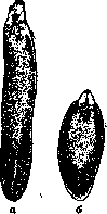
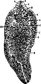
Figure 1.
Figure 2. Figure 1. Liver flukes of man: a-Fasciola gigantica; б-Fasciola hepatica. Figure 2. Fasciola hepatica: 1 -oral sucker; 2-genital pore; 3-intestine; 4-uterus; 5-ovary; 6-Mehlis' body; 7 and 9-seminal vesicles; S-yolk glands; 10-yolk ducts; 11-ventral sucker; 12-bursa cirri; 13- cirrus. usks of Limnaea natalensis and others. The development cycle (fig. 5) occurs with a change of hosts; in definitive hosts (mammals) fasciolas in the biliary ducts of the liver lay eggs, which are excreted into the intestine along with bile, and from the latter with feces - into the external environment. Within 2-4 weeks, a miracidium matures in the egg, which, when the eggs get into water, hatches into the light. The miracidium - penetrates into the body of the intermediate host (mollusk), in which further development takes place (sporocyst, redia, daughter redia, cercaria, see Trematodes), which is completed in 45-70 days. Then cercariae exit from the mollusks into the water, where they encyst, attaching to underwater objects (vegetation, etc.) or remaining free in the water (on the surface, on the bottom). This stage of development is called adolescaria. Infection of animals and man occurs by swallowing adolescariae with water or grass. The adolescarium that has entered the digestive tract of a mammal penetrates into the thickness of the intestinal wall and penetrates into the liver either hematogenously or by migration of fasciola larvae into the liver through the abdominal cavity. For the development of fasciolas in the definitive host to the sexually mature state, 10-14 weeks are required.-The pathogenic effect of fasciolas is manifested in the form of mechanical and toxic strokes. The first is produced by young fasciolas during their migration in the liver parenchyma, as a result of which extensive or less extensive destructions are observed
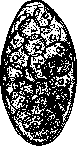

Figure 3.
Figure 4. Figure 3. Egg of Fasciola hepatica. (After Skryabin and Schultz.) Figure 4. The small pond snail-Limnaea truncatula-intermediate host of the fasciola. of tissues; similar phenomena may also be observed in other organs. Upon penetration into the bile ducts, the parasites cause trauma to the walls with the spines covering the cuticle; when accumulated in large numbers, fasciola obstruct the bile ducts and ducts. At the same time, there is also a toxic effect from the toxic substances secreted. Migrating fasciola usually carry representatives of the microbial flora, which can complicate the main process. In humans, F. is quite rare, with only about 200 cases described in the literature, half of which were recorded in the USSR. However, some of those cases, the diagnosis of which is based on coprological research, as determined by Vogel (1922), Strom (1927) and others, belong to the so-called false fascioliasis, in which eggs of fasciola contained in eaten products (liver in various forms) are found in the feces. Symptoms of F.: enlarged liver, colicky pains, nausea, sometimes vomiting, dyspeptic phenomena, mild jaundice, irregular increases in temperature, chills, sweats, dizziness, headaches, increased nervous excitability. In severe cases, shortness of breath, progressive exhaustion and fatal outcome are observed. Sometimes there is "aberrant localization" of fasciola: in the eye, in the subcutaneous tissue, in the lungs, in the blood vessels, etc. Diagnosis can be made by finding the parasite's eggs in the feces, duodenal contents (taking into account the possibility of finding "transit eggs" from eaten fasciola-infected livers).- Therapy in veterinary practice is carried out with carbon tetrachloride, hexachloroethane and preparations of the rhizome of male fern. Emetin has been successfully tested on humans (Vasil'eva, 1927; Semenov and Kogan, 1927; Kirikov, 1927; Kryukov 1927).- Public prevention consists primarily in organized control of F. of animals, which are the main carriers of fasciola: land reclamation, chemical control of mollusks-intermediate hosts, strict rejection and destruction of affected livers, deworming of animals. Personal prevention of humans: abstaining from drinking raw water from swamps, ponds, arykhs, in which intermediate hosts may live, and from eating raw meadow grasses (sorrel), vegetables, which are watered from standing basins.
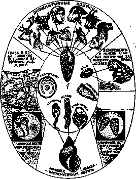
Figure 5. Diagram of the fasciola development cycle. (After Skryabin and Schultz.)
and ducts. At the same time, there is also a toxic effect from the toxic substances secreted. Migrating fasciola usually carry representatives of the microbial flora, which can complicate the main process. In humans, F. is quite rare, with only about 200 cases described in the literature, half of which were recorded in the USSR. However, some of those cases, the diagnosis of which is based on coprological research, as determined by Vogel (1922), Strom (1927) and others, belong to the so-called false fascioliasis, in which eggs of fasciola contained in eaten products (liver in various forms) are found in the feces. Symptoms of F.: enlarged liver, colicky pains, nausea, sometimes vomiting, dyspeptic phenomena, mild jaundice, irregular increases in temperature, chills, sweats, dizziness, headaches, increased nervous excitability. In severe cases, shortness of breath, progressive exhaustion and fatal outcome are observed. Sometimes there is "aberrant localization" of fasciola: in the eye, in the subcutaneous tissue, in the lungs, in the blood vessels, etc. Diagnosis can be made by finding the parasite's eggs in the feces, duodenal contents (taking into account the possibility of finding "transit eggs" from eaten fasciola-infected livers).- Therapy in veterinary practice is carried out with carbon tetrachloride, hexachloroethane and preparations of the rhizome of male fern. Emetin has been successfully tested on humans (Vasil'eva, 1927; Semenov and Kogan, 1927; Kirikov, 1927; Kryukov 1927).- Public prevention consists primarily in organized control of F. of animals, which are the main carriers of fasciola: land reclamation, chemical control of mollusks-intermediate hosts, strict rejection and destruction of affected livers, deworming of animals. Personal prevention of humans: abstaining from drinking raw water from swamps, ponds, arykhs, in which intermediate hosts may live, and from eating raw meadow grasses (sorrel), vegetables, which are watered from standing basins.
Related articles
Mentioned in
Cite this page
“Fascioliasis.” Soviet Medical Encyclopedia. English translation of Bolshaya Meditsinskaya Entsiklopediya, 1st ed. (Moscow, 1928–1936), ed. N. A. Semashko. https://sovietmedicalencyclopedia.pages.dev/article/fascioliasis/