TREMATODES
Historical document, translated for reference. It reflects medical knowledge of the 1920s–30s and is not medical advice.
Summary
Trematodes, or flukes, are parasitic flatworms characterized by their dorsoventrally flattened bodies and attachment organs. The class is divided into monogenean flukes, which are mostly ectoparasites of fish, and digenean flukes, which are endoparasites of vertebrates and have complex life cycles involving intermediate hosts.
Encyclopedia article (1928–1936)
TREMATODES, or flukes (Trematodes Rud. 1808), a class of flatworms (Platodes; syn. Trematoides Eichw, 1829; Pendularia Owen 1855 and others). General characteristics of the class T. Parasitic flatworms whose bodies are flattened to a greater or lesser degree in the dorsoventral direction; forms with ribbon-like bodies are comparatively rare. They have attachment organs, which are most often located at the anterior end, on the ventral surface, or at the posterior end of the body. The attachment organs may be of muscular or chitinous nature. The body surface is lined with a cuticle, which is a derivative of scattered cells located between diagonal muscles or inward from them, while the true epithelium is preserved only on the surface of the larval forms of T. (miracidia). The nervous system consists of ganglia, from which three pairs of longitudinal nerves extend backward, connected to each other by a system of transverse commissures. The mouth opens terminally or subterminally. The intestine is either a single sac or bifurcated; in the latter case, it may form an extensive branching system. The reproductive system is most often hermaphroditic, less often dioecious, and usually of very complex structure. There are two or more testes; less commonly, there is a single testis. Development is direct (in monogenean T.), accompanied by a rather complex metamorphosis (in digenean trematodes), with the participation of an intermediate host, in whose body the larvae often multiply parthenogenetically (metagenesis). Very often, in addition to the intermediate host, there is an additional host. Definitive hosts of T. are as a rule vertebrate animals, in whose various organs and tissues the marita (sexually mature individual) lives. Intermediate hosts are usually mollusks; additional hosts are both fish and various Arthropoda. The class T. includes at present several thousand species, united in several hundred genera, belonging to more than 80 families. T. are conventionally divided into 2 subclasses: monogenean T. (Monogenea) and digenean T. (Digenea), which differ from each other in the following main characteristics.--A. Monogenean T. are for the most part ectoparasites, inhabiting the gills, oral cavity, and skin coverings of fish. Some are localized on cephalopod mollusks and on parasitic crustaceans of marine fish. There are endoparasites that inhabit the urinary bladder of frogs, the pharynx, nasal cavities, lungs, and intestines of turtles. One species parasitizes under the eyelid of the hippopotamus. Monogenean T. are characterized by the presence of special attachment organs in the form of one large or several pairs of suckers, mostly symmetrically arranged, and complex chitinous structures. Sometimes there are eyes in the number of 2-4. The pharynx is surrounded by peripharyngeal pockets; the intestine is mostly branched. The excretory system opens dorsally with two openings. Hermaphrodites. The uterus contains one or a very limited number of eggs, which are mostly provided at the poles with special appendages, or filaments. Development is direct, without an intermediate host, with or without metamorphosis (Fig. 1). B. Digenean T. occur in the mature state almost exclusively in vertebrates, most often in the digestive tract and its glands, less often in the lungs, kidneys, circulatory system, etc. A large number of Digenea species occur in humans, domestic and commercial animals, causing various diseases (so-called trematodoses), and therefore have great sanitary and economic significance. Digenean T. are for the most part small flatworms of leaf-like shape, from 0.1 to 10 mm in length and rarely up to 100-150 mm. The body is covered with a cuticle, sometimes armed with chitinous structures: spines, hooks. Inside the cuticle is located the muscular system, consisting of smooth muscle fibers; at the anterior end of the body is mostly located the so-called oral sucker. In most T. there is also a ventral sucker, located on the ventral surface of the body. In some T. there is also a third sucker, the so-called "genital", into which the genital openings open. On the other hand, there are also
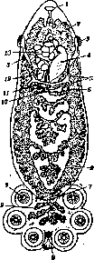
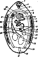
Figure 1. Monogenean trematoda Poly-stomum integerrimum (according to Zeller): 1-oral opening; 2-intestine; 3-entrance to vagina; 4-ovary; 5-longitudinal bile ducts; 6-yolk-intestinal canal; 7-small hooks on disk; 8-large hooks on disk; 9-suckers on disk; 10-common sperm duct; 11-transverse bile ducts; 12-ootype; 13-uterus with eggs.
Figure 2. A digenetic trematode (diagram after Skryabin and Schultz, 1931): 1-oral sucker; 2-pharynx; 3-esophagus; 4-intestine; 5-female genital opening; 6-ventral sucker; 7-uterus; 8-yolk glands; 9-spermatheca; 10-bile duct; 11-ootype; 12-Laurens' canal; 13-ovary; 14-testes; 15-blind ends of intestinal branches; 16-excretory bladder; 17-excretory pore; 18-vas deferens; 19-Melis' body; 20-common sperm duct; 21-seminal vesicle; 22-genital bursa; 23-male genital opening. Such trematodes, in which the ventral sucker is absent and there is only an oral sucker, and even more rarely there are no suckers at all, as they have been reduced. The digestive system begins with the oral opening, located at the bottom of the oral sucker and leading into the pharyngeal cavity and through it into the esophagus; from the latter, in most cases, two intestinal branches extend backward and end blindly; in most trematodes there is no anal opening, only in a few forms there is one or even two anal openings. In some trematodes, the intestinal branches have lateral branches; in other cases, the intestinal branches connect with each other, forming an intestinal arch (Fig. 2). Sensory organs in mature trematodes, so-called marita, are mostly undeveloped. The excretory system has the structure of protonephridia. The unpaired tube of the excretory system opens at the posterior end of the body. Most trematodes are hermaphrodites. The male reproductive apparatus most often consists of two testes, from which one vas deferens extends each, which unite into a single sperm duct; the latter is usually enclosed in a special muscular organ-the male genital bursa (bursa cirri) and opens with the male genital opening on the ventral surface of the parasite. Vas deferens, sometimes forming a seminal vesicle in the bursa, is here most often surrounded by the so-called prostate glands. The female genital opening usually opens next to the male one and leads into the uterus, which connects with the ootype, where the formation of eggs occurs; with the ootype are connected via the oviduct the ovary, then the spermatheca and paired yolk glands; the yolk glands envelop the egg with nutritive material and in addition form the shell; with the ootype is also connected a special organ-Melis' body, which secretes a substance that washes the ootype and uterus and facilitates the movement of the egg along the genital tract. Finally, to the ootype in many trematodes is attached a small canal, opening on the dorsal surface of the parasite-Laurens' canal, serving to remove excess yolk cells that interfere with egg formation, sometimes playing the role of a vagina during copulation. However, in most cases, copulation occurs by the introduction of the cirrus into the opening of the adjacent uterus. As a rule, cross-fertilization occurs in trematodes. Biology of digenetic trematodes.
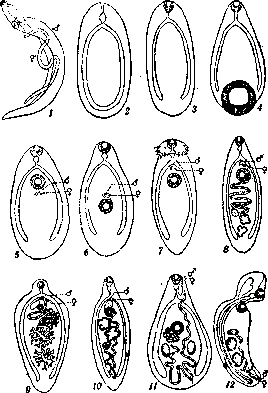
Figure 3. Characteristics of the main families of digenetic trematodes (diagram after Skryabin and Schultz, 1934): I- Schistosomatidae; 2-Cyclocoeliidae; 3- Notocotylidae; 4-Paramphistomatidae; 5-Troglo-trematidae; 6-Heterophyidae; 7-Echiuostomatidae; 8-Opisthorchidae; 9-Fasciolidae; 10-Dicrocoeiii-dae; 11-Plagiorchidae; 12-Strigeidae.
Trematodes with separate sexes, represented by males and females, family Schistosomatidae. B. Hermaphroditic trematodes: I. Ventral sucker is absent. Only oral sucker is present. a) Intestinal branches at the posterior end of the body fuse, forming a single arch-family Cyclocoeliidae. b) Intestinal branches do not fuse at the posterior end, maintaining the independence of two blind ends-family Notocotylidae. II. Both oral and ventral suckers are present. a) Ventral sucker is located at the posterior end of the body-family Paramphistomatidae. b) Ventral sucker is located on the ventral surface of the body. 1. Genital opening opens behind the ventral sucker-family Troglotrematidae. 2. Genital opening opens in front of the ventral sucker. a) There is a genital sucker into which the genital openings open-family Heterophyidae. b) No genital sucker is present. ax) At the anterior end there is an adoral disc with spines-family Echinostomatidae. bx) No adoral disc is present. aa) The uterus has only an ascending branch. a3) The ovary is in the posterior part of the body. Genital bursa is absent-family Opisthorchidae. ba) The ovary is in the anterior half of the body. Genital bursa is present-family Fasciolidae. ba) The uterus forms a loop, first descending and then ascending. a3) The ovary is behind the testes-family Picrocoeliidae. b3) The ovary is in front of the testes-family Plagiorchidae. For more specific characteristics of individual families, genera and species of human trematodes, see separate articles.
Related articles
Mentioned in
Cite this page
“TREMATODES.” Soviet Medical Encyclopedia. English translation of Bolshaya Meditsinskaya Entsiklopediya, 1st ed. (Moscow, 1928–1936), ed. N. A. Semashko. https://sovietmedicalencyclopedia.pages.dev/article/trematodes/