Pseudophyllidea
Historical document, translated for reference. It reflects medical knowledge of the 1920s–30s and is not medical advice.
Summary
Pseudophyllidea is an order of tapeworms characterized by the absence of muscular suckers on the scolex and the presence of primitive attachment organs. These parasites have complex life cycles with two intermediate hosts and infect mammals, birds, reptiles, and fish.
Encyclopedia article (1928–1936)
PSEUDOPHYLLIDEA (Cams; 1863), an order of tapeworms. The scolex of pseudophyllidean tapeworms is characterized by the absence of muscular suckers and the presence of more primitive attachment organs, usually in the form of two sucking grooves or so-called bothria. Strobilization is usually sharply expressed. Part of the reproductive system is built on the trematode type; thus, vitellaria in the form of numerous follicles are arranged in two symmetrical groups along the edges of the proglottid; the coiled uterus opens with an aperture for egg discharge on the ventral side of the segment; the eggs are covered with a dense shell, in most cases with an operculum. However, there are also organs of typically cestode character: numerous testes, an ovary located medially at the posterior edge of the proglottid; a vagina, the aperture of which opens near the male genital pore. As a result of the combination of trematode and cestode characteristics, P. are characterized by the presence of 3 genital apertures on the ventral surface of each segment: one male and two female. In some species, the entire reproductive apparatus of each segment is duplicated (genus Diplogonoporus) and then there are 6 genital apertures. - Pseudophyllidean tapeworms, whose development cycle has been studied, develop with two intermediate hosts (see below - broad tapeworm). The latter are usually inhabitants of freshwater basins, along the shores of which P. are distributed, inhabiting in the adult state in the intestines of mammals, birds, reptiles, and fish. The order P. is divided into 4 families. A number of representatives parasitic in humans belong to the family Diphyllobothriidae (syn. Bothriocephalidae), which is assigned to the subfamily Diphyllobothriinae (Lühe) and to its two genera: Diphyllobothrium (Cobbold; 1858) and Diplogonoporus (Lönnberg; 1892); of these, only the genus Diphyllobothrium has wide distribution and practical significance, the representatives of which parasitize in humans both in the adult state in the small intestine [D. latum (figs. 1 and 2), D. minus, D. cordatum], and in the larval stage, namely in the plerocercoid stage, in the connective tissue (D. mansoni, D. proliferum). - 1. Small tapeworm, Diphyllob. minus (Cholodkovsky; 1916) (syn.: Dibothriocephalus minor, Bothriocephalus minor; figs. 4 and 5), described by Cholodkovsky from a single specimen obtained from the human intestine in Transbaikalia. The length of the strobila is 10-26.5 cm with a maximum width of mature proglottids of 6 mm. The sucking grooves on the scolex widened anteriorly. The uterus begins to form at a distance of 30 mm from the scolex, its loops run parallel to the posterior edge of the proglottid. The size of the eggs is 0.07 mm in length and 0.04 mm in width. Recently, it was thoroughly reinvestigated by Talyzin, who confirmed its specific independence and noted in infected fishermen anemia of the mucous membranes, nervous excitability, frequent diarrhea, and loss of appetite. - 2. Manson's tapeworm, Diphyllobothr. mansoni (Cobbold; 1889) (syn.: Dibothriocephalus mansoni, Bothriocephalus mansoni). In the mature tapeworm form, it parasitizes in dogs. Man is the second intermediate host, in which the plerocercoid under the name Sparganum mansoni parasitizes in the connective tissue of various organs, e.g., under the skin of the eyelid, under the peritoneum, in the urethra, in the thoracic and abdominal cavities, etc., and reaches a length of 8-60 cm. Plerocercoids of Dibothriocephalus mansoni are also found in the intramuscular tissue of frogs and some other animals. The first intermediate host of D. mansoni, in which the procercoid lives, is the copepod Cyclops leuckarti. D. mansoni is distributed in Japan and China. Sparganum often causes non-inflammatory tumors, sometimes painful, and in rarer cases may lead to suppuration. Removal can be done surgically.
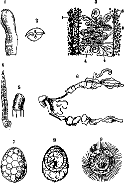
Figure 1 and 2. Diphyllobothrium latum. Scolex.
Side view (1) and cross-section (2). Figure 3. Diphyllobothrium latum: 1- yolk glands; 2-tt st s; 3- bursa cirri; 4- ovaries. Figures 4 and 5. Diphyllobothrium minus. Strobila and scolex. Figure 6. Diphyllobothrium proliforum. Larva. Figures 7, 8 and 9. Egg of Diphyllobothrium latum in various stages of development and coracidium. Broad tapeworm, Diphyllobothrium latum (L.; 1748) (syn.: Dibothriocephalus latus, Bothriocephalus latus), parasitizes in the small intestine of humans, dogs, and cats. It is distributed extremely widely across the globe, characterized by focal distribution. In the USSR, it is found mainly in the north, along the shores of large rivers. In the lower Amur region, the Nivkh, Ulchi, and Golds, according to helminthological expedition data (1928), are significantly infected with this parasite. The natives of the lower Ob River and its tributaries (Khanty, Mansi, Samoyeds, Komi) are especially heavily infected (helminthological expedition to the Urals region, 1929). The highest percentage was found among the Mansi (76.6%) on the tributary of the North Sosva. According to Yeselovich (1928), out of 1,563 residents of Tomsk district examined, 76 were infected with the broad tapeworm. In Arkhangelsk (Kolpikova; 1925), infection with the broad tapeworm accounts for 50% of all helminthiases with an overall infection rate of 80% of the adult population. In general, the north of the USSR is almost unexplored. Other regions of the USSR give individual figures of infection with the broad tapeworm. Thus, in 1925, only 3 cases were found in the Donbas out of 7,234 examined; in Central Asia (1926)-2 cases out of 3,000 examined; in the middle and southern regions of the European part of the USSR, as well as in the Transcaucasian republics, various researchers either do not find the broad tapeworm at all or indicate single findings. In Western Europe, foci of diphyllobothriasis are certain areas of French Switzerland, northern and central Italy, East Prussia, southern Sweden, Finland, Latviag Estonia, Romania. Among Asian states, the broad tapeworm is especially common in Japan.-In humans, from 1 to 106 specimens of the broad tapeworm have been found. The lifespan of the tapeworm is up to 35 years. The length of the strobila can exceed 9 m; the number of proglottids-several thousand. The width of proglottids usually exceeds the length, and only the terminal part of the strobila has square and rarely slightly elongated proglottids. The scolex of the parasite (fig. 2), flattened from the lateral sides, is equipped on the ventral and dorsal sides with two slit-like bothria. Mature proglottids (fig. 3) are equipped with numerous testes and yolk glands occupying the lateral edges outward from the excretory vessels running along; the bilobed ovary resembles butterfly wings in shape; the uterus is twisted, rosette-shaped and lies in the central part of the proglottid. On the uncolored grayish strobila, the uterus of individual proglottids stands out sharply as dark central spots, the pigmentation of which depends on the accumulation of eggs. Eggs 0.068-0.071 mm in length and 0.045 mm in width, with a rather thin double-contour shell, grayish-brown in color; equipped with a lid on one pole (fig. 7 and 8). On the opposite pole, the shell has a small tubercle, which is clearly distinguishable only when the egg is in a strictly horizontal position. The development cycle of the broad tapeworm proceeds with two intermediate hosts. The egg excreted through the uterine opening of the parasite into the intestinal cavity enters the external environment with feces, where it matures in the presence of moisture. A mature egg contains an embryo, the so-called coracidium (fig. 9), which represents an oncosphere with six embryonic hooks, clothed in a ciliated coat. When a mature egg falls into water, the lid opens, the coracidium emerges and swims with the help of cilia. For further development, it must enter an intermediate host-a copepod (Cyclops strenuus, Diaptomus grae-lis, Diaptomus graciloides), in whose digestive tract it sheds its cilia and, with the help of hooks, penetrates through the intestinal wall of the copepod into its body cavity, where over a period of approximately 3 weeks it transforms into the so-called procercoid. The latter is an elongated larva with a spherical six-hooked appendix at the posterior end of the body. Further development of the procercoid occurs in the second intermediate host-in a fish (mainly pike, perch, burbot, ruff, whitefish, trout, etc.), into which it enters when the copepod is swallowed. In the digestive tract of the fish, the copepod is digested, and the freed procercoid penetrates into the thickness of the intestinal wall and from there into various organs of the fish (muscles, liver, etc.), as well as into the roe, where it grows and transforms into a plerocercoid. The latter is characterized by the presence of bothria on the scolex and a small strobila.-Recently, it has been established that in adult predatory fish, plerocercoids are always present in much larger numbers (up to 250 specimens) than in young fish, whereas infection of fish with procercoids from copepods can only occur in early youth, while the small fish feed on plankton. There are three explanations for this phenomenon: it is possible that the plerocercoids of the broad tapeworm, like related forms (Sparganum), have the ability to reproduce asexually in the body of fish. On the other hand, the accumulation of plerocercoids can occur due to adult fish feeding on small fish whose stomachs contain infected copepods. And finally, thirdly, when a fish eats another fish infected with plerocercoids, it is possible that the latter do not perish in the stomach but settle in the organs of their new host. Perhaps the parasite uses all three of these possibilities. When swallowed with raw or insufficiently fried or boiled meat of an infected fish, the plerocercoid is released in the intestine of the final host, attaches to the wall of the small intestine with bothria and begins to grow rapidly. The pathogenesis of diphyllobothriosis consists of two moments-toxic and mechanical. The mechanical role of the broad tapeworm is of secondary importance. However, cases of intestinal obstruction by parasites, exit of the worm per os with the danger of suffocation, etc., should be pointed out. The toxic effect of the broad tapeworm can cause severe disorders in the host. The main and most important moment of intoxication in the broad tapeworm is its effect on the blood and blood-forming organs, due to which anemia develops from mild forms to pernicious anemia inclusive. The picture of malignant anemia on the basis of diphyllobothriosis is typical. Even before establishing the dependence of the development of pernicious anemia on the broad tapeworm, there were indications in the literature of finding this worm in autopsies of those who died from malignant anemia as accidental coincidences. First, Botkin in his lectures in 1883-84 points to the worm and in particular to the broad tapeworm as one of the etiological moments of malignant anemia. Then numerous case reports appear, and a whole controversy arises regarding the dependence of pernicious anemia on diphyllobothriosis. Finally, in 1913, Ragoz analyzes both a series of his own observations and the mass of accumulated literature on this issue, bringing complete clarity to it.-The main moments of action of the broad tapeworm are-hemolysis, a decrease in the number of red blood cells, a change in the color index, dissolution of neutrophils and distortion of the activity of the bone marrow. All these phenomena can be noted in almost all cases of parasitism by tapeworms; at first weakly expressed, only hinted at, they gradually intensify, creating a picture of one of the most formidable blood diseases-malignant anemia (Ratosa). In the few fatal cases of diphyllobothriosis anemias, the same patho-anatomical picture is described as in pernicious anemia. There are also known acute cases with severe hemorrhagic diathesis, in which the bone marrow was found aplastic. The nature of diphyllobothriosis toxins has not yet been precisely clarified; it can be assumed that they belong to lipoids. It is very interesting and still not clarified why in some cases there is severe intoxication up to a fatal outcome, while in others there is an absence of noticeable clinical signs. An opinion was expressed that intoxications arise in connection with the death of the parasite and the absorption of decomposition products; and indeed, it was repeatedly possible to find the parasite clearly altered. However, there are also numerous cases of severe intoxication with unchanged parasites. Furthermore, individual variations in the toxicity of different individuals of the parasite are possible, or the toxicity of the parasite depends on the duration of its stay in the intestine; perhaps the degree of permeability of the intestinal mucosa to toxins changes; the question of the constitution and individual resistance or idiosyncrasy of the host was raised. Ragoz is right, who writes that the degree of anemia is the result of a dynamic equilibrium between the destructive and distorting forces of the parasite's poison and the compensatory activity of the bone marrow; the virulence of the worm, the degree of its decomposition, the energy of the blood-forming organs, etc.-factors that tend to tip the balance one way or the other. The presence of helminthic intoxication and the constantly caused peculiar distortion of the hematological formula are the main agents in the pathogenesis of the named form; constitutional predisposition plays only a subordinate role.
The symptomatology of diphyllobothriosis is very diverse, ranging from cases with barely noticeable symptoms of anemia, sometimes with insignificant gastrointestinal and nervous phenomena, to the picture of a pronounced pernicious anemia. Even in the mildest cases, the blood represents a 'forme fruste' of malignant anemia (Ragoza). In severe cases, some facial edema, marked pallor; the number of red blood cells may fall significantly below 1 million per 1 mm3; anisocytosis, poikilocytosis, polychromatophilia, nucleated red blood cells; hemoglobin content may reach 25%; F.I. usually exceeds unity. Severe weakness, mental depression, sometimes inability for independent movement. Disorders of coordination of movements, neuritis, paralysis, complete loss of consciousness. In children, seizures with tonic and clonic convulsions, opisthotonus, rolling of the eyes, pupil dilation. Seizures of varying frequency from several times a month to 3-6 per day, lasting from 4-5 minutes to 2-3 hours, sometimes with loss of consciousness, which can last from half an hour to entire days. Such phenomena may proceed with high temperature, chills, delirium and produce the impression of a very severe illness, for example, tuberculous meningitis. The tongue is coated, vomiting up to several times a day; achylia, constipation, diarrhea, sometimes alternating; pains most often in the epigastrium. Edema of the extremities or the whole body, dropsy of cavities, erythematous rash, sometimes hemorrhages on the skin, on the fundus of the eye. The liver is often significantly enlarged, usually protruding 1-2 fingers beyond the edge of the ribs; sometimes the liver is painful. Slight enlargement of the spleen is usually only confirmed by percussion. The diagnosis of diphyllobothriosis is made by the presence in the feces of eggs of Diphyllobothrium latum, which in most cases are found in large quantities and are easily detected by microscopy even in a simple smear. The prognosis, with correct and timely recognition and subsequent removal of the parasite, is usually favorable. Even very severe nervous symptoms can quickly disappear after removal of the parasite. Gastrointestinal phenomena also disappear, blood is restored. The patient's condition usually improves remarkably quickly; sometimes complete recovery occurs within a week. However, a few far-advanced cases have been described that ended fatally despite removal of the parasite. Death, in Ragoza's opinion, is the exception to the rule; it is explained either by exhaustion of the bone marrow or by fatty degeneration of internal organs. Therapy consists of expelling the worms, for which the best remedy is Extr. Filicis maris aethereum. The usual dose for an adult when administered orally is 8.0, for a child-0.5 per year of life. The entire dose is prescribed in 10 gelatin capsules, which are taken in the morning on an empty stomach within x/г hour and washed down with black coffee. 1-2 days before treatment, a liquid diet is prescribed, the evening before-a saline laxative, in the morning before taking the extract-an enema. 2 hours after taking Extr. Fil. maris-another saline laxative. If there is a long delay in bowel movement or if the parasite is not excreted with the first stool, an enema is prescribed immediately. Extr. Fil. maris can be administered through a duodenal tube, which allows the dose to be somewhat reduced (4.0-6.0 for an adult). The extract is administered in this case either heated, which achieves its liquefaction, or in the form of an emulsion. Prevention. Smoking and salting fish does not kill plerocercoids, and only fish sufficiently exposed to high temperature (well-boiled or fried) is safe for consumption. It is very important to protect water bodies from contamination with human feces, which can serve as a source of invasion of intermediate hosts. To prevent cats and dogs from spreading the parasite's eggs, they should not be fed raw fish. 4. Diphyllobothrium cordatum (Leuck.; 1863) (syn.: Dibothriocephalus cordatus, Bothriocephalus cordatus) is significantly shorter than the broad tapeworm, reaching only 115 cm in length. The scolex has a heart-shaped form, the neck is rudimentary, the number of segments is about 600. The developmental cycle is not clarified, although the 2nd intermediate host is assumed to be fish, as the parasite is found in ichthyophagous (fish-eating)-seals, walruses; it has also been confirmed in dogs and more rarely in humans. Diphyllobothrium cordatum is known in Greenland and on the island of Iceland. Clinical observations are not available. 5. Diphyllobothrium proliferum (Ijima; 1905), like D. mansoni, occurs in humans in the plerocercoid stage under the name Sparganum proliferum (fig. 6). The latter represents small forms 1-12 mm in length, capable of reproducing by division, for which reason they are found in large numbers. The developmental cycle is not clarified. Man is probably a facultative (accidental) 2nd intermediate host; the obligatory 2nd intermediate host, as well as the 1st intermediate and definitive hosts are unknown. Sparganum proliferum has been found in individual cases in Japan and Texas. Sparganum proliferum forms a huge number of small cysts in the connective tissue of various organs, as well as the skin, which is affected by nodules resembling acne or small tumors. Each small cyst contains several parasites, which are easily shelled off. The number of parasites per 1 dm2 can reach several hundred. Sparganum proliferum apparently also secretes hemolytic toxins, since persons affected by it are anemic and cachectic.
Related articles
Cite this page
“Pseudophyllidea.” Soviet Medical Encyclopedia. English translation of Bolshaya Meditsinskaya Entsiklopediya, 1st ed. (Moscow, 1928–1936), ed. N. A. Semashko. https://sovietmedicalencyclopedia.pages.dev/article/pseudophyllidea/