Teniidosis
Historical document, translated for reference. It reflects medical knowledge of the 1920s–30s and is not medical advice.
Summary
Teniidosis refers to diseases caused by mature stages of parasitic worms from the Taeniidae family that infect the small intestine of certain mammals. This article describes two types affecting humans: teniasis (pork tapeworm) and teniarinchosis (beef tapeworm), including their morphology, life cycles, clinical manifestations, diagnosis, treatment, and prevention methods.
Encyclopedia article (1928–1936)
TENIIDOSIS, diseases caused by mature stages of parasitic worms belonging to the family Taeniidae (see Cestodes) and parasitizing in the small intestine of certain mammals. In humans, two types of T. occur: teniasis and teniarinchosis. 1. The causative agent of teniasis is the pork tapeworm, or armed tapeworm (Taenia solium, 1758)-reaches 2 m in length and is characterized by the following features. The scolex is equipped, in addition to four suckers, with a rostellum with 22-28 hooks of 2 different sizes (Fig. 1), where the larger ones (0.16-0.18 mm) alternate with the smaller ones (0.11-0.14 mm), forming a crown of hooks. The proglottids (Fig. 2) are characterized by an additional (3rd) lobe located between the longitudinal axis of the segment (along which the trunk of the developing uterus is formed) and the vagina. Mature proglottids of the pork tapeworm are characterized by the number of branches of the main trunk of the uterus (7-10) and their coarse configuration. The oncosphere (see Cestodes) is most often spherical; its dimensions are 0.031-0.030 mm. The oncosphere membrane is thick, radially striated, and has 3 pairs of hooks. Man is the only definitive host of the pork tapeworm, and consequently the only source of contamination of the external environment by its proglottids and oncospheres, which are scattered by man along with feces. Intermediate hosts are the pig (obligatory host) and more rarely man, dog, cat, camel, wild boar (facultative hosts). The oncosphere swallowed by the intermediate host is freed from the radially striated membrane, penetrates the intestinal wall with the help of its hooks, enters the bloodstream, and with the help of the circulatory system can be carried to various organs and tissues of the intermediate host. After 2½-4 months of infection, larvae, so-called finnae, or cysticerci (Cysticercus cellulosae), are formed in the body of the pig, located mainly in the intermuscular connective tissue, and also often in the central nervous system.
Figure 1. Scolex of Taenia solium from above; four suckers and hooks are visible.
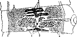
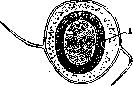
Figure 2. Hermaphroditic proglottid of Taenia solium; 1-ovary (3 lobes); 2-yolk cells; 6-seminiferous tubules; 4-developing uterus.
The causative agent of teniarinchosis is the beef tapeworm, or unarmed tapeworm (Taeniarhynchus saginatus, Goeze, 1782, syn. Taenia saginata, T. medicanellata). This parasite reaches 10 m in length and occasionally even more. The scolex of the parasite, besides suckers, is usually equipped with a rudimentary rostellum, but the chitinous armament is absent (Figs. 3 and 4). The ovary has only 2 lobes. In mature proglottids (Figs. 5 and 6), the uterus has 18-30 lateral branches from the central trunk, which, in contrast to the branches of the uterus in the pork tapeworm, are very delicate and closely spaced. The oncosphere 0.03-0.04x0.02-0.03, often spherical, is practically indistinguishable from the oncosphere of the pork tapeworm (Fig. 7). The definitive host is man. The intermediate host is cattle, in whose intermuscular connective tissue, 3-6 months after swallowing the oncosphere, a finna, or cysticercus (Cysticercus bovis) is formed. Both of the above-described parasites are cosmopolitan; both teniasis and teniarinchosis can occur wherever pork or beef meat is consumed. In various regions of the USSR, accurately diagnosed cases of teniasis usually do not exceed tenths of a percent. In the central regions of the USSR, in the Donbas, in the Far East, and some other regions, the prevalence of teniarinchosis, although somewhat higher than that of teniasis, is also insignificant. Man becomes infected with teniidosis from raw or inadequately cooked or fried finna-infected pork meat (teniasis) or cattle meat (teniarinchosis). The pork and beef tapeworms attach themselves to the uppermost part of the small intestine, often in the duodenum, occurring as single specimens and, more rarely, in large numbers (up to several dozen in individual cases).-The pathological effect of the parasites consists mainly of two factors: 1) mechanical influence due to the fixation of the parasites to the intestinal mucosa by suckers,
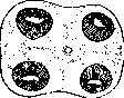
Figure 3. Scolex of Taeniarhynchus saginatus from above; 4 suckers; no hooks.
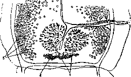
Figure 4. Hermaphroditic proglottid of Taeniarhynchus saginatus: 1-ovary (2 lobes); 2-yolk cells; 3-seminiferous tubules; 4-uterus in the very initial period of development.
in the pork tapeworm and by hooks, which leads to local circulatory disorders and disruption of the integrity of the mucosa. The movement of the parasites, and in more rare cases their obstruction of the intestinal lumen, can also have a pathological effect. 2) On the other hand, as a result of the vital activity of the parasite, substances are secreted that have a toxic effect on the host's body and cause chronic poisoning of it. Clinical manifestations in T. do not fit into the picture of a characteristic symptom complex on the basis of which a precise diagnosis of the disease could be made. Most often, the pathological phenomena consist of a number of disorders of the gastrointestinal tract, often together with certain changes from the nervous system, more rarely-from the blood-forming organs. The range of these phenomena in different patients can be very wide-from complete health to severe diseases, even with fatal outcomes in individual cases. One usually has to deal with symptoms of mild and moderate severity, such as: disorders of appetite, nausea, vomiting, disorders of stool, headaches, dizziness, sometimes to the point of fainting. Apathy, loss of working capacity, irritability, and sometimes pronounced symptoms of anemia are often observed. However, the characteristic picture of pernicious anemia occurs much less frequently than in diphyllobothriasis. Teniasis more often than teniarinchosis occurs with weakly expressed pathological phenomena, but it is much more dangerous than teniarinchosis, as it can lead to cysticercosis (see Cysticercosis). One of the symptoms of the disease, which characterizes teniarinchosis to a greater extent, is the ability of mature proglottids, detached from the strobila of the parasite, to exit through the anus and crawl for some time along the thighs and other parts of the host's body.
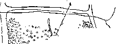
Figure 5. Mature proglottid of Taenia solium with developed uterus.
The diagnosis of T. can be made based on the structure of the proglottids or oncospheres excreted by the patient. Neither the clinical picture nor immunobiological reactions are specific. The absence of oncospheres in feces does not give the right to deny the presence of parasites. To differentiate teniasis from teniarinchosis, it is possible only by mature or hermaphroditic proglottids and the scolex. Attempts to distinguish the beef tapeworm from the pork tapeworm based on the structure of the oncosphere always lead to numerous errors due to the absence of sharp distinctive features in the structure of the oncospheres.-The prognosis in T. is favorable in most cases. The best therapeutic agent is the extract of male fern, which gives good results in adults, while in children even a repeated course of treatment often remains ineffective. The treatment method-see Lepechinia. Prevention of T. should be carried out in two directions. 1. Protection of man from the possibility of infection from finna-infected meat. For this purpose, veterinary-sanitary control over cattle and pork meat has been organized in slaughterhouses and meat inspection stations, followed by the destruction of the entire carcass in generalized finniasis or the release of conditionally suitable meat (with the use of boiling, salting, freezing). However, in addition to the above public health measures, due to the imperfection of methods for diagnosing finniasis in its mild degrees and the insufficiency of veterinary-sanitary supervision, personal prevention is also necessary. The latter comes down to consuming cattle and pork meat exclusively in well-boiled and fried forms. 2. Protection of cattle and pigs from infection with finniasis. For this purpose, it is necessary to carry out extensive sanitary education of the population, and in particular to explain the harmfulness of the custom of scattering feces everywhere, especially in barns, yards, streets, and other places accessible to animals. It is necessary to ensure the provision of rational closed latrines to which domestic animals that could eat or scatter feces and thereby contaminate the surrounding soil with them and the elements contained in them would not have access.
V. Podzial'skaia. TENOSIN (Tenosin), a preparation consisting of histamine and tyramine and used in medical practice as a substitute for ergot. T. is a sterile liquid containing in 1 cm³ 0.000125 g of histamine and 0.00625 g of tyramine, the pharmacodynamic properties of which T. owes its use to. Its action sets in faster than with ergot, but ceases more quickly. Dosage: orally 20 drops three times a day; subcutaneously or intramuscularly 1 cm³ several times a day. Side effects are rare: headache, nausea, vomiting. Intravenous administration is unacceptable.



Related articles
Mentioned in
Cite this page
“Teniidosis.” Soviet Medical Encyclopedia. English translation of Bolshaya Meditsinskaya Entsiklopediya, 1st ed. (Moscow, 1928–1936), ed. N. A. Semashko. https://sovietmedicalencyclopedia.pages.dev/article/teniidosis/