Echinococcus
Historical document, translated for reference. It reflects medical knowledge of the 1920s–30s and is not medical advice.
Summary
Echinococcus is the larval stage of the tapeworm Echinococcus granulosus, causing a parasitic disease in humans and various mammals. The adult parasite resides in the intestines of definitive hosts like dogs, while humans serve as intermediate hosts where the larvae develop into hydatid cysts.
Encyclopedia article (1928–1936)
ECHINOCOCCUS, the larval stage of the tapeworm Echinococcus granulosus (Batsch, 1786) (fig. 1). A helminthic disease of humans and various domestic and wild mammals, resulting from the settlement of E. in various organs and tissues (especially frequently in the liver and lungs), is called echinococcosis. Man serves as an intermediate host for this cestode. The mature stage of the parasite inhabits the small intestine of dogs (fig. 2), wolves, and jackals, which are the definitive hosts of Echinococcus granulosus. A small white cestode, it reaches only 2-6 mm in length and consists of a scolex, neck, and 3-4 segments. On the scolex there are 4 muscular suckers and a rostellum armed with a double crown of hooks (fig. 3), numbering from 36 to 38. Exceptionally, the number of hooks may decrease to 28 and increase to 50. The large hooks reach a length of 0.040-0.045 mm, while the small ones are 0.030-0.038 mm. The first 2 segments are asexual, the 3rd contains a hermaphroditic system of organs, while the last, mature segment, the largest, exceeding the length of the rest of the strobila, contains the uterus, elongated along its length and characterized by the presence of lateral diverticular outgrowths. In the uterus of the mature segment, 400 to 800 eggs are localized. The egg shell lacks filaments. The oncosphere (fig. 4) is covered with a thick radially striated shell with a diameter reaching 0.030-0.036 mm. The oncospheres of E., excreted with the feces of dogs and other definitive hosts, possess considerable resistance. In water they maintain viability for 12 days; at 0° they did not perish for 116 days (Deve). Definitive hosts, mainly dogs, disseminate mature segments, resp. eggs and oncospheres of Echinococcus granulosus with their excrement. The latter, when swallowed by intermediate hosts (including humans), are freed from their shell and with the help of their 6 hooks penetrate into the thickness of the intestine, from where by two paths—either entering the blood vessels or using the lymphatic system—they enter the circulatory system. Very often the oncosphere is detained in the blood vessels of the liver, which is one of the
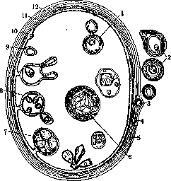
Figure 1. Echinococcal cyst (schema according to Blanchard): 1, 8, 9 and 10—formation of internal daughter cysts; 2, 3, 4 and 5—formation of external daughter cysts; 6—transformation of the scolex into a cyst; 7—scolex; 11—embryonic membrane, 12—cuticular membrane.
most frequent sites of localization of Echinococcus. Oncospheres that have passed through the portal circulation or used the lymphatic flow can penetrate through the right heart and the pulmonary artery into the lungs, where they can be trapped by pulmonary capillaries. Finally, individual oncospheres pass from the lungs into the left heart, and from there through the aorta into the systemic circulation and can be carried to any organ and any tissue, where they gradually transform into the vesicular form—the larval type of Echinococcus. Thus, all organs and all tissues can become sites of localization of E. The formation of echinococcal cysts from oncospheres in the body of the intermediate host proceeds very slowly. Deve (1910) and Dew ess


<P^
Figure 2.
Figure 2. Echinococci from the intestine of a dog (tapeworm stage). Figure 3. Hooks of Echinococcus from the sputum of a patient. The most frequent sites of localization of Echinococcus. Oncospheres that have passed through the portal circulation or used the lymphatic flow can penetrate through the right heart and the pulmonary artery into the lungs, where they can be trapped by pulmonary capillaries. Finally, individual oncospheres pass from the lungs into the left heart, and from there through the aorta into the systemic circulation and can be carried to any organ and any tissue, where they gradually transform into the vesicular form—the larval type of Echinococcus. Thus, all organs and all tissues can become sites of localization of E. The formation of echinococcal cysts from oncospheres in the body of the intermediate host proceeds very slowly. Deve (1910) and Dew ess
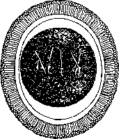
Figure 4. Oncosphere of Echinococcus. (1925) observed the development of the larva in the liver of a pig and describe this process as follows. Oncospheres, swallowed by the intermediate host, concentrate in the liver after 3-12 hours, usually getting stuck in the lumen of an intralobular capillary. Extremely quickly the parasite is surrounded by mononuclears, which hide it from the observer's eyes. After 28 hours the mononuclears are displaced by lymphocytes, forming a follicle with eosinophils on the periphery. After 60 hours the parasite becomes visible as a small protoplasmic round body, barely reaching 0.020 mm, located in the center of a focus of reactive inflammation of relatively large size (about 0.2 mm). After 4 days in the center of this, still very small parasite (approx. 0.030-0.040 mm in diameter) vacuolization is observed—the first hint of the formation of an internal cavity. After 7 days the parasite takes on a proper spherical shape, reaches 0.060-0.070 mm in diameter, and acquires the basic elements of the structure of the future Echinococcus cyst; inside there is a cavity filled with fluid; this fluid bathes the internal germinal membrane, consisting of granular, nucleated protoplasm, which is surrounded by a very delicate cuticular membrane. According to Du, the parasite takes on a cystic structure by the end of the second week. The further growth of the parasite proceeds very slowly. According to Dve after a month it has a size of only about 1 mm, after 3 months about 1.5-2.0 mm, after 5 months it averages about 5 mm in diameter. The larva of the parasite (the cyst of Echinococcus). In the body of the intermediate host the larva of Echinococcus can have 2 morphological modifications, presenting itself either as a unilocular (Echinococcus unilocularis) or multilocular (E. multilocularis) echinococcus. a) Echinococcus unilocularis, unilocular echinococcus. The most common type of larva, consisting of a cyst filled with fluid. In humans the cyst can reach the size of a child's head, in domestic animals the cyst is usually of much smaller size. Analyzing the structure of the cyst, we see that it consists of a number of elements. The wall of the cyst consists of 2 membranes: the outer cuticular and the inner germinal. The cuticular membrane is usually of a milky-white hue and consists of many concentrically arranged lamellated plates. According to Lulike, the cuticular membrane is close to a chitinous substance. The germinal membrane, lining the inside of the cyst cavity, is very rich in glycogen; reaching a thickness of 0.010-0.025 mm, it contains both muscle fibers and calcareous bodies, as well as excretory vessels. In turn, two layers can be distinguished in it: an inner layer consisting of large cellular elements, and an outer layer of smaller cells. The cavity of the cyst is filled with echinococcus fluid. This fluid, being a derivative of the blood of the intermediate host, has a slightly yellowish tint, a neutral or weakly acidic reaction, and a specific gravity of 1.009-1.015. In this fluid a considerable amount of NaCl, grape sugar, tyrosine, inositol, albumin, and succinic acid can be found. Sometimes hematoidin is encountered, and in kidney echinococcosis—uric acid salts. In a number of cases, especially in echinococcosis of domestic animals, cysts can be observed consisting of the 3 elements mentioned above: the cuticular and germinal membranes and fluid. Such cysts have received a special name—acephalocysts, because they do not contain scolices inside them. Such cysts must be considered sterile, as they do not contain invasive elements for the definitive host. Such acephalocysts are most common in ruminants (in cattle in 80%, in sheep in 75%, according to Lichtenheld). In humans, on the contrary, acephalocysts are extremely rare. In addition to such 'sterile' cysts, cysts of a different structure are observed (the following classification of echinococcus cysts is a pathological-anatomical classification, but by no means a zoological one). On the germinal membrane, brood capsules, scattered without any definite order, may be found. These capsules are clothed with the same membranes as the maternal cyst, only they are arranged in reverse order: the cuticle, not lamellated, is on the inside, and the germinal layer is on the outside. On the walls of the brood capsules scolices develop, either filling the internal cavity of the capsule or protruding outward. The number of scolices in the brood capsules varies within wide limits. Sometimes scolices detach from the capsule and are found freely suspended in the cavity of the maternal cyst, forming the so-called hydatid sand. This form of echinococcus is called E. veterinorum, as it is encountered especially frequently in domestic animals: pigs, sheep, cattle. In contrast to this form, the form E. hominis, which is especially common in humans, is encountered, when so-called daughter cysts form in the cavity of the maternal cyst. Daughter cysts are formed in the thickness of the maternal cyst wall from small areas of the germinal membrane; they grow either outward (exogenously) or inward (endogenously) and may completely bud off from the maternal cyst. In their exogenous growth they push into the space between the cuticular membrane of the maternal cyst and the connective tissue protective capsule: this type, formed by the host, is called E. hydatidosus exogenus. In their endogenous growth the daughter cysts penetrate into the cavity of the maternal cyst, where they can be found free and sometimes in considerable numbers—E. hydatidosus endogenus. The arrangement of the layers in the membrane of the daughter cysts completely coincides with that of the maternal cyst, and their cuticular membrane also has a lamellated structure. In the cavities of daughter cysts, as in the maternal cyst, brood capsules with scolices can form. Daughter cysts can develop endogenously or exogenously and also granddaughter cysts. Endogenous daughter cysts can also have a different origin: the classic experiment of Deve established that scolices suspended in the fluid of the maternal cyst can undergo a peculiar metamorphosis and transform into a daughter cyst. It has been experimentally established that the transplantation not only of daughter cysts and individual scolices, but also of individual fragments of the cyst wall to experimental animals causes the formation of echinococcus cysts in them, developing both brood capsules and scolices, which is important not to forget during surgical treatment of E.) b) Echinococcus multilocularis, s. alveolaris, multilocular, or alveolar echinococcus (Fig. 5). This form, which occurs relatively rarely in humans and quite frequently in cattle, is characterized by the fact that the internal cavity of the larva
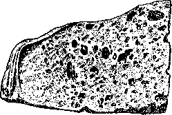
Figure 5. Multilocular echinococcus of the human liver (schematic).
is filled not with fluid, but with a conglomerate of many small, compressed, irregularly shaped bubbles, which are united by connective tissue substance [see separate table (pp. 415-416), figs. 3 and 4]. On a cross-section, the parasite acquires a honeycomb structure. In these bubbles (not in each one) scolices are localized. The question of the zoological relationship between the multilocular and unilocular echinococci is interpreted differently by various authors. Leuckart, Possel (1906, 1910), and among Russian researchers Melyshkov-Razvedenkov (1901) and his students consider that the multilocular echinococcus is an independent species, independent of the unilocular one. The main morphological criterion for supporters of the dualistic trend is the difference in the structure of the hooks on the rostellum of the scolex: in the unilocular echinococcus, the ratio of the total length of the hook to its handle is 1:4.6, while in the multilocular it is 1:2.7. There are also supporters of the monistic view, which speaks of the zoological unity of the unilocular and multilocular echinococci. This question should still be considered unresolved for now. Frequency of finding E. in humans. In Germany, one clinically detected case of human echinococcosis occurs per 1,056 inhabitants (in the Rostock area), and in the southern provinces per 23,685 inhabitants (Mosler and Peiper). The autopsy material gives a higher percentage of E. (in Rostock-2.43%). In the USSR, the most unfavorable areas for E. are Western Siberia, the North Caucasus, and Transcaucasia. According to the data of Mirolubov (1927), in Tyumen, out of 3,820 autopsies, 58 bodies were found to be echinococcosis, which is 1.54% according to autopsy material. In humans, echinococcosis most often develops between the ages of 30 and 50 years. Frequency of finding E. in domestic herbivores. According to Turin's data, the highest percentage of herbivore animals infected with E. falls on the southern and southeastern regions of the European part of the USSR. Cattle are most often infected with E., followed by pigs, goats, and sheep. In horses, echinococcosis is registered very rarely (0.43%). Frequency of echinococcosis spread in dogs. According to data from the All-Union Institute of Helminthology, the average percentage of dogs affected by echinococcosis in different regions of the USSR reaches 13% with a clear predominance in the south. Epidemiology of E. Already a priori it is clear that the factors favorable to the spread of E. should be: a) abundance of dogs, b) shortcomings in slaughterhouse operations (insufficiency of the network, defects in inspections), c) high degree of infection of carriers of echinococcosis virus-dogs and slaughter animals, d) insufficient familiarity of the population with this disease. Human invasion occurs when segments of the tapeworm or individual eggs, resp. oncospheres, excreted by the dog from the intestine, enter his digestive tract. The dog is in a privileged position compared to other domestic animals: it has access not only to the yard but very often also to the interior rooms of human dwellings, right up to the bedroom and kitchen. By constantly licking itself, particularly the anal area, and thus contaminating its muzzle and losing the detached segments on the floor or on the ground, by its bed, on its bedding, the dog can collect all the scattered material again on its fur, on its paws, on its muzzle. An echinococcosis-infected dog is always a great threat to those around it, moreover, even every uninfected dog, not isolated from contact with others, also represents a danger. Communication of a person, especially children, with a dog leads to this infection. The relative frequency of lesions of individual human organs by E. is evident from the table (according to Teichmann, 1898) (see p. 661). Prevention: slaughter of animals should be carried out in slaughterhouses or in places where there is veterinary-sanitary supervision. No organ affected by echinococcal cysts (liver, lungs, kidneys) should be released beyond the slaughterhouse. Affected organs should either be destroyed or at least boiled to kill the echinococcus larvae. In addition, dogs should not be allowed into slaughterhouses, not only in the places of slaughter but also into the slaughterhouse yards. To protect animals and humans from infection with E., it is first necessary to mercilessly destroy stray dogs. A mandatory tax on dogs will be an auxiliary measure here. Those dogs that have a specific purpose and value (service dogs in state kennels, sheepdogs in sheep farms, hunting dogs) should be subjected to regular periodic deworming. For this purpose, during treatment, dogs should be kept on a leash, and excrement should be burned or buried in the ground. In view of the fact that an echinococcosis-infected dog is a constant threat of human infection, simple and easily implementable measures of personal prevention should always be observed and promoted, which are as follows: 1) do not kiss dogs, 2) do not sleep in the same bed with a dog, 3) do not feed dogs from common dishes, 4) do not allow dogs into rooms where food supplies are stored, 5) do not allow children to play with dogs, 6) protect food supplies from being licked and sniffed by dogs, 7) wash hands before each meal.
which scribbish. The clinical picture of echinococcosis has been developed poorly. Only when the parasite reaches significant size or undergoes pathological changes (rupture), the reaction from the parasite carrier becomes clear to the clinician, and the latter establishes the nature of the disease. Changes in the carrier's body in the earlier periods of parasite development are poorly expressed, insufficiently characteristic, and require special research. Therefore, as a rule, recognition of echinococcosis in the clinic is established late. This explains why in most cases echinococcosis is observed in the 8th and 4th decades of life, whereas the invasion mostly belongs to childhood. Only the localization of the parasite, associated with the appearance of noticeable symptoms at small sizes, e.g. in the brain or in the orbit, allows one to make, if not exact recognition, at least to give reason for surgical intervention, which will reveal the essence of the disease relatively early, even in childhood. With the most frequent localization of the parasite (in the liver), the disease proceeds for a long time with little noticeable, difficult to detect and insufficiently evaluated symptoms of chronic echinococcal intoxication. These include loss of appetite, weakness, weight loss, skin pigmentation, shortness of breath, palpitations, pains of various localization and intensity, sometimes increasing to the degree of attacks of varying duration, eosinophilia in the blood, urobilinuria. All these phenomena disappear after removal of the parasite. These phenomena of chronic echinococcal intoxication are even less expressed in alveolar echinococcosis. Nevertheless, one cannot deny echinococcal intoxication and it must be taken into account in clinical observation. This is indicated by the nature of acute echinococcal intoxications and changes in the blood. Substances coming from the parasite into the carrier's body cause the appearance of antibodies as antigens. Hence the possibility of developing anaphylactic phenomena in echinococcosis. Of these, urticaria is the most noticeable, by its frequency and prominence for the patient representing a very important symptom in the clinic of echinococcosis, which can be detected in the anamnesis. It is sometimes local and sometimes diffuse and is accompanied by itching of varying intensity. Urticaria appears either without visible causes or more often when the integrity of the echinococcal cyst is violated (rupture, puncture, operation). It can be accompanied by extremely severe disorders of the cardiovascular, respiratory and nervous systems: the patient becomes restless, temperature rises, breathing and pulse quicken, sometimes the pulse drops to disappearance, convulsive contractions appear in various muscle groups, leukocytosis increases in the blood. In most cases, patients recover more or less quickly, but the matter can also end in death. These pictures of anaphylactic shock are characteristic of echinococcosis. Often in echinococcosis, a decline in nutrition is observed, and in young individuals, also weakness of physical development. The deposition of fat in the connective tissue is usually small, especially when the echinococcus has undergone necrosis, even if not suppurated. In alveolar echinococcosis, on the contrary, the nutrition of patients is preserved for a long time, which cannot but be connected with the density and power of connective tissue proliferation. Much more pronounced are the manifestations of intoxication in the suppuration of the parasite, but then they have a clearly septic character. In the blood in echinococcosis, the number of neutrophils, monocytes and especially eosinophils increases. In view of the fact that tissue eosinophilia is usually noted around the parasite, the idea of a connection between it and peripheral blood eosinophilia suggests itself. Research by N. Napalkov showed that there is no parallelism between tissue and peripheral eosinophilia. The number of eosinophils in the blood sometimes increases very significantly, up to 50% and higher, and sometimes little or no eosinophils at all. Shiltov tried to connect this with the intensity of the reproduction processes in the cyst. One should not lose sight of the fact that eosinophilia is also characteristic of other helminthiases. Therefore, its presence does not speak for echinococcosis, and its absence against it. After removal of the echinococcus, it decreases, sometimes quickly, sometimes slowly. Its persistence after removal of the echinococcus arouses suspicion of the presence of overlooked cysts during the operation, of course if other helminths are not found in the intestine. Eosinophilia is often accompanied by a low (5-10%) monocytosis. Specific features of the blood serum in echinococcosis are revealed in the form of precipitation reaction and complement fixation reaction. The precipitation reaction for clinical purposes is unreliable: it is often positive in the absence of echinococcosis and negative in its presence. In addition, one must keep in mind that the appearance of precipitates may also depend on bacterial contamination. All this prevented the Kraus reaction (appearance of specific precipitates) in echinococcosis from entering clinical practice. The complement fixation reaction has greater clinical significance. It was especially propagated in France by Weinberg. According to Weinberg, it is positive in 65% of cases of echinococcosis, according to Kreuter - in 50%. It turned out that its results strongly depend on the technique of its application, which is also far from simple. Therefore, this reaction did not receive widespread use. The greatest application was found by the Casoni skin anaphylactic reaction: 0.2 cm3 of transparent centrifuged echinococcal fluid, obtained from the slaughterhouse or during operation from a person, free from scolices, particles of the germinal layer and leukocytes, is injected into the thickness of the skin of the palmar surface of the forearm. For comparison, the same amount of saline solution is injected into a place distant from the injection. After the introduction of the fluid into the thickness of the skin, a white dense nodule appears, which disappears after 1-2 hours if saline solution was introduced or if the reaction is negative. In case of a positive reaction, red spots appear around the site of injection of the echinococcal fluid, which merge into a continuous redness spreading over a significant area of skin. The reddened skin swells and becomes dense. Two types of reaction are distinguished: early and late. The early one is detected in the first half hour (Chizhova) in the form of a papule with a red border and lasts 2-3 hours. It is not as conclusive as the late one, which appears 2-3 hours after injection and lasts a day or more. The intradermal reaction gives a positive result in more than 90%. It is very demonstrative, simple in technique and does not require laboratory conditions. Local manifestations of echinococcosis are much more studied. Among them, first place is occupied by the echinococcal tumor. The latter is characterized by round contours, smooth surface and tense elastic consistency. In alveolar echinococcosis, the tumor differs in board-like density and a finely bumpy surface. The size of the tumor is always more or less significant, since small tumors escape observation due to their asymptomatic nature. The concept of size is of course very conditional: a tumor that remains asymptomatic in size in the liver already causes obstructive jaundice in the bile ducts, in the brain - paralysis, and in the heart - perforation of the wall with fatal hemorrhage. The sphericity of the tumor conditioned by its liquid contents under the influence of local mechanical conditions may lose its correctness, but even after rupture, at least partial roundness of the contours remains. Due to strong tension, fluctuation in the tumor is rarely palpable, only with a thin and weak abdominal wall. The so-called hydatid vibration noted by old authors is extremely rare. In the case of calcification of the E. tumor, a parchment-like crunch may be obtained on pressure. The same happens in echinococcosis of bones, especially tubular ones. Besides the tumor formed by the parasite itself, sometimes there is swelling of the affected organ, e.g. the liver, due to disorders of its blood circulation, parenchymal degeneration and vicarious hypertrophy. In echinococcosis of abdominal organs, the spleen often enlarges. Enlargement of the liver and spleen is especially characteristic of alveolar echinococcosis. Usually one tumor is palpable, but this does not mean that singularity is characteristic even for hydatid E., sometimes called unilocular (this name should be abandoned). Multiple cysts are very common, not less than in 50% of cases and even more, but they are often overlooked, even during surgery. Very often some time after surgery the patient again comes with a cyst located close to the site of the removed cyst, even if the removal was performed without its opening, therefore without seeding the operative field with the contents of the cyst. Often neighboring cysts merge together, and sometimes during surgery a neighboring cyst has to be opened through the cavity of an already opened cyst. Usually neighboring cysts are of different sizes, and after removal of the larger cyst, the overlooked smaller one begins to grow rapidly. More often, multiplicity is secondary due to rupture of the cyst and autotransplantation of daughter vesicles, scolices and particles of the germinal layer to a new place in the serous cavity or in the connective tissue.
Usually with this, there are various manifestations of anaphylactic shock, and indications of them can sometimes be found in the patients' medical history. Such contamination can occur during surgery and especially after a trial puncture. Therefore, during surgery, it is always necessary to consider the possibility of contamination and take appropriate measures to prevent it, and trial punctures in echinococcosis should be completely abandoned. Secondary cysts can also develop by exogenous growth, but it is possible to establish the exogenous origin of their origin only by histological examination of the fibrous capsule of the host. Cysts that grow into a neighboring organ can lead to confusion with multiple cysts, so that one cyst simultaneously lies in two organs, for example, in the liver and the lower lobe of the right lung. In this case, such a cyst can rupture into both the bronchus and the bile duct, as a result of which the patient will expectorate bile-containing sputum, and daughter vesicles may be found in the bile ducts. As a rule, echinococcosis is multiple in bones. Due to unfavorable spatial relationships in the bone, the parasite produces abundant growth of small daughter vesicles, which fill the spongy part of the bone, expand and thin its compact part. A change in the spatial conditions of parasite growth immediately affects the nature of growth: on both sides of the affected bone, parosteal cysts develop, which are already voluminous in size. We have similar conditions of development in the so-called alveolar echinococcus, which gives a mass of small vesicles among the powerful growth of dense connective tissue. An echinococcal tumor can be detected radiologically. This is very easily achieved in air-containing organs (lungs) and when gases develop in the suppurating echinococcal cyst. From this arose the idea of using the introduction of air into the abdominal cavity to detect echinococcosis of abdominal organs, including the liver. Especially favorable conditions for radiodiagnosis of echinococcosis are created when the parasite is seeded, both hydatid and alveolar. In bone echinococcosis, a characteristic picture is also obtained. It is generally believed that the tumor is the first manifestation of echinococcosis, but this does not correspond to reality. Usually preceding it are pains of varying intensity and quality, sometimes intensifying to the degree of painful attacks. The sources of pain may be the serous membrane, which is stretched when the volume and position of the affected organ changes and reacts to toxic and infectious influences, or the bile ducts when their patency is impaired. Pain may also be associated with the rupture of a cyst into neighboring serous cavities or into the cellular tissue. In this case, they are accompanied by anaphylactic phenomena. In bone echinococcosis, pains can have a tearing and boring character and are intensified by pressure. For alveolar echinococcosis, a painless course is characteristic. Pain sensations may arise in the liver periodically as a result of disorders of liver blood circulation and swelling of the hepatic parenchyma under the influence of hepatotropic echinococcal intoxication. Despite the periodicity of pains and often accompanying jaundice of the sclera, they cannot be attributed to lesions of the extrahepatic bile ducts, because they occur much more often than echinococcal cholangitis, while the hepatic parenchyma always suffers, not only in the vicinity of the parasite but also far from it. As a result, a deficiency in the detoxifying function of the liver develops, followed by a deficiency in kidney function and nephrotic changes in them. The clinical manifestations of echinococcosis are highly modified due to the so-called rupture into neighboring tissues and organs. Ruptures occur frequently; for example, liver echinococcosis gives ruptures in no less than 10%. Lung echinococcosis gives ruptures even more often. Ruptures occur into adjacent serous cavities (peritoneum, pleura), into loose cellular tissue (retroperitoneal, perirenal), into tubular organs (bronchi, bile ducts, intestine, ureter), into blood vessels (into the portal vein, even into arteries). Alveolar echinococcus ruptures significantly less frequently than hydatid. Ruptures are accompanied by anaphylactic phenomena and extremely diverse symptoms depending on the direction of the rupture: when rupturing into serous cavities, symptoms of irritation (peritoneum, pleura) appear; in the cellular tissue, a painful swelling similar in speed of appearance to a hematoma develops; through tubular organs (bronchi, intestine, ureter), the characteristic components of the contents of the echinococcal cyst (hooks, daughter vesicles, and fragments of the chitinous sac) are expelled outward, or these formations cause obstruction of the tubular organ (bile duct, ureter) with subsequent obstruction. After rupture, the emptied cyst undergoes scarring; sometimes suppuration develops in it. The contents that have leaked into the serous cavity or loose cellular tissue can give rise to the development of a secondary echinococcus. Bone echinococcosis can rupture into a joint. In addition to rupture, the growth of echinococcus into a neighboring organ is also observed. Thus, parasites on the convex surface of the liver or spleen, developing towards the diaphragm, grow through it into the lower lobe of the lung. Bone echinococcus can grow through the joint cavity into a neighboring bone. Changes in the clinical course are also introduced by the death of the parasite. Upon death, the chitinous membrane becomes more permeable and the toxic influence of the parasite, both local and general, becomes more pronounced. Dead echinococci always give an adhesive process around them, with a tendency to seeding, indicating reduced tissue viability. Then on the radiograph, sharp contours of the cyst are obtained. Echinococcal intoxication upon death of the parasite becomes more pronounced: decline in nutrition and blood formation, decreased resistance to infectious influences, disruption of liver protective functions. A dead parasite often becomes a focus of pus-forming infection and then gives mixed intoxication. Even after encapsulation and seeding, it long retains dormant infection that can cause a new outbreak. The most serious changes in the clinical picture arise with the development of suppuration in the parasite. The most frequent cause of suppuration is the bacillus coli, but other types of pus-forming infection are not excluded. A damaged parasite, for example after rupture, and a dead one are particularly prone to suppuration. Suppuration often occurs in a cyst examined during surgery, located near the removed or drained cyst. It can be assumed that degenerative processes in the parasite increase the amount of carbohydrates and proteins in the cyst contents and thus make it a more favorable nutrient medium for infection. With the development of suppuration, pain and feverish condition appear. The tumor increases rapidly, especially if suppuration is accompanied by gas development, which in hydatid echinococcosis, in contrast to alveolar, is often the case, because the bacillus coli gives gas formation in the presence of carbohydrates in the medium. If the integrity of the parasite's membranes is preserved, the tumor retains its round shape, and on the radiograph, a voluminous air bubble is seen above the horizontal level of the fluid. When a suppurated cyst with gas formation ruptures, the roundness of the tumor is lost, because then the cavity into which the suppurated parasite ruptures is involved in the process. This most often occurs in the pleura; in such a case, the physical manifestations give a picture of gas-containing purulent pleurisy, and only the suddenness of its development may suggest its specific nature. Of course, in the case of simultaneous excretion with sputum of hooks, fragments of chitin, and vesicles, the question is clarified precisely. When a suppurated cyst ruptures into a serous cavity or cellular tissue, septicopyemic condition always develops, and often even before rupture, in case of gas-containing suppuration of the parasite: chills, fever with large ranges, sweats, diarrhea, extreme exhaustion. The body weakened by echinococcal intoxication fights poorly with purulent infection, and upon rupture, a fatal outcome often occurs. This must be taken into account in urgently necessary surgical treatment of a suppurated echinococcus. In the clinical manifestations of echinococcosis, much depends on the localization of the parasite (see Liver, Lungs, Spleen, Bone, Thyroid gland, Brain). The affected organ leaves its imprint on the picture of the disease. For example, bone echinococcosis can give a picture of a spontaneous fracture of a tubular bone. Moreover, the position of the parasite in the organ is also of great importance. Parasites lying in the center of the liver, lung, give much fewer symptoms than those lying close to the surface of these organs. The clinic of alveolar echinococcosis receives a special imprint due to its patho-anatomical features: extremely extensive development of connective tissue with small vesicles in its loops. A picture is obtained of a neoplasm developing without pain, without fever, without signs of a cyst. The abundance of connective tissue is revealed by the density of the tumor, the unevenness of its surface, and compression of the bile ducts, as a result of which severe jaundice often occurs.
The growth of connective tissue proceeds along the bile ducts, leaving the venous vessels patent, which is why ascites is absent. The alveolar echinococcus differs from malignant tumors in its resistance to nutrition, which is not disturbed for a very long time. Recognition of echinococcosis is based on the symptomatology described. The decisive moment in diagnosis can only be the finding of hooks, scoleces, vesicles, and fragments of the chitinous membrane. This can only occur when the E. ruptures into hollow and tubular organs: into the stomach, intestine, into the ureter, bladder, into the bronchus. Otherwise, these characteristic parts of the parasite can only be obtained surgically. In former times, attempts were made to achieve this by trial puncture. At present, in view of the danger of anaphylactic shock and dissemination, puncture has been rejected. Among specific reactions, the Casoni skin reaction provides solid support in recognition; although its reliability is not absolute, it is nevertheless very great. In most cases, only the presence of a cystic tumor is recognized, and only a probable assumption is made about the possibility of its echinococcal nature, based mainly on its localization. Thus, in the liver, due to the rarity of non-parasitic cysts and the high frequency of echinococcal cysts, recognition of a cyst itself decides the question in favor of echinococcosis. In the lungs, the question is usually decided by X-ray examination, which reveals the round contours of the cyst. The multiplicity of small round vesicles on the X-ray of the bone distinguishes bone echinococcosis from solitary bone cysts. Rare localizations of the parasite are not recognized in most cases, even in such accessible organs for examination as the thyroid gland. In general, the possibility of echinococcosis must be kept in mind in order to suspect it based on the results of physical examination; then the assumption of echinococcosis can be supported by establishing eosinophilia in the blood and anaphylactic skin reaction. Beyond this, recognition can only proceed in cases of open echinococcus, when its characteristic elements are excreted to the outside. - The prognosis in echinococcosis is much more serious than is generally thought. It should not be forgotten that echinococcal cysts are often multiple, are located in vital organs, and represent foci of parasite reproduction, that chronic echinococcal intoxication undermines nutrition and the body's resistance, that the parasite itself undergoes processes dangerous for its carrier (rupture, dissemination, transfers, suppuration) and has an unlimited capacity for growth, that even its death does not always lead to recovery, and often increases the danger due to the development of suppuration. Treatment of echinococcosis is possible in two directions: to achieve the death of the parasite, counting on its encapsulation and even sterilization, or to remove the parasite from the affected organ surgically. Attempts have been made to achieve the death of the parasite by various methods: by introducing various medications into the affected organism (mercuric chloride, iodine, extract of male fern, salvarsan, quinine), by X-ray irradiation, by electrolysis, by introducing medications into the parasite's sac (mercuric chloride, formalin). However, the death of the parasite does not yet mean recovery if the dead parasite remains inside the organism. A significant amount of dead organic matter, moreover toxic to the surrounding tissues and to the whole organism, can lead to suppuration, dangerous for the patient, especially in such organs as the liver, lung, spleen, kidney, brain. Therefore, the only reliable method of treatment can be the removal of the parasite. The removal of the parasite must be complete, free from the danger of dissemination, and not leave long-unhealing and suppurating cavities. From these points of view, the numerous proposals for operative technique must be evaluated. In view of the frequent multiplicity of cysts, access to them must be sufficiently wide not only to remove the recognized cyst but also to widely examine the adjacent tissues and organs for any other cysts that may have escaped recognition. In this respect, surgical practice still leaves much to be desired. If possible, the parasite should be removed entirely, together with the fibrous capsule, in order not to leave not only adjacent cysts but even embryos. Even better for this purpose, if possible, is the resection of part of the affected organ. If resection or removal together with the fibrous capsule is impossible, sometimes it is possible to remove the chitinous sac without opening it, which protects against dissemination. Nevertheless, after removal of the unopened chitinous sac, the walls of the remaining fibrous capsule must be treated with some antiparasitic substance to eliminate the danger of the preservation of viable parasite particles. For this purpose, pouring ether into the sac with removal of the excess after 2-3 minutes can be recommended. Ether damages the walls of the sac less than formalin, which severely disrupts the subsequent healing of the cavity, and is less toxic to the whole organism than mercuric chloride or carbolic acid. If the chitinous sac of the parasite, after exposure, still has to be punctured or incised, its circumference must be carefully surrounded by gauze compresses, which should be removed only after pouring ether on them, so that viable particles and formations (scoleces, vesicles) are not introduced into the serous cavity or cellular tissue when they are removed. For the same purpose, before opening, some antiparasitic substance should be introduced into the sac, after first aspirating part of the fluid through a trocar; and in this case, the introduction of ether is appropriate. After removal of the parasite, the sterile cavity should be destroyed by suturing its walls with string, starting from the bottom. If this is not possible, then after cutting off as much as possible of the fibrous capsule, its edges should be sutured to the edges of the abdominal wall incision and the remaining cavity should be drained (marsupialization). Hermetically suturing the fibrous sac by Bobrov's method is not theoretically justified and is practically dangerous, often fails, and often gives so-called recurrences, which in reality are the growth of parasites overlooked during the operation. In cases of suppurated parasites, of course, the only question can be about drainage. - Alveolar echinococcosis can be cured only by resection of the entire affected part of the organ. Unfortunately, the possibility of resection is not often present, due to the involvement of the central parts of the organ, for example, the liver. The treatment of echinococcosis of the lungs, bones, and other organs in general is based on the principles stated. In the postoperative course, one often has to deal with persistent bile leakage from the remaining postoperative fistula. Research by Massalitinov proved that this is caused by the wide bile ducts in the thickness of the fibrous capsule of the parasite. This also provides a reason for removing it together with the parasite. With long-lasting fistulas after removal of E. in bile-duct fistulas, a cancerous new formation can develop. It will reveal itself by repeated persistent hemorrhages.
Related articles
Mentioned in
Cite this page
“Echinococcus.” Soviet Medical Encyclopedia. English translation of Bolshaya Meditsinskaya Entsiklopediya, 1st ed. (Moscow, 1928–1936), ed. N. A. Semashko. https://sovietmedicalencyclopedia.pages.dev/article/echinococcus/