Eye
Historical document, translated for reference. It reflects medical knowledge of the 1920s–30s and is not medical advice.
Summary
This article from the 1928-1936 Great Medical Encyclopedia discusses the anatomy and physiology of the human eye, detailing its membranes, protective and supportive structures, refractive media, and vascular tract.
Encyclopedia article (1928–1936)
EYE, the most important of the sense organs, the main function of which is the perception of light rays and their evaluation by quantity and quality (through its agency about 80% of all sensations of the external world are received). This ability belongs to the retina, which represents, as development shows, a division of the brain. All other parts of the eye perform only auxiliary work for the proper functioning of the retina and consist of 1) the supporting or protective apparatus—the sclera and cornea, 2) the nourishing vascular tract, and 3) the refracting—cornea, lens, and vitreous body. Anatomy and physiology of the eye. The eye (bulbus oculi) has the shape of an imperfect sphere (ovoid; see figure 1). The anteroposterior diameter of the eye is 24 mm, the horizontal 23.5 mm, and the vertical 23 mm. In women and children, all these dimensions are somewhat smaller. By likening the eyeball to a sphere, it becomes possible to apply geometric concepts to it. Thus, a distinction is made between the anterior pole—the center of the cornea—and the posterior pole—the point diametrically opposite to it. The line connecting the poles is called the geometric axis of the eye. A plane perpendicular to
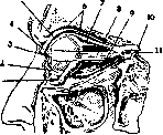
Figure 1. Sagittal section through the eyelids and orbit: 1—m. obliq. inf.; 2—palpebra inf.; 3—cut edge of conjunctiva bulbi; 4—palpebra sup.; 5—fornix sup.; 6—m. levat. palp. sup.; 7—m. rect. sup.; 8—m. rect. ext.; 9—m. rect. inf.; 10—n. opticus; 11—opening for the passage of nerves.
the axis and equidistant from the poles is named the equator and divides the eye into anterior and posterior halves. Circles drawn through the poles are called meridians. Of these, the vertical divides the eyeball into temporal and nasal halves, and the horizontal into upper and lower halves. The circumference of the equator of an adult eye is about 77 mm. The weight of the eyeball averages 7-8 g. The outer membrane of the eye is the s c l e r a (sclera, tunica fibrosa)—the white fibrous coat, passing anteriorly into the transparent cornea. Externally it is intensely white, internally brownish. Its thickness at the posterior pole is 1 mm, at the corneal margin 0.6 mm, and at the site of muscle attachment 0.3 mm. The superficial layer (episclera) consists of loose connective tissue rich in blood vessels. The main layer is formed by dense, long connective tissue bundles having a regular arrangement in the meridional and equatorial directions. At their periphery, elastic fibers are encountered, entering particularly abundantly into the composition of the internal layer (lamina fusca sclerae). The latter layer received its name due to the presence of pigment cells in it. In the equatorial region, the sclera is pierced by the emissary veins of the vortex veins, and posteriorly, around the papilla of the optic nerve, by small openings for the passage of arteries and nerves. These openings connect the suprachoroidal and Tenon spaces. There are few blood vessels in the sclera tissue, while the nerves give numerous branches ending densely in the region of the limbus and ciliary body. On the inner edge of the sclera there is a groove (sulcus sclerae internus)—the site of attachment of the ciliary muscle, and on the outer edge lies the sulcus sclerae externus. Posteriorly, the sclera passes into the hard sheath of the optic nerve, and the opening in it is filled by a thin cribriform plate (lamina cribrosa) intended for the passage of the optic nerve fibers. This cribriform plate is formed at the expense of the pia mater of the optic nerve. Anteriorly, the sclera passes into the transparent cornea, inserted like a watch glass. At the site of transition of the sclera into the cornea, a grayish corneoscleral rim is formed, bearing the name limbus corneae (see figure 2). The third membrane
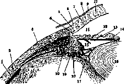
Figure 2. Angle of the anterior chamber (sagittal section): 1—retina; 2—choroidea; 3—sclera; 4—can. Schlemmi; 5—limbus corneae; 6—conjunctiva; 7—endothelium; 8—substantia propria; 9—epithelium; 10—cornea; 11—camera ant.; 12—dilatator pup.; 13—iris; 14—sphincter pup.; 15—camera post.; 16—хрусталик [lens]; 17—zonula Zinnii; 18—proc. ciliares; 19—corpus ciliare; 20—aa. ciliares.
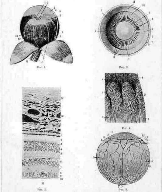
the conjunctiva, takes part in the formation of the limbus. Its submucosal tissue ends in the limbus, and the stratified squamous epithelium assumes an extremely regular arrangement and covers the cornea anteriorly, on account of which it is sometimes called conjunctiva corneae. The main layer of the cornea (substantia corneae propria) occupies 90% of its entire thickness and consists of 60-100 transparent plates of dense connective tissue arranged parallel to the surface. Between them is enclosed a system of juice canaliculi and clefts in which flat branching cells of the cornea are located. The surface of the cornea facing inward is covered with endothelium. Since the proper functioning of the eye is possible only with complete transparency of the cornea, the outer layer of which is exposed to the harmful influences of the external environment, for the best protection of the cornea between its own tissue and the covering epithelium there is a structureless membrane (Bowman's membrane), a derivative of the substantia propria. The endothelium, arranged in a single layer, cannot protect the corneal parenchyma from the penetration of fluid from the anterior chamber into it; therefore, between the parenchyma and the endothelium, a thin but dense, structureless membrane (Descemet's membrane) of the same origin is laid down. The cornea has no blood vessels, being nourished partly at the expense of the limbal network, but mainly by the lymphatic fluid circulating through its juice canaliculi. Nerves abundantly supply the cornea, being located in various planes. Their thin trunks penetrate through the openings of Bowman's membrane, spreading between the epithelial cells and forming plexuses. The shape of the cornea is ellipsoidal, slightly flattened. The vertical diameter of its base is 10 mm, the horizontal 11-12 mm. Its thickness at the periphery is about 1 mm, and in the center somewhat less. Its radius of curvature is 7-8 mm. The inner wall of the cornea at the base gradually loses its regular structure. Its plates, consisting of collagenous and elastic fibers and covered with endothelium, form the supporting framework of the angle of the anterior chamber of the eye and gradually pass into the root of the iris. The gaps between the trabeculae are named Fontana's spaces. Upon removal of the sclera and cornea, the nourishing apparatus of the eye is exposed—the vascular tract. It consists of three divisions: a) the choroid (choroidea), occupying the entire posterior section, b) the ciliary body (corpus ciliare), and c) the iris. The vascular tract (see separate table, fig. 1), tractus uvealis, is a hollow sphere with two openings: anteriorly—for the passage of light rays—the pupil (pupilla) and posteriorly—for the exit of the fibers of the optic nerve. The choroid is thin, brown in color, and occupies the space from the optic nerve opening to the ora serrata (see separate table, fig. 2). It consists of a connective tissue stroma permeated with pigment cells. A large number of blood vessels are embedded in the stroma. The larger ones are located near the sclera, then gradually decrease in caliber, and the layer facing the retina carries only extremely thin capillaries (choriocapillaris) serving for the nutrition of the outer layer of the retina. The vascular layer is separated from the retina by a thin and structureless membrane (lamina basalis), and from the sclera by plates of connective tissue between which narrow lymphatic clefts are placed (lamina suprachorioidea). The arteries passing near the sclera run unbranched to the ciliary body, participating in its nutrition. From the majority of the large venous vessels, vortex veins are subsequently formed (venae vorticosae). The next division of the vascular tract—the ciliary, or muscular body (corpus ciliare)—extends from the anterior edge of the choroid to the limbus. It has the shape of a ring 5-6 mm wide, appearing as a triangle in cross-section; ciliary processes (processus ciliares), about 70 in number, depart from its inner surface in a meridional direction. The posterior, gradually thickening part of the ciliary body—from the choroid to the ciliary process—is named the orbiculus ciliaris, and the anterior is the corona ciliaris (see figure 2 and separate table, fig. 3). In its thickness, from outside inward, is located the ciliary muscle (m. ciliaris), then the continuation of the choroid, devoid of a capillary layer in this place.
The ciliary muscle as a whole, viewed in a sagittal section, repeats the contours of the ciliary body, i.e., the shape of a triangle with its acute angle directed posteriorly, with the greater part of this triangle being occupied by the meridional and radial bundles, the so-called Brücke's muscle. The meridional bundles are located along the outer side of the triangle, i.e., parallel to the sclera. These bundles are most powerful in their anterior section, narrowing somewhat only at the place of their attachment, at the border of the cornea and sclera. At the posterior end of the meridional part of the muscle, its bundles, forming in part anastomoses among themselves, are lost in the thickness of the suprachoroidal lamina. The radial part is located inward from the meridional. The direction of the fibers is fan-like and oblique posteriorly and inwardly, and the closer to the front, the less the muscle bundles deviate posteriorly. In the radial part, there is more interstitial connective tissue, through which vessels and numerous nerves pass. The anterior end of the bundles is connected with the pectinate ligament, and the connective tissue base of the trabeculae of this angle passes into the interstitial tissue of the muscle. The other end of the radial bundles is directed toward the vascular layer of the ciliary body, where the interstitial tissue of the muscle passes into the connective tissue basis of the vascular layer. Thus, both the name given to the ciliary muscle by Brücke—m. tensor chorioideae—and Hess's name—m. protractor corporis ciliaris—are correct. Inward from the described muscles, in the projecting angle of the ciliary body, lies the circular Müller's muscle. In the ciliary corona, ciliary processes protrude into the cavity of the eye. Each of them contains a glomerulus (see separate table, fig. 4) constructed mainly of venous capillaries; the glomeruli represent a kind of corpora cavernosa. The ciliary body is abundantly supplied with nerves ending both in the muscles (oculomotor nerve) and in the processes (ciliary nerve). All the indicated parts of the ciliary body are a continuation of the choroid, and its covering, consisting of a pigmented and non-pigmented layer of epithelium, is formed at the expense of the modified retina. Anterior to the ciliary body lies the iris, a thin ring-shaped plate bordering the pupil. The choroid participates in its construction, forming its stromal leaf with a large number of vessels, and the retina—by two layers of pigment epithelium (see figure 2). The innermost layer, located posteriorly, was formed by the transition of the non-pigmented epithelium of the ciliary body into pigmented. The second layer, developed from the pigment epithelium, is a radially arranged muscle expanding the pupil (m. dilator pupillae), innervated by the sympathetic nerve. In the thickness of the vascular layer, not far from the pupil, lies the circular muscle narrowing the pupil (m. sphincter pupillae) and innervated by branches of the oculomotor nerve. When viewed in a plane from the front, the iris appears folded into diverse folds resembling rhombuses. The trabeculae forming the folds are called trabeculae. Vessels pass through most of them. The depressions between the trabeculae are called crypts. At the bottom of the latter, the pigment leaf shines through. Pigment cells are located in the stroma of the iris, upon the greater or lesser accumulation of which the color of the iris depends. Its surface facing the anterior chamber does not have an endothelium, as was previously thought. The purpose of the iris is to retain a greater or lesser amount of rays or to serve as a diaphragm of the eye. The play of the pupil provides the optimum light for the retina. For the combination of rays on the retina, the light-refracting apparatus of the eye serves, which consists of the cornea, the fluid of the anterior chamber, the lens, and the vitreous body (see figure 2).
K. Orlov, A. Pokrovsky. Aqueous humor (humor aqueus) is a fluid filling the anterior and posterior chambers of the eye. The chemical composition of the aqueous humor in the horse and bull, as more accessible for study, is seen from the following tables [composition (in %) of the aqueous humor and blood serum of the horse (according to Duke-Elder) and the bull (according to Tron)].
Dry substances (in the horse).
99.6921
1.0869
0.0201
0.0078
0.0123
0.004
0.028
0.029
0.002
0.0983
Content of inorganic substances (in the bull).
Aqueous
Serum
0.339
0.0190
0.0082
0.00105
0.437
0.0012
0.0047 Thus, all substances found in blood serum have been detected in the aqueous humor, albeit in different proportions. First of all, this refers to protein colloids and fats, which are found in the aqueous humor in significantly smaller quantities than in blood serum. This also explains the small amount of dry residue in the aqueous humor. Crystalloids are also found in the aqueous humor in different ratios than in blood serum, specifically anions (chlorine) in a greater amount, and cations (potassium, calcium, and magnesium) in a smaller amount than in the serum. In addition to the above-mentioned substances, a number of enzymes and antibodies have also been detected in the aqueous humor in concentrations significantly lower than in the serum. The chemistry of human aqueous humor has been less investigated, which is explained by the difficulty of obtaining a sufficient amount of material. However, the same characteristic features have been established here as well: poverty in protein (0.02% versus 7-8% in serum) and an excess of chlorine compared to blood serum. In general, the aqueous humor in its chemical composition differs strongly from blood serum and approaches cerebrospinal fluid very closely. The question of the nature of the aqueous humor still remains open, and in this regard, there are two diametrically opposed views. Some authors (Seidel and his school) regard the aqueous humor as a secretion of the ciliary epithelium, while others (Meesmann, Baurmann, Duke-Elder, Tron) consider it an ultrafiltrate of the blood. The first view is based mainly on the fact that the magnitude of blood pressure in the intraocular vessels is not great enough for fluid to filter from them into the interior of the eye, as well as on certain cytological data of the ciliary epithelium cells. The second view is based on the fact that the differences in the chemical composition of the aqueous humor and blood serum can be quite satisfactorily explained by a number of physicochemical factors, and the peculiarities by Donnan equilibrium without the participation of any secretory activity of the cells. Thus, the insignificant content of proteins, enzymes, and antibodies in the aqueous humor (according to the ultrafiltration theory) is explained by the fact that all these substances, as colloids, possess a large molecule and therefore are retained by the vascular wall. The reduced content of cations and the excess of anions in the aqueous humor compared to the serum fit entirely within the framework of Donnan equilibrium. According to the secretory theory, all these differences are explained by the secretory activity of the ciliary epithelium. The ultrafiltration theory has the advantage that it cohesively explains a whole series of features in the composition of the aqueous humor, relying on data from precise chemical studies. Its general acceptance is hindered only by data on the magnitude of blood pressure in the vessels of the eye, which, in the opinion of the supporters of the secretory theory, make the filtration of fluid from the vessels of the eye impossible. However, these arguments are not yet entirely convincing, since the determination of pressure in the vessels of the eye still yields very contradictory results among different authors. If a puncture of the anterior chamber is performed and the aqueous humor is evacuated, the chamber is restored again after 15-30 minutes due to the accumulation of new aqueous humor. This newly formed secondary aqueous humor differs strongly in its composition from normal aqueous humor. The general character of the changes occurring in the aqueous humor after puncture is characterized by the fact that its composition approaches that of the serum to a significant degree. In first place is the increase in the amount of protein, which after puncture can reach 3-4%. Simultaneously with this, an increase in enzymes and antibodies occurs in the secondary aqueous humor. Regarding inorganic constituents, a decrease in the content of anions and an increase in the concentration of cations are observed in the secondary aqueous humor. Some time after the puncture, the aqueous humor gradually acquires its normal composition again. Changes entirely analogous to those observed in the aqueous humor after puncture also occur in it after a whole series of irritations, such as after subconjunctival injections of NaCl, instillation of dionin. Atropine and pilocarpine also exert a certain influence on the protein content in the aqueous humor.
e. Lens. The crystalline lens is a transparent body having the shape of a biconvex lens. The center of its anterior surface is called the anterior pole, and the center of the posterior surface is the posterior pole. The plane perpendicular to the line connecting both poles is called the equator of the lens. Its outer membrane is dense, almost structureless, strongly refracts light, and is called the capsule. Only anteriorly beneath it is located a low cylindrical epithelium, while the entire cavity of the lens is filled with long prismatic fibers having the appearance of a flattened hexagon in cross-section and formed from the epithelium located along the equator. Young fibers are layered on the periphery, while the central ones lose their nuclei and sclerose. The process of fiber formation takes place until deep old age. The fibers connect in the region of sutures, visible on the anterior and posterior surfaces and having in embryonic life the shape of a star with three rays, wherein the rays of the anterior star always fall in the spaces between the rays of the posterior one. Subsequently, this star takes on an increasingly branched appearance. The lens is held in place by means of the zonule of Zinn (zonula Zinnii s. ciliaris; see figure 2 and separate table [pp. 275-276], fig. 3). The latter consists of the finest structureless fibrils directed toward the lens capsule from the ciliary processes and to a lesser extent from the flat part of the ciliary body. The lens and the zonule of Zinn play a prominent role in the act of accommodation. - The vitreous body occupies the entire posterior section of the eye, being located between the lens and the retina. It is a transparent gelatinous mass. Anteriorly, it has a depression for the lens (fossa patellaris). When removed from the eye, the vitreous body retains its spherical shape. When its integrity is violated, a transparent fluid begins to flow out of it. The described phenomenon finds its explanation in the fact that the vitreous body has a fine reticular framework, in the meshes of which the intraocular fluid is enclosed. Its fibrous framework, of ectodermal origin, attaches at the ora serrata and is more condensed near the retina and the lens, whereas in the center it is extremely loose. The condensation of fibers at the periphery is called the membrana hyaloidea. From the exit of the optic nerve to the posterior capsule of the lens along the course of the embryonic art. hyaloideae, or canalis Cloqueti, there is a lymphatic space. The fluid of the vitreous body is the same as that of the anterior chamber—the space bounded by the cornea, the angle of the anterior chamber, the iris, and the central region of the lens. The same fluid also fills the posterior chamber located between the posterior surface of the iris, the processes of the ciliary body, the fibers of the zonule of Zinn, and the lens (see figure 2). The space enclosed between the fibers of the zonule of Zinn is called the canal of Petit. In a biological sense, the aqueous humor of the posterior chamber is closer to blood serum than the aqueous humor of the anterior chamber. The vitreous body serves as a support for the retina, enabling it to adhere to the choroid throughout its entire extent.
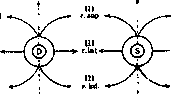
The retina is a continuation of the brain. It is a thin, delicate, and during life transparent membrane, which quickly (after half an hour) becomes turbid after death. It occupies the space from the ora serrata to the exit of the optic nerve from the eye cavity. At 3 mm outward from the optic nerve lies the place of best vision—the macula lutea (yellow spot) with a central depression—the fovea centralis [see separate table (pp. 303-304), fig. 4 and 5]. In a functional respect, the retina can be divided into 2 main layers: the cerebral and the neuroepithelial (rod and cone cells). The first faces the vitreous body, and the second faces the vascular coat, specifically its choriocapillary layer [see separate table (pp. 275-276), fig. 2]. Adjoining the latter is the layer of pigment epithelium (1), consisting of low hexagonal cells, sharply pigmented. Their thin processes are directed toward the rods and cones located in the second layer of the retina (2). Rods are thin cylindrical formations with a longer outer segment enclosing visual purple. In cones, the inner segment is significantly thicker than the outer. The region of best vision—the central pit—is occupied exclusively by cones as the most differentiated elements. In the direction of the periphery, the number of cones decreases, and the number of rods increases. The membrana limitans externa (3) separates the rods and cones from their thread-like bodies with nuclei located in the outer nuclear layer (4). The cells terminate in thin fibrils passing into the outer reticular (plexiform) layer (5). Processes of the cells of the next (6) inner nuclear layer approach the endings of the cones and rods. There lie the nuclei of bipolar, horizontal, and amacrine cells and Müller's supporting fibers. The processes of these cells intertwine in the inner reticular (plexiform) layer (7) with the dendrites of ganglion cells (8). The latter are large multipolar cells analogous to those of the cerebral substance. Axon-cylindrical processes extending from them gather in the nerve fiber layer (9) and pass through openings in the lamina cribrosa of the sclera. Horizontal and amacrine cells are association cells: horizontal cells, whose processes branch in a single plane, serve to connect the rod and cone cells among themselves; amacrine cells direct their processes into the inner reticular layer and connect the dendrites of ganglion cells. Müller's supporting fibers pass through the entire thickness of the retina from the membrana limitans interna, against which their expanded feet rest, to the membrana limitans externa, giving off lamellar processes in the nuclear layers that form baskets. The retina is separated from the vitreous body by means of the membrana limitans interna. The site of exit of the nerve fibers is called the papilla nervi optici (the intraocular part of the optic nerve). After passing through the lamina cribrosa, the fibers become covered with a myelin sheath and, uniting, form the trunk of the optic nerve. In it, the fibers occupy a position corresponding to that of the retina. Only the bundle coming from the region of the macula lutea is at first located in the lower-outer quadrant, then in the outer quadrant, and later still, much later, passes into the center of the optic nerve trunk. The fibers of the optic nerve have the same character as in the white matter of the brain. Thinner fibers are considered strictly visual, while thicker ones are pupillary. The optic nerve is covered by three membranes: dura, arachnoid, and pia mater. The latter penetrates into the thickness of the optic nerve, forming trabeculae between its bundles. Between the membranes are lymphatic spaces: subarachnoid and subdural. In the optic nerve, depending on the position, intraocular, orbital, canalicular, and cranial sections are distinguished. In the orbital cavity, the optic nerve is curved like the letter S. Its length is 28-29 mm. The eyeball is located in the anterior section of the orbit and is separated from its contents by a thin fibrous plate—Tenon's capsule, which begins from the dura mater of the optic nerve, extends forward, surrounds the eyeball, merges with the fasciae of the muscles, and ends near the limbus and in the fascia tarso-orbitalis. The space between it and the sclera is filled with delicate connective tissue pads. The eyeball is set in motion by three pairs of external muscles: four recti and two oblique (see figure 1). All of them, with the exception of the inferior oblique, originate at the apex of the orbit from the tendinous ring dividing the superior orbital fissure into two parts. The recti muscles attach anterior to the equator of the eye, as a result of which their movements correspond to their name. The oblique muscles attach posterior to the equator, and the point of application of their force is directed toward the posterior section of the eye, as a result of which the cornea moves inversely to their name. The superior oblique, beginning at the muscular funnel, goes to the upper-inner angle of the orbit, loops its tendon through the pulley, and then heads toward the posterior-outer section of the eye. The inferior oblique originates at the anterior edge of the orbit on the inner side and attaches to the eyeball posteriorly and outwardly. The function of the external and internal recti is to turn the eyeball inward and outward, respectively. Upward and downward movement is achieved by the joint actions of the corresponding rectus muscle and the reverse oblique (as can be seen from the attached diagram). The superior and inferior recti are adductors, and the external ones are abductors. By the joint action of all the recti originating deep in the orbit, the eyeball is retracted.
Diagram of eye muscle movement. inwards (backwards), and by the action of the obliques, which have a fixed point in front, the eyeball is protruded from the orbit. - The superior oblique is innervated by the IV pair (n. trochlearis), the external rectus by the abducens (n. abducens), and all the others by the III pair (n. oculomotorius). All motor and sensory nerves enter the orbit from the cranial cavity through the superior orbital fissure. The sensory nerve of the eye is the first branch of the trigeminal nerve (n. trigemini) - the ophthalmic nerve (n. ophthalmicus). It penetrates the orbit in three branches: the frontal nerve (n. frontalis), supraorbital nerve (n. supraorbitalis), and nasociliary nerve (n. nasociliaris). The latter sends 2-3 branches of long ciliary nerves into the eyeball. In the vicinity of the optic canal (foramen n. optici), between the trunk of the optic nerve and the external muscle, lies the ciliary ganglion (ganglion ciliare), which receives roots from the nasociliary nerve, the oculomotor nerve (n. oculomotorii), and from the sympathetic plexus (plexus carot.). From the ganglion depart 6 nerve trunks, which divide along their path and enter the eyeball in the number of 20, containing motor, sensory, and sympathetic fibers. The blood supply to the eye and orbit is provided by the ophthalmic artery (a. ophthalmica), a branch of the internal carotid artery (a. carotis int.) (see figure 3). It enters the orbit together with the optic nerve through the optic canal (foramen n. optici), gives branches to the muscles, lacrimal gland, eyelids, conjunctiva, and eyeball, and terminates in the vascular network of the face with three arteries: the supraorbital artery (a. supraorbitalis), frontal artery (a. frontalis), and nasociliary artery (a. nasociliaris). The eyeball receives blood through 5 systems of arteries: 1) The central retinal artery (art. centralis retinae) enters the thickness of the optic nerve at a distance of 10-15 mm from the eyeball and nourishes the inner cerebral layer of the retina [see color table (articles 303-304), Fig. 3]. It terminates in thin-walled capillaries, having no
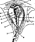
Figure 3. Arterial system of the orbit: 1 - optic nerve (n. opticus); 2 - central retinal artery (a. centr. retinae); 3 - short posterior ciliary arteries (aa. ciliares post. breves); 4 - vortex vein (v. vorticosa); 5 - muscular artery branch (ramus a. muscularis); 6 - anterior ciliary artery (a. ciliar. ant.); 7 - long posterior ciliary artery (a. ciliar. post. longa); 8 - ophthalmic artery (a. ophthalmica).
anastomoses. This same artery gives branches to the central parts of the optic nerve. 2) Short ciliary arteries, about 20 in number, penetrate through openings in the sclera near the optic nerve, branching in the choroid, forming its choriocapillary layer and nourishing the outer neuroepithelial layers of the retina (see sep. table, Fig. 5). 3) Long posterior ciliary arteries, usually two, pass through the same openings in the sclera, but run in the outer layers along the horizontal meridian of the choroid, without giving off branches until the ciliary body, where they participate in the formation of the vascular system of the ciliary body and iris. 4) An auxiliary role in the nutrition of the anterior section of the uvea is played by the anterior ciliary arteries, branches of the muscular arteries. 5) The peripheral parts of the anterior section of the optic nerve and its sheath are nourished by the Zinn's zonule, formed by the posterior ciliary arteries, while the posterior section receives nutrition from the recurrent central retinal artery (a. centr. retinae recurrens). The conjunctiva in the region of the transitional folds receives blood from the arteries of the eyelid, and the conjunctiva of the sclera around the limbus partly from the arteries of the eyelid, partly from the ciliary arteries. The lacrimal gland is nourished by the lacrimal artery (arteria lacrimalis); the lacrimal sac receives nutrition from the arteries of the eyelids. The eyelids are supplied with numerous arteries - branches of the ethmoidal and lacrimal arteries (art. ethmoidalis et lacrimalis). - Venous blood is removed from the eyeball as follows
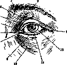
Figure 4. Front view of the eye: 1 - lateral palpebral angle (angulus oc. lat.); 2 - eyebrow (supercilium); 3 - head of the eyebrow (caput supercilii); 4 - orbitopalpebral sulcus (sulcus orbito-palpebralis); 5 - upper eyelid (palpebra sup.); 6 - semilunar fold of conjunctiva (plica semilunaris conjunct.); 7 - medial palpebral angle (angulus oc. med.); 8 - lacrimal caruncle (caruncula lacrimalis); 9 - lower eyelid (palpebra inf.); 10 - palpebromalar sulcus (sulcus palpebro-malaris); 11 - intermarginal space (spatium intermarginale); 12 - of the inner layers of the retina and from
corneal limbus (limbus corneae).
of the central sections of the optic nerve, blood collects into the v. centralis retinae. 2) From the vascular tunic, the ciliary body, and partly the iris, into the vortex veins (4–6), passing through the emissaria of the sclera at the equator of the eye. [See separate table (pp. 275–276), figs. 1 and 5, and color table (pp. 303–304), fig. 3.] 3) Part of the blood is carried off by the anterior ciliary veins, which connect with Schlemm's canal, a venous network located annularly around the limbus in the thickness of the sclera (venous sinus; see figure 2). From the conjunctiva and the lacrimal apparatus, blood flows through small venous trunks that empty into the v. ophthalmica. Numerous veins of the eyelids partly flow into the facial veins and partly, passing between the fibers of the m. orbicularis oculi, into the v. ophthalmica superior. All the veins of the orbit, connecting together, form two venous trunks: the v. ophthalmica superior et inferior. They, separately or merging into a single vein, pass through the superior orbital fissure, empty into the cavernous sinus, and provide anastomoses to the deep veins through the inferior orbital fissure. - The lymphatic vessels of the eyelids and conjunctiva empty into the preauricular lymph node, and the vessels of the lower eyelid are connected with the submandibular nodes. Most authors believe that in the anterior section of the eye, intraocular fluid passes from the posterior chamber into the anterior one and is removed from the eye through Schlemm's canal. From the posterior sections of the eye, the outflow of fluid goes through the lymphatic space in the vitreous body into the lymphatic system between the membranes of the optic nerve. Anteriorly, the eyeball is protected by the eyelids (see figure 4), upon the closure of which complete separation from the external world occurs. The posterior surface of the eyelids is lined with a mucous membrane, the conjunctiva of the eyelids. Topographically, the conjunctiva is divided into 3 sections (see figure 1): the conjunctiva of the cartilage, of the transitional fold, and of the eyeball. The conjunctiva is a delicate mucous membrane, tightly fused with the cartilage and limbus. The conjunctiva passes onto the sclera (see). The conjunctiva is moistened and washed by means of lacrimal fluid. - The lacrimal glands (see Lacrimal passages) are located in the outer corner of the orbit and are divided by the fibers of the tendon of the m. levatoris superioris into orbital and palpebral parts. They pour their secretion through numerous punctate openings in the upper-outer corner of the vault of the conjunctival sac. Comparative anatomy of the eye, see Visual organs. Pathology of the eye. The pathology of the eyeball as a whole is characterized by a change in its position and size. The eye is located in the anterior section of the orbit, moreover closer to its outer edge. The eyeball may move forward in the anteroposterior direction, which causes protrusion of the eye (exophthalmus), or backward, which causes its retraction (enophthalmus). Movement of the eyeball to the sides gives lateral displacements. - For developmental malformations of the eye, see Ablepharia, Albinism, Amblyopia, Aniridia, Ankyloblepharon, Anophthalmus, Ox eye, Heterochromia, Epicanthus, Cataract, Coloboma, Cryptophthalmus, Microphthalmus, Staphyloma, Cyclopia, etc. See also the individual membranes of the eye. Disease of the eyeball itself can be expressed either in an increase or a decrease in its size. A uniform increase in all dimensions of the eyeball occurs in the juvenile form of glaucoma, when, under the influence of increased intraocular pressure, the elastic membranes stretch ad maximum. By analogy with a bladder filled with water, such a condition is called hydrophthalmus, and by similarity with the eye of an ox, buphthalmus (see Ox eye, Glaucoma). A significantly smaller and not entirely uniform increase in the eyeball occurs with high myopia, under the influence of gradually increasing or recurrent inflammatory-degenerative phenomena in the vascular tunic and sclera. A sharp increase in the eyeball can be caused by an intraocular tumor due to an increase in intraocular pressure and direct ingrowth of the eye tissues. Finally, inflammation of all membranes of the eye (panophthalmitis) causes stretching of the outer wall of the eye. The eyeball, with intact membranes, is completely protected from the introduction of microbes from the outside. Only when the integrity of the membranes is violated can microorganisms penetrate inside the eye and cause inflammation there to one degree or another. Most microorganisms, with the preservation of the diaphragm of the eye, limit their influence to its anterior segment, and only the most virulent ones, such as pneumococci or streptococci, lead to inflammation of all membranes of the eye. If access to the vitreous body is open, then even weakly virulent microorganisms can cause panophthalmitis (Bacillus subtilis, Bacterium coli, Bacillus xerosis, the Koch-Weeks pneumococcus, the Morax-Axenfeld diplobacillus, Bacillus zur Neddeni, etc. [see color table (pp. 303–304), fig. 2]. The etiology of panophthalmitis can be exogenous or endogenous. In exogenous infection, the portals of entry are: an operation wound or scar (usually with adhesion of the iris), in particular a trepanation opening of the sclera with a thinned conjunctival cushion, penetrating ulcers of the cornea, then its outcomes—leukomas and staphylomas, traumatic injuries of the eye with the penetration of infected foreign bodies into its cavity. The clinical picture depends on the site of introduction of the infection and its character. The eye responds to it with an inflammatory reaction. The edges of the wound are infiltrated, pericorneal hyperemia appears, the conjunctiva swells, the fluid of the anterior chamber becomes turbid, a grayish reflex appears in the pupil area, acquiring an increasingly yellowish tint. Purulent infiltration of the tissue occurs in the iris. In later stages, the iris has the appearance of a yellowish plate impregnated with pus. The ciliary body, the vascular tunic, and the vitreous body are also involved in the process. Intraocular pressure rises sharply, the eye becomes hard as stone. The outer membrane of the eye stretches as much as age conditions permit. In exceptional cases, the eye can reach the size of an apple, so that the eyelids are not able to close it. In old age, when the capsule of the eye loses its elasticity, the compression of the nerves reaches maximum strength, which causes excruciating pain and burdensome phenomena of phosphenes. The surrounding tissues of the eye respond with edema, sometimes infiltration. The eyeball, thanks to the edema of Tenon's capsule, yields forward, causing protrusio bulbi. Its mobility is sharply limited, sometimes disappearing completely. The edematous conjunctiva is elevated in the form of a yellowish-red roller. The eyelids turn red and swell severely. The organism as a whole reacts to this disease of the eye with an increase in temperature, general malaise, and sometimes even vomiting. The duration of the course depends on the moment of opening of the capsule. Pus clears a path outward. The sclera in the anterior section becomes thinned until its perforation occurs, after which the pain subsides quickly. Panophthalmitis of traumatic origin with a wide opening of the capsule proceeds faster and with less pain, since the purulent contents have an outlet through the wound. The duration of the course (2–3 weeks) can be sharply reduced by performing an appropriate operation. - The second path of infection is endogenous, when certain microorganisms penetrate into the eye: a) by means of embolism (the septic principle is carried from a purulent focus into the terminal capillaries of the retina), b) upon the flooding of the bloodstream with microorganisms (the latter are detained in the wide-meshed network of the vascular tunic and ciliary body), c) the process can spread from back to front in orbit phlegmon or in thrombophlebitis of the veins, d) inflammation of the meninges can also cause the occurrence of panophthalmitis. Among general diseases, panophthalmitis is most often caused by: pyemia, especially postpartum, ulcerative endocarditis, scarlet fever, erysipelas, influenza, typhoid and relapsing fevers, smallpox, diphtheria, Weil's disease. In children, the occasion for panophthalmitis can be suppuration of the navel or smallpox vaccination, as well as pneumonia. Panophthalmitis of metastatic origin proceeds less turbulently than panophthalmitis of exogenous origin. At the very beginning of the disease, a decrease in visual acuity is noticed, then a slight pericorneal injection and weak turbidity of the aqueous humor appear, deposits on Descemet's membrane, hypopyon, and barely noticeable posterior synechiae. A yellowish reflex is barely outlined in the pupil area. In the future, a typical picture of panophthalmitis may develop, but without particularly severe pain (endophthalmitis septica). Panophthalmitis as a complication of meningitis proceeds especially sluggishly. With it, the cornea and anterior chamber retain their transparency. The yellow reflex from the fundus of the eye due to suppuration in the vitreous body resembles the picture of amaurotic cat's eye (pseudoglioma), sometimes giving rise to diagnostic errors (glioma retinae). Panophthalmitis ultimately leads to the melting of all membranes of the eye and the wrinkling of its capsule, i.e., to atrophy of the eyeball. The more turbulent the course of panophthalmitis, the more chances to expect the complete end of the process without a focus remaining in the eye that could later give exacerbations. In most cases, panophthalmitis guarantees the eye against sympathetic inflammation.
Taking the latter into consideration, old authors, by introducing a "seton" into the eye, provoked panophthalmitis with the aim of preventing sympathetic inflammation. - Treatment of incipient panophthalmitis consists of general and local measures. The first consists of the use of osmotherapy (infusions of a 25% grape sugar solution), proteinotherapy (intramuscular injections of milk), autohemotherapy, intravenous injections of 3% collargol, and dry-air baths. Subcutaneous morphine and pantopon are used as analgesics. Locally: mydriatic agents, subconjunctival injections of mercuric cyanide, warming with a thermocautery, counterirritants (leeches, heat, diathermy, etc.). In clearly expressed cases of panophthalmitis, opening of the sclera, preferably evisceration or exenteration of the eyeball by emptying the contents of the eye with subsequent cauterization of the posterior segment (the site of entry of the optic nerve and the openings for the passage of nerves and vessels). The most radical operation is considered to be enucleation, which, however, is recommended to be performed only when the integrity of the outer coat of the eye is preserved. A special contraindication against performing enucleation is the acute period of panophthalmitis (panophthalmitis florida) due to the fear of introducing infection into the orbit and the cranial cavity. Usually, after the removal of the eyeball, an iodoform gauze pack is inserted into Tenon's capsule. Some authors (Fuchs) call the maximum shrinkage of the membranes of the eye after panophthalmitis phthisis bulbi (phthisis bulbi has nothing in common with tuberculosis of the eye and with phthisis bulbi essentialis, a disease accompanied by a periodic decrease in visual acuity and intraocular pressure), whereas the outcome of plastic inflammation of the uvea is described by them as atrophy of the eye (atrophia bulbi).
K. Orlov. Circumpapillary atrophy (atrophia circumpapillaris) is characterized by atrophic changes in the vascular tunic of the eye around the optic disc and is observed 1) in old age (atrophia senilis) as a result of choroiditis circumpapillaris, 2) in glaucoma (halo glaucomatosus), and 3) in myopia (staphyloma posterior). In the affected area, the following atrophic changes occur in the peri- or parapapillary parts of the choroid and the adjacent pigment layer: the pigment layer gradually begins to disappear, which in the end is completely destroyed. Then atrophy of the stroma of the choroid and choriocapillaris develops: they lose their structure, taking on the appearance of a thin connective tissue sheet, while in this area remnants of blood vessels can be distinguished with difficulty here and there. The basal lamina is usually preserved; for the most part, the retina covering the atrophied area of the choroid is not affected either. The process consists chiefly in the degeneration of capillary vessels. Disease of the veins probably also plays a significant role. In some cases, broad atrophy of the disc is observed with anterior choroiditis. In myopia, first of all, the zone adjacent to cas on the temporal side atrophies, and from here the atrophy can already spread along the entire periphery of the disc. Sometimes circumpapillary atrophy is one of the types of congenital malformation of the scleroretinal-choroidal canal and the optic nerve. A. Lyutkevich. Atrophy of the eyeball. An atrophic eye is a blind eye, reduced in volume and softer to the touch than normal. From the pressure of the four rectus muscles, such an eye reveals flattening, and in severe degrees, even depressions along the course of the muscle tendons. The cornea is reduced, flattened, often cloudy, leukomatous, sometimes completely transparent, vesicle-like protruded or folded. The anterior chamber is either completely absent or of varying depth. The iris is cloudy, shrunken; often not even traces of it can be found in the dense scar tissue. Sometimes the retina is detached, sometimes together with the choroid, and forms a funnel-shaped cord extending from the optic disc to the ora serrata and enclosing the remains of the shrunken vitreous body. Development of bone is frequently found in the eye. Upon pressure, the eye is painful or insensitive. Shrinkage of the eye (of varying degrees) is especially severe after panophthalmitis. In such cases, the eyeball completely loses its spherical form and turns into a shapeless lump of shrunken outer capsule with complete emptiness of the cavity, where only remnants of uveal tissue can be found among the mutually contacting inner walls of the sclera, which is shrunken into folds and strongly thickened. Atrophy of the eyeball is the outcome of severe internal infections entering the eye in various ways: 1) in acute and chronic general infectious diseases, such as tuberculosis, syphilis, typhus, influenza, leprosy, smallpox, etc.; this also includes metastatic ophthalmia in various septic diseases; 2) in perforating wounds (including operations), after which not only may the wounded eye perish amidst the phenomena of developing atrophy, but sometimes a picture of sympathetic inflammation arises, which also usually ends in atrophy of the other eye; 3) in destructive processes in the cornea leading to the penetration of infections into the interior of the eye, such as glaucoma, retinal detachment, cysticercus, intraocular tumor (sarcoma). The external appearance of the eye and the degree of its shrinkage depend on the underlying process that caused the atrophy. Thus, after endogenous uveitis and endophthalmitis, the cornea remains transparent, and the decrease in the size of the eye is often weakly expressed; the same is observed in sympathetic ophthalmia. On the contrary, the cornea presents a solid leucoma or is shrunken beyond recognition together with the anterior part of the sclera (phthisis bulbi anterior) after severe purulent processes in it ending in its destruction. The same is observed after panophthalmitis. - Therapy. If the atrophic eye is constantly painful (atrophia dolorosa) or if pain appears in attacks, periodically, together with phenomena of irritation, such an eye is subject to enucleation as harboring danger to the other eye (sympathetic inflammation). Painless atrophic eyes serve as objects of therapy only for cosmetic reasons: tattooing of the leucoma, applying a prosthesis over the atrophic eye, enucleation with simultaneous transplantation of fat or cartilage into the place of the removed eye and subsequent placement of a prosthesis—these are the main therapeutic techniques for atrophy of the eyeball.
S. Ochapovsky. Eye apoplexy, hemorrhage resulting from the rupture of an artery, can take place in all membranes and parts of the eye, while it is usually lost among other symptoms of the disease picture. Only subconjunctival apoplexies attract the attention of patients; in themselves, they are innocuous and pass without a trace, but etiologically they have different diagnostic and prognostic significance. In this regard, one must distinguish: 1) apoplexies easily arising in arteriosclerotics from any physical exertion and sudden rise in blood pressure [lifting heavy objects, bending, coughing, vomiting, sneezing (sometimes in sleep), tight stool, etc.]; 2) massive apoplexies in pertussis even in children with good blood vessels; 3) subconjunctival apoplexies of traumatic origin. The later the ecchymoses appear under the conjunctiva, the deeper is their origin. Fractures of the base of the skull often produce subconjunctival hemorrhages, especially in the lower part of the eyeball, ONLY on the 3rd-4th day.
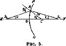
M. Averbach. Tumors of the eye, since they always originate from one or another of its membranes, will be considered in the description of the pathology of the latter (see Cornea, Sclera, etc.). Eye injuries caused by factors of various characters constitute a very practically important section of eye pathology; the traumatization of eyes in various industries should be specially noted from the point of view of labor protection (for details on eye injuries, see the pathology of individual membranes). Parasites of the eye. Among protozoa (Protozoa), the eyelid can be affected by leishmanias (Leischm. tropica, see Leishmaniases). Among parasitic worms, the most dangerous is the cysticercus (bladderworm) of the pork tapeworm (Taenia solium), since cysticercosis of the eye often leads to the loss of this organ; cysticerci can be in the anterior chamber of the eye, in the deep parts of the eye, as well as deep in the orbit, in the conjunctiva, cornea, or in the thickness of the eyelid. Among other larval forms, a parasite of the conjunctiva and eyelids in Japan and Indochina is the Sparganum plerocercoid of the broad tapeworm Diphyllobothrium Mansoni. In the orbit, as in the eye itself, the echinococcus (see) is sometimes localized. Cases of finding the fluke Monostoma lentis in the lens of a woman's eye and Agamodistomum ophthalmobium also in the lens removed for congenital cataract have been described. Agam. ophth. is a young form of some fluke (liver fluke?). Stray lung flukes [Paragonimus Ringeri (Japan, Formosa)] are sometimes encysted in the eyelids or in the tissues of the orbit. Nematodes, in turn, can live in the human eye and in the muscles serving the eye. Trichinae are sometimes encysted in the ocular muscles; under the skin of the eyelids in Sierra Leone, Guinea, Uganda, the filarial worm Loa loa is found, causing the so-called "Calabar swelling" or oedemes ambulants; Filaria conjunctivae encysts in the eyelids and in the eye in certain regions of Europe; a case of extraction of Filaria extraocularis from a tumor in the corner of the eye (in Krasnodar) is known; other poorly studied filarias of the eye are also known. Microfilariae are also found in this organ—both in hypertrophied connective tissue and in the vessels of the choroid and retina, causing thrombosis and hemorrhages; cases of elephantiasis of the eyelids [pathogen—Wuchereria (Filaria) Bancrofti] are known, as well as an example of exceptional localization in the nasolacrimal duct of an ascarid that penetrated here from the nasal cavity, where the ascarid got during vomiting. Arthropods are also parasites of the eye. The itch mite (Sarcoptes scabiei) was found in keratitis in cauliflower-like growths, while there was no scabies on the patient's body. The hair follicle mite (Demodex folliculorum, see) is found in the follicles of the eyelash glands and in the Meibomian glands. The pubic louse (Phthirius inguinalis) is sometimes localized on the eyebrows and edges of the eyelids, causing their inflammation; its nits can also be glued here; under the skin of the eyelids, the larvae of the botfly (Hypoderma) and Dermatobia hominis (South America) sometimes parasitize. In the thickness of the epidermis of the eyelids, the larva of the stomach botfly (Gastrophilus equi, see) burrows passages, causing a disease known under the name of "hair," creeping disease, Larva migrans (see). In the eye, the Wohlfahrt fly (see), the sheep botfly, and the large white-headed fly give birth to their larvae. In the anterior chamber of the eye, the larva of the botfly (Hypoderma) has been found. False parasites also occur in the eye; for example, the fly Calliphora vomitoria lays eggs in the eyes of children; the larva of the beetle Necrobia in one case capsulated in tumor-like growths of the sclera, and so on. The fauna of parasites of the human eye in the USSR has been studied very little. E. Pavlovsky. Dioptrics. The projection of external objects onto the light-sensitive layer of the retina is subject to the laws of physics. From this point of view, the eye is considered as an optical apparatus consisting of several refracting media with different refractive indices. These media are bounded by spherical surfaces whose centers are located on a single straight line called the optical axis; in other words, the eye represents a centered optical system. The properties of such a system are determined by the so-called optical cardinal points, with the help of which one can determine both the position and the size of the image of an object located at any distance from the given system. The simplest case, from which the basic properties of centered systems can be derived, is a system consisting of two media separated from each other by a spherical surface. This case will be considered first. Let two media with refractive indices $n_1$ and $n_2$ be given, delimited by a spherical surface PQ (see figure 5), having a center C and radius $r$. From the light source $S_1$, lying on the optical axis XY,
let us take ray $S_1A$, forming a very small angle $\alpha$ with the axis XY and upon meeting surface PQ forming an angle of incidence $\beta$. Upon transition into the second (right) medium after refraction, ray $S_1A$ takes the direction $AS_2$, intersecting the optical axis at point $S_2$ and forming an angle of refraction $\gamma$ with radius CA. The second ray $S_1B$, coinciding with the optical axis, passes into the second medium without refraction. Based on the law of physics, we have:
Denoting further $OS_1$, the distance of the point from the refracting surface, by $f_1$, $OS_2$, the distance of the image, by $f_2$, the radius $OC = AC$ by $r$, from $\Delta CS_1A$ and $\Delta CS_2A$ we have: $\frac{f_1 + r}{S_1A} = \frac{\sin \beta}{\sin \alpha}$, $\frac{f_2 - r}{S_2A} = \frac{\sin \gamma}{\sin \beta}$. Dividing the 1st equation by the 2nd, we get: $\frac{(f_1 + r)S_2A}{(f_2 - r)S_1A} = \frac{n_2}{n_1}$.
. . With very small angles $\alpha$ and $\beta$, one can assume $S_1A = f_1$ and $S_2A = f_2$; then equation (a) takes the form: $\frac{f_1 + r}{f_1} \frac{n_1}{n_2} = \frac{f_2 - r}{f_2}$, which after transformation and division of both parts of the equation by $f_1f_2$ takes the form: $\frac{n_1}{f_1} + \frac{n_2}{f_2} = \frac{n_2 - n_1}{r}$ (1).
(2). This formula determines the position of the principal focus in the second medium. Rays traveling in the first medium parallel to the optical axis converge in it. In the case of $f_2 = \infty$, we have $\frac{n_1}{f_1} = \frac{n_2 - n_1}{r}$, from where $f_1 = -\frac{n_1 r}{n_2 - n_1}$,
(3). the expression for the principal focus $F_1$ in the first medium. This will be the point from which rays that have passed into the second medium will take in it a direction parallel to the optical axis. Equation (b) can be presented in the form: $\frac{n_1}{f_1} + \frac{n_2}{f_2} = \frac{n_2 - n_1}{r}$, and upon dividing both parts of the equation by $n_2 - n_1$, we will get: $\frac{n_1}{n_2 - n_1} \frac{1}{f_1} + \frac{n_2}{n_2 - n_1} \frac{1}{f_2} = \frac{1}{r}$. Inserting the quantities $F_1$, equal to $-\frac{n_1 r}{n_2 - n_1}$, and $F_2$, equal to $\frac{n_2 r}{n_2 - n_1}$, we will have: $-\frac{F_1}{f_1} + \frac{F_2}{f_2} = 1$ (4). This formula makes it possible to determine the position of the focus of a point if the principal focal lengths $F_1$ and $F_2$ of the given
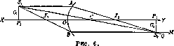
system and the distance of the point $f_1$ or $f_2$ from the refracting surface are known. Let us turn to the case (see figure 6) when $S_1$ is outside the optical axis XY, and find its focus in the second
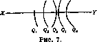
medium. 1st method: let us draw $S_1A \parallel XY$ and the straight line $AQ$ through $A$ and $F_2$, then $S_1B$ through $F_1$ and from $B$ let us draw the line $BN \parallel XY$. The intersection point of these straight lines in the second medium will give the desired focus $S_2$ of point $S_1$. 2nd method: the ray from $S_1$ passes without refraction through the center of the spherical surface (nodal point). The intersection with $BN$ or $AQ$ will give the image of point $S_1$ in the second medium. Considering $\perp S_1P_1$ to the axis $XY$, it is easy to show by construction that any point on $S_1P_1$ will have its focus on the line $S_2P_2$ perpendicular to XY. Denoting the size of the object $S_1P_1$ by $G_1$, and the size of its image by $G_2$, from similar triangles $\Delta P_1CS_1$ and $\Delta P_2CS_2$ we find: $\frac{G_2}{G_1} = \frac{f_2}{f_1}$, i.e., the sizes of the object and its image relate to each other as their distances from the center of the system. Let us turn to the important case when a very thin bundle of rays passes through several media bounded by a series of spherical surfaces $P_1Q_1, P_2Q_2 \dots P_nQ_n$ (see figure 7), the centers of which are located on a single straight line XY. To solve such a problem, it is necessary to know: 1) the radii of the refracting surfaces, 2) the distances of these surfaces from each other, 3) the refractive indices of the media entering the system. Using these "optical constants," mathematical physics provides a method for finding the cardinal points, namely—2 principal, 2 focal, and 2 nodal, which determine the path of any ray in a complex system. The properties of these points are as follows. Principal points. Let a complex system consisting of 4 media with 3 refracting surfaces be given (see figure 8). Its principal foci are $F_1$ and $F_2$. Ray $PX_1$ is incident parallel to the optical axis. After refraction, it corresponds to ray $X_1F_2$, passing through the 2nd principal focus. Obviously, these rays, if continued, will have some intersection point $S_2$. Further, let us take the continuation of ray $PX_1$—the line $OY_1$ as the incident ray of the last medium.
dys. After refraction, it corresponds to the ray Y2FX passing through the anterior focus Ft. Obviously, these rays, when continued, will also have a point of intersection. Let this be point Sx. As seen from Fig. 8, points Sx and Sa are connected by a parallel to the axis XY, and consequently lie at an equal distance from the optical axis. Rays PXX and FjY2, traveling in the first medium, converge at point Sx, and their corresponding rays in the last medium converge at S2. Thus, if point S-l is considered as the source of rays, then S2 will be its image in the last medium, and vice versa. If planes _L perpendicular to the optical axis H-Jii and H2h2 are drawn through Sx and S2, then
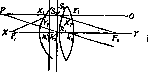
Figure 8.
in view of the aforesaid, each point St in the plane Hrkr will have a corresponding image in Дв/(4, and moreover, this image will be at the same distance from the axis XY as Sx; in other words, an object which is in the plane B.XЬ,X will correspond to an image in the plane Hzh2, equal to it in magnitude and direction. These planes are called the principal planes, and the points Hr and h2 are called the principal points of the optical system. In a simple system, these principal planes merge into one—into the plane of the refracting surface, since under the condition Д=0, formula (4) yields /2=0, i.e., the object and its image are in the plane of the refracting surface.--Principal focal points. Rays passing through the first focal point become parallel to the optical axis after refraction. Rays parallel in the first medium pass through the 2nd focal point after refraction. These points correspond entirely to the principal focal points in a simple system.-Nodal points kx and k2 (see Figures 8 and 9) are at a distance from the corresponding principal planes along the path of the ray equal to the difference of the focal lengths Fx-F2 and are characterized by the fact that a ray (Pkt) in the first medium directed towards the first nodal point (fcj) travels in the last medium parallel to its i, h, h *i^----~f-~ ~2 ')*» -\Ъ A,, "~"--Л * *a ^=^i; ^г e; Figure 9. initial path and is directed towards the second nodal point. From triangles A1P1к1 and A2P2кг (see Figure 9) we have: --= тф , i.e., Ста -ЛзЧд the magnitude of the object relates to the magnitude of the image as the distance of the object from the 1st nodal point to the distance of the image from the 2nd. Comparing formula (5) with the one just obtained, it is easy to understand that in a simple system these points merge into a single nodal point C—the center of the refracting surface. Using the property of cardinal points, it is easy to construct an image in a complex centered system. Object A1P1. From point Pt we draw the line Px^i | [ of the optical axis to the first principal plane. From here the ray must go along SXS2 perpendicular to Л^ and H2h2, since the second principal plane is the optical image of the first. From S2 the ray goes through the second focus F2 and takes the direction S2Q. The second ray P1e1, passing through the first focus Flt intersects H1h1 in point eг, whence, remaining all the time parallel to the axis XY, it takes the direction e2N. The intersection point of these two rays P2 will be the image of Px, and the image of the object (as was derived in drawing 2) will be obtained in A2P2. The light-refracting apparatus of the eye consists of three spherical surfaces: the cornea, the anterior and posterior surfaces of the lens, separating 4 media—air, aqueous humor, *ю lens, and vitreous body. Optical constants obtained by direct measurement gave the following average figures: Refractive index of air.....
1 »
»
cornea ....
1,377 »
» aqueous humor...
1,337 »
» lens (total) .
1,437 »
» vitreous body .
» The first focal point lies at..........13,75 » in front of » The second focal point lies at..........22,79 » behind » Since the focal lengths of a complex system are counted from the first principal plane, in the schematic eye F1 = - l,75+(-13,75) = = - 15,5 mm, whence its refractive power = 64,4 D. This power for the cornea will be equal to 43,2 D, since .Fi=23,l mm (see formula 3). Consequently, in the schematic eye, out of the total refractive power equal to 64,4 D, the cornea accounts for 43,2 D. As can be seen from the above figures, the principal points in the schematic eye lie so close to each other (0,34 mm) that without a large error they can be replaced by a single common one; the same can be said about the nodal points. Adopting such simplifications, we obtain a system consisting of two media: the anterior with a refractive index иx=1 and the posterior п2=1,33; with a radius of the dividing surface = =5 mm, we obtain a simple dioptric system with Fj = -15 mm and F2 =20 mm. The refractive power of such a reduced eye is 66,7 D. Image construction in the reduced eye—see Figure 6, where OP2 is the longitudinal axis of the eye, and the image P2S2 coincides with the retina. In the emmetropic eye, the distance from the cornea to the light-sensitive layer of the retina must be in a definite relation to the refractive power of the eye. The reduced eye of an emmetropic person with a refractive power of 60 D has a longitudinal axis = 22,17 mm; 63 D—longitudinal axis = 21,11 mm; 66 D—longitudinal axis = 20,15 mm.
Related articles
Cite this page
“Eye.” Soviet Medical Encyclopedia. English translation of Bolshaya Meditsinskaya Entsiklopediya, 1st ed. (Moscow, 1928–1936), ed. N. A. Semashko. https://sovietmedicalencyclopedia.pages.dev/article/eye/