Choroid
Historical document, translated for reference. It reflects medical knowledge of the 1920s–30s and is not medical advice.
Summary
This article from the 1928–1936 Soviet Medical Encyclopedia details the anatomy, histological layers, pigmentation, and physiological role of the choroid coat of the eye, highlighting its vascular structure and significance for nourishing the retina.
Encyclopedia article (1928–1936)
CHOROID of the eye (chorioidea) represents the posterior section of the vascular tract and is located posteriorly from the ora serrata of the retina to the opening of the optic nerve (Fig. 1). This section of the vascular tract is the largest and encompasses two thirds of the circumference of the eye. The choroid is brown in color; its thickness ranges from 0.05 to 0.08 mm, whereas at the posterior pole it reaches 0.1-0.2 mm. The outer side of the choroid only touches the sclera, and the two membranes are separated by a lymphatic cleft. The choroid is rather tightly fused with the optic nerve at the site of its entry into the eye cavity. There is also some connection between the choroid and the sclera at the sites where the latter is pierced by the vortex veins. Along its entire remaining extent, the choroid is easily separated from the sclera. The inner surface of the choroid is covered by a layer of heavily pigmented epithelium, which embryologically belongs to the retina (Fig. 2). Anteriorly, the choroid passes directly into the flat part of the ciliary body.

Figure 1. Choroid (located posteriorly from the ora serrata to the optic nerve opening) 1-cornea, 2-iris,
va) 7-cornea; 2-iris;
*
»
3-ciliary body; 4-ora serrata; 5-choroid; 7-sclera; 8-Tenon's capsule; 9-central vessels; 10-rectus inferior muscle.
[Note: OCR fragmentation of figure caption and text]
Histologically, 5 layers are distinguished in the choroid (Fig. 2): 1. The suprachoroidal layer (suprachorioidea, s. membrana fusca), bordering the sclera and consisting of 5-6 lamellae lying one above the other. In turn, each of these lamellae consists of elastic fibers on which endothelial cells and chromatophores are located. 2. Haller's layer (tunica vasculosa Halleri), formed by large vessels, mainly veins, giving numerous anastomoses and closely adjacent to each other. Between the veins lie connective tissue and elastic fibers and chromatophores. 3. The layer of medium and small vessels (Sattler), containing medium and small vessels. 4. Ruysch's layer (membrana Ruyschii, s. chorio-capillaris), formed almost exclusively by capillaries arranged in a dense network. This network is especially dense in the region of the macula lutea. 5. The glassy lamina (membrana vitrea, s. elastica, s. membrana Bruchii, s. lamina basalis)—a thin homogeneous membrane covering the inner surface of the choroid and separating the choriocapillary layer from the pigmented epithelium of the retina. The choroid has a dark brown color due to its rich pigment content. The carriers of the latter are chromatophores (Fig. 3), branched pigment cells in which the pigment consists of small amorphous lumps. Chromatophores penetrate all layers of the choroid except the choriocapillary layer and the glassy lamina; the intervascular space is almost entirely filled by them. Together with the pigment epithelium of the retina, the pigment of the choroid prevents too much scattered light from passing to the retina, playing the role of the dark lining of a camera obscura. However, the presence of pigment does not make the wall of the eyeball completely impermeable to light, which makes it possible to perform diascleral transillumination. An even more important physiological significance of the choroid lies in the fact that it nourishes the retina and vitreous body. The choroid is abundantly supplied with vessels belonging to the system of ciliary vessels, short posterior ciliary arteries (aed. ciliares post. breves) [see vol. VII, sep. table (p. 275-276), fig. 5 and col. table (p. 303-304), fig. 3]. These vessels form anastomoses, thanks to which circulation disorders in the choroid are smoothed out rather quickly. Veins
blood from the choroid is drained by the vortex veins (vv. vorticosae); an insufficient number of these and their oblique course through the sclera in narrow channels are unfavorable factors for the circulation in the choroid. The ligation of the vortex veins leads to severe circulatory disorders and an increase in intraocular pressure. Due to the fact that the outer layers of the retina are nourished by the choriocapillary layer of the choroid, a close functional connection exists between these two membranes, although embryologically they are completely distinct. Such a close connection between the choroid and the retina leads to the fact that when the former is diseased, the latter also suffers, and in an anatomical sense, there is no such thing as pure choroiditis; rather, chorioretinitis usually occurs, in which the outer layers of the retina are always diseased. Between the sclera and the vascular membrane there is a supra- or perichoroidal space. The perichoroidal space communicates through perivascular lymphatic clefts with Tenon's space and the intervaginal space of the optic nerve. To examine the choroid, the method of ophthalmoscopy is used. When deciding the question of in which membrane a given change in the fundus should be localized, the following rules are guided by: 1) retinitis is characterized by delicate, smoky opacities or white spots which may alternate with hemorrhage; 2) in choroiditis, a polymorphism of the ophthalmoscopic picture is observed: fresh foci appear as yellowish spots, and alongside them, old ones can be seen in the form of white spots. Depending on the duration of the process, pigment deposition occurs at the periphery of the spot or along its extent. This association of white choroidal spots with pigment is the main factor for localizing pathological changes in the choroid; 3) a very important differential sign is considered to be the relation of the central vessels to the spots of the fundus. Spots located in the retina cover the central vessels, whereas these vessels always pass over spots belonging to the choroid. Pathology of the vascular membrane. Incomplete closure of the embryonic ocular cleft leads to the formation of the so-called typical coloboma of the choroid (coloboma chorioideae). Usually, the latter begins downwards from the optic disc and, gradually expanding, goes to the periphery. Upon ophthalmoscopy, a vast white field is visible at the bottom of the eye, where the choroid is absent, and the retina is also often absent or, if preserved, in a sharply altered state, being devoid of pigment. The translucency of the sclera at the site of the coloboma imparts a white color to it. Worthy of mention is the atypical macular coloboma of the choroid, in which there is a defect of round or oval shape in the region of the yellow spot; its bottom is depressed, the edges sharply outlined and pigmented (see Coloboma). Atrophic changes in the choroid form a ring-shaped white field around the optic disc—circumpapillary atrophy (atrophia circumpapillaris) (see separate table to the article Typhus, Fig. 4). It is observed in myopia, glaucoma, and in old age (see Eye). A peculiar atrophy of the choroid is represented by atrophia gyrata chorioideae. In it, complete atrophy of the choroid takes place, occupying the greater part of the fundus, leaving free only the belt around the disc or the region of the yellow spot. In senile age, degenerative changes are observed, known under the name of drusen of the vitreous lamina (see separate table to the article Typhus, Fig. 1). Upon ophthalmoscopy, these drusen look like small shiny white foci, are a thickening of the vitreous lamina of the choroid, and cause atrophy of the pigment epithelium at the site of their formation. Also in senile age, very subtle degenerative changes occur in the region of the yellow spot with an irregular arrangement of the pigment epithelium, which leads to visual impairment. Inflammation of the choroid is named choroiditis (see). Contusions and wounds of the eye are accompanied by various damages to the choroid. In these, hemorrhages from individual vessels of the choroid can be observed, visible upon ophthalmoscopy as subretinal extravasates. The retinal vessels run over these foci without changing their course. After the resorption of hemorrhages, white plaques form on the fundus. With abundant hemorrhages from the vessels of the choroid, blood detaches the retina, sometimes tearing it and penetrating into the vitreous body. Such hemorrhages often lead to loss of vision. Expulsive hemorrhage as a result of the rupture of one of the large vessels of the choroid must be specially noted. It is observed in individuals suffering from vascular sclerosis, elevated blood pressure, and occurs during operations associated with the opening of the ocular cavity and a sudden drop in intraocular pressure (cataract extraction). With strong blows to the eye with a blunt instrument, a rupture of the vascular membrane occurs, initially covered by escaped blood. Upon resorption of the hemorrhage, a typical ophthalmoscopic picture of choroidal rupture is observed: usually, on the temporal side of the fundus, a yellowish, subsequently white stripe is visible, located concentrically to the optic disc and resembling a crescent moon in shape (see separate table to the article Typhus, Fig. 2). The retinal vessels pass over this area without changing their course. Over time, the escaped blood is completely resorbed, and pigment appears along the edges of the rupture. In case of hemorrhages and ruptures of the choroid, complete rest for the eye, bandages, resolvent agents, and subconjunctival injections of sodium chloride are recommended. Cataract extraction and scleral trepanation can sometimes cause detachment of the choroid: the anterior chamber is not restored or is very shallow, intraocular pressure is lowered, and under side illumination or examination in transmitted light, a yellowish or brownish mass protruding into the vitreous body is visible. The performance of the indicated operations may be accompanied by tears of the iris root, and through these tears, aqueous humor penetrates into the subchoroidal space, detaching the choroid. A pressure bandage on the eye promotes the apposition of the vascular membrane. Tuberculosis of the choroid proceeds in several forms: 1) miliary tubercles in the choroid are observed in general miliary tuberculosis and tuberculous meningitis. Upon ophthalmoscopy, they look like yellowish-white plaques, located in the peripapillary region or around the yellow spot; 2) solitary tubercles have the appearance of a grayish-yellow, grayish-red, smooth or nodular tumor; gradually increasing in size, these tubercles lead to retinal detachment, elevated intraocular pressure, and perforation of the sclera; 3) diffuse infiltration of the choroid by tuberculous masses gives the picture of disseminated tuberculous choroiditis with the transformation of the choroid into granulation tissue; in this form, tuberculosis of the choroid proceeds in the presence of inflammatory phenomena and the participation of the ciliary body and iris in the process; 4) disseminated choroiditis (see separate table to the article Typhus, Fig. 3) most often owes its origin to tuberculosis. It is of course out of the question to speak of therapy for the first form of tuberculosis of the choroid in view of the hopelessly grave general condition of the patient. For the second and third forms of choroidal tuberculosis, enucleation of the eye is indicated. Disseminated tuberculous choroiditis responds to tuberculin therapy, as a result of which pigmented atrophic plaques remain at the site of the tubercles. Syphilis of the choroid manifests in the following typical forms: 1) Disseminated peripheral chorioretinitis (chorio-retinitis disseminata peripherica) is observed in congenital syphilis. The entire periphery of the fundus is densely covered with small and black foci, as if sprinkled with pepper and salt; 2) Central recurrent chorioretinitis (chorio-retinitis centralis) is accompanied by the appearance of yellowish-white foci around the yellow spot; 3) Diffuse chorioretinitis (chorio-retinitis diffusa) is characterized by cloudiness of the fundus, whitish foci in the region of the yellow spot, and pigment deposition at the periphery of the fundus. At the same time, there is a smoothing of the boundaries of the optic disc and dust-like cloudiness of the vitreous body; 4) Disseminated choroiditis (chorioiditis disseminata), in which individual white spots are visible first at the periphery of the fundus. The spots are bordered around the circumference by pigment and, with the gradual development of the disease, spread from the periphery closer and closer to the region of the macula lutea and the optic disc; 5) Circumpapillary choroiditis (chorioiditis circumpapillaris), in which inflammatory foci are located near the disc itself. Energetic specific treatment of syphilitic lesions of the choroid yields very good results. Among congenital benign tumors of the choroid, neevi choroidae are very rarely encountered.
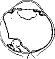
Fig. 4. Sarcoma of the choroid with chorioideae. Upon ophthalmoscopy, they are detected in the form of spots with indistinct margins; their size reaches the diameter of the optic disc. Sometimes one can observe a melanoma of the choroid (melanoma chorioideae) in the form of sharply demarcated, somewhat elevated black or mottled foci, the size of the disc and larger. - Among malignant tumors of the choroid, s a r c o m a should be noted, most frequently having the character of a melanosarcoma. In the development of choroidal sarcoma, 4 stages are distinguished: 1) At the beginning of its development, the sarcoma proceeds unnoticed by the patient due to its small size, producing only a small defect in the visual field. Then a localized detachment of the retina ensues, which takes on the appearance of a tightly stretched sail beneath which the tumor can be observed. With further growth of the tumor, a large serous detachment of the retina develops, preventing visualization of the tumor (Fig. 4). 2) The second period of development of choroidal sarcoma proceeds under the sign of increased intraocular pressure. 3) In the third period, the tumor perforates the membranes of the eye and spreads outward along the optic nerve or along the retinal membrane, passing through the scleral vessels and nerves. 4) The fourth stage is characterized by the onset of metastases, especially to the liver, which rapidly leads the patient to death. Metastases to other organs can also occur in the first periods of development of the tumor. Necrosis of the choroidal sarcoma, although rarely, causes acute iridocyclitis, in which the eye is hard and the pupil is dilated. Sarcoma of the choroid usually takes the form of a localized formation growing into the cavity of the eye. However, there is also the so-called flat sarcoma, spreading along the plane of the choroid. Choroidal sarcomas consist of spindle-shaped, round, and polygonal cells with a round or oval nucleus. Choroidal sarcomas are rich in blood vessels, and bleeding from the latter can serve as the cause for a sudden attack of glaucoma. - For the diagnosis of choroidal sarcoma, indications of trauma and myopia are taken into account, in which serous detachment of the retina occurs. The latter proceeds with decreased intraocular pressure, whereas in intraocular tumors this pressure is increased. Diagnosis is facilitated by the method of transillumination through the sclera of the eye cavity using a special Sachs electric lamp. If a dense tumor is located behind the sclera, which does not allow light to pass through during transillumination, the pupil does not light up with a red light. When the tumor is located at the posterior pole, Golovin recommends retrobulbar transillumination: a special rod equipped with a small electric light bulb is inserted through an incision in the conjunctiva and pressed against the posterior section of the eye. With tumors in the posterior section of the eye, the pupil is darkened; otherwise, it lights up with a red light. For the diagnosis of choroidal sarcoma, some recommend the examination of subretinal fluid obtained by trial puncture in order to find sarcomatous cells, but such an examination is considered unsafe. The examination of urine for the presence of melanin has a certain diagnostic value. The prognosis for sarcomas of the choroid is unfavorable: left to itself, the tumor progresses, usually leading to a fatal outcome. Patients often die from metastases to internal organs. Treatment consists of early enucleation of the eyeball; if the tumor grows into the orbit, exenteration of the latter.
11. Arkhangelsky.
Related articles
Mentioned in
Cite this page
“Choroid.” Soviet Medical Encyclopedia. English translation of Bolshaya Meditsinskaya Entsiklopediya, 1st ed. (Moscow, 1928–1936), ed. N. A. Semashko. https://sovietmedicalencyclopedia.pages.dev/article/choroid/