Ophthalmia
Historical document, translated for reference. It reflects medical knowledge of the 1920s–30s and is not medical advice.
Summary
A historical overview of the term "ophthalmia," noting its shift from a general term for eye inflammation to a specific designation for conditions like sympathetic ophthalmia. It details the pathology, clinical presentation, and the specific risk of sympathetic inflammation following penetrating eye injuries.
Encyclopedia article (1928–1936)
OPHTHALMIA (ophthalmia), a term that, with the addition of the appropriate adjective, was very often used by ophthalmologists as late as the 19th century to denote more diffuse inflammatory diseases of the eyes; at the present time, it is applied to a very limited circle of such processes. Thus, this name designates eye diseases caused by the prolonged or intense action of various kinds of radiant energy (ophthalmia electrica). The terms ophthalmia aegyptica, bellica, militaris, contagiosa, which were widely used in the 19th century to denote trachoma, have now completely gone out of use (see Trachoma). Sympathetic ophthalmia, or sympathetic iridocyclitis (more correctly, uveitis), emerged as an independent nosological form at the beginning of the 19th century. The main essence of this disease is the sequential involvement of both eyes, whereby the disease of the first (sympathizing) eye is in the overwhelming majority of cases causally linked to a previous penetrating wound of its membranes, especially in the region of the ciliary body. However, there are observations in the literature where this inflammation arose after a subconjunctival rupture of the sclera or during the disintegration of an intraocular sarcoma. The presence of a foreign body in the eye does not in itself lead to sympathetic inflammation, which in the primarily affected eye proceeds clinically in the form of iridocyclitis (see Iritis), leading to the gradual shrinkage of the eyeball. It is necessary to stipulate that not every traumatic iridocyclitis or uveitis leads to sympathetic inflammation, but only one having a specific pathological-histological character, peculiar precisely to this form of the disease. Thanks to the work mainly of Fuchs, it has been established that the main specific process plays out in the vascular tract, most constantly affecting the posterior segment of the choroid—predominantly its large-vessel layer. This entire membrane already macroscopically appears thickened many times over (Figure 1), and these thickenings
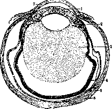
Figure 1. 1—iris; 2—ciliary body; 3—vitreous body; 4—retina; 5—choroid; 6—sclera; 7—cornea.
have an uneven, focal character. In the sympathizing eye, there are usually two concurrently occurring processes: an ordinary, more or less acutely or chronically proceeding septic traumatic iridocyclitis—uveitis, and a specific sympathetic one, for which the focality of the infiltrate is characteristic, consisting of lymphocytes, epithelioid cells, and giant cells of the Langhans type (Figure 2). The infiltrate is constructed in the same way in the ciliary body, and, spreading between the muscle bundles and then filling the ciliary processes, it has a tendency to extend beyond the limits of the pigment epithelium into the interior of the eye. In the iris, the histological structure of the inflammatory process is less characteristic and approaches the ordinary type
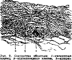
(endarteritis).
of diffuse parenchymatous iritis with characteristic focal formations (Fig. 3). In general, the process can be briefly designated as a proliferative inflammation of the vascular tract with secondary changes in the retina, optic nerve, and vitreous body. Externally and subjectively, the disease does not differ in any way from ordinary septic endophthalmitis or plastic iridocyclitis and can drag on for many years, being limited only to the primarily affected eye, and then, without any visible cause, it passes to the other eye—sympathetic inflammation. This transition can be very early, however, not earlier than 14 days after the eye injury, or, conversely, very late—after years; rare cases have been described where sympathetic ophthalmia arose after 42, 43, and 50 years. However, the most dangerous in terms of the possibility of the occurrence of sympathetic inflammation is the first year. According to Nettleship, out of 200 cases of sympathetic inflammation, in 18 the disease manifested in the first 4 weeks, in 170—within the first year, and in 12—after a year.
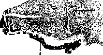
Figure 3. Iris: 1—tuberculoid
nodule with a giant cell; 2—accumulation of epithelioid cells; 3—pigment layer. Not so long ago, sympathetic inflammation was identified with sympathetic irritation, which was considered as a kind of initial form of the former, but now sympathetic irritation is considered a non-independent process, sometimes accompanying inflammation, which is manifested in three clinical forms. 1. Plastic uveitis (uveitis plastica sympathica). It often begins without subjective pain with a simple weakening of vision, which progressively intensifies. Objectively: pericorneal injection and sluggish iritis with early clouding of the aqueous humor and the appearance of exudate on the cornea, in the pupil, and in the vitreous body; the iris gives the picture of ordinary iritis: change in color, loss of luster, blurring of the relief, narrowing of the pupil, posterior synechiae. The course is distinguished by sluggishness, and the disease, subsiding and exacerbating, can drag on for many years. Often the process is complicated by an increase in intraocular pressure to such an extent that it is difficult to decide why vision is being lost. Ultimately, however, the process ends in the shriveling of the eyeball. 2. Serous uveitis, expressed by symptoms characteristic of any serous iritis in general, is encountered significantly less frequently than the first form. The disease is prolonged, persistent, and often turns into the above-described plastic uveitis with its usual outcome. 3. Inflammation of the optic nerve and retina. The rarest form, initially manifesting only by a decrease in vision and an ophthalmoscopic picture of neuritis, with swelling of the optic disc, clouding of the retina, and small hemorrhages. The course of this form is more favorable than the first two, however, here too, uveitis with its sad outcome may subsequently join. The pathological-anatomical process in the sympathized (subsequently diseased) eye proceeds basically the same as in the sympathizing one: the same character of cellular infiltrate, having a proliferative, focal character and involving the entire vascular tract. The chronic course with exacerbation and subsidence of the process affects the pathological-anatomical picture by the formation of strands and membranes of various ages. The possible pathways for the transition of the inflammatory process from the primarily diseased eye to the other—healthy—one, indicated as early as the middle of the 19th century by Mackenzie, are as follows: 1) ciliary nerves, 2) intersheath spaces of both optic nerves and the chiasm, and 3) the circulatory system. In accordance with the views prevailing in medicine in general at that time, the greatest attention was paid by ophthalmologists to the first pathway—the ciliary nerves, the irritation of which in the sympathizing eye is transmitted to the other eye and causes inflammation in humans, and in experiments in animals, only irritation (Müller's theory). As bacteriology developed, the neuro-ciliary theory was replaced by the new Leber-Deutschmann theory, explaining the transition of the disease to the other eye by the transfer of microorganisms (presumably specific) there along the intersheath space of the optic nerve of the first eye, then along the chiasm, and again along the optic nerve itself into the second eye. Deutschmann admitted that not only microorganisms but also chemical bodies could pass into the healthy eye by this path. Attributing, however, the role of causative agents of sympathetic inflammation exclusively to microorganisms, he proposed a new name for this disease—ophthalmia migratoria, clearly emphasizing by this the method of the disease's transition. However, this theory, which found ardent and active followers, was soon shaken in its foundations, and in its place was put forward the metastatic theory, which later found an ardent supporter in the person of Römer, according to whom an as-yet-unknown causative agent, possessing sharp specificity for the vascular tract of the eye, is transferred from the sympathizing eye to the other eye by the general bloodstream via metastasis. Of the cited theories, this latter one best explains the clinical facts, but its weak point is the elusiveness of the causative agent under the given conditions. Motais introduced a correction to this theory: instead of the general bloodstream, unknown microorganisms pass into the other eye via veins connecting the venous systems of both orbits through the nasal cavity. At approximately the same time, Belliarminov and Zelenkovsky experimentally developed a bacteriotoxic theory, which explained, as the name itself shows, the genesis of the disease by the transfer not of microbes, but of their toxins along the same intersheath spaces of the nerves and the chiasm. This theory acquired many supporters, however, it did not receive general recognition, as it also has weak points. In accordance with new trends in pathology, Golovin put forward a cytotoxic theory, the essence of which boils down to the fact that during trauma to the ciliary body, its epithelial covering is destroyed, and the products of decay, especially of pigment cells, possess poisonous properties in relation to the normal coverings of the ciliary body of the healthy eye. Getting into the general bloodstream, these poisonous products are carried into the healthy eye and there cause sympathetic inflammation. Elschnig came forward with an anaphylactic theory: the tissue of the vascular tract destroyed during trauma or intraocular sarcoma is an antigen that sensitizes the healthy eye, as a result of which, in the presence of suitable conditions in the body, sympathetic inflammation flares up. It is necessary to note that the pathogenesis of sympathetic inflammation cannot be considered solved even to the present time. Under such conditions, it is not surprising that the problem of treating sympathetic inflammation is also among the very weakly substantiated ones. All treatment rests on empirically obtained data: general mercury treatment (rubbing or intravenous injections) has been used since olden times, as well as sodium salicylate, collargol or electrargol, grape sugar, and neosalvarsan in the same form. As for local treatment, it does not differ from that of iridocyclitis in general (see Iritis). With such a sad state of therapy, the striving of ophthalmological thought toward prophylaxis is natural, which is undoubtedly on the right path, since it is only to it that one can attribute the fact that the imperialist war yielded a negligible number of sympathetic inflammations compared to previous ones, despite the huge number of eye wounds. At the present time, the only true prophylactic measure is the timely removal of the dangerous eye. In general, the indications for enucleation can be formulated as follows: a blind eye is subject to removal if, within two weeks after a penetrating wound, there are phenomena of irritation and no noticeable clear reduction in inflammatory phenomena. Such already atrophic eyes in which inflammatory phenomena have flared up again are also subject to removal, as well as those in which bone plates have developed. All operations proposed as a substitute for enucleation have proven untenable and can be used where there are no grounds to expect sympathetic inflammation (eyes lost from purulent panophthalmitis) (see Enucleation). Metastatic Ophthalmia represents a lesion of the deep membranes of the eye due to the introduction of infection here by the bloodstream during various general and local infectious processes: streptococci-staphylococci in erysipelas, phlegmon, puerperal disease, scarlet fever; pneumococci in pneumonia, meningococci, gonococci, causative agents of typhoid fever, dysentery, whooping cough, influenza, etc. The pathogenic agent (embolus) is carried into the retina or vascular tract, where it produces an inflammatory-purulent focus, limited or rapidly spreading to other membranes of the eye and passing into panophthalmitis. In the first case (introduction into the retina), upon early examination, one can find on the fundus of the eye a white focus of retinal clouding and hemorrhages (Roth's septic retinitis), but more often, as with metastasis to the choroid, clouding of the vitreous body develops (yellowish reflex from the fundus of the eye), toxic iritis accompanied by hypopyon, pericorneal injection, edema of the conjunctiva, edema and hyperemia of the eyelids, especially the upper one. The prognosis is always very serious, since in the best case, persistent clouding of the vitreous body remains (sclerosis of the vitreous body, pseudoglioma in children), but more often the matter ends in the gradual shriveling of the eyeball if, at the height of the process, perforation of the membranes—cornea or sclera—has not occurred, usually in the area of muscle attachment. Ophthalmia scrofulosa—scrofulous lesion of the eyes in its various forms. The diverse manifestation of scrofulosis in the form of phlyctenular inflammations of the cornea and conjunctiva, often arising simultaneously with inflammation of the eyelid margin (blepharitis scrofulosa), eczema of the eyelids, constitutes the picture of scrofulous Ophthalmia. Its pathogenesis and treatment—see Keratitis, Conjunctivitis. Electric Ophthalmia (ophthalmia electrica) arises under the influence of a Voltaic arc (short circuit, electric welding, prolonged filming with strong electric lamps—Jupiter, quartz lamp, etc.). Snow Ophthalmia (ophthalmia nivalis)—inflammation of the anterior segment of the eye under the influence of reflected sunlight from snowy surfaces—is encountered in mountainous regions, in the Arctic. Solar Ophthalmia (ophthalmia solaris)—the same as the previous one, but due to reflection from vast water surfaces or direct action, as, for example, when observing a solar eclipse with an unprotected eye.
In addition, thermal Ophthalmia is observed when working near open-hearth furnaces, etc. (see Radiant energy). Ophthalmia nodosa occurs when hairs covering the bodies of certain caterpillars enter the conjunctival sac. This disease usually appears in the spring and summer during work in gardens, when, while picking and destroying caterpillars, the latter can easily fall onto the upturned faces of the workers. Immediately after work, such individuals develop a severe inflammatory process of the conjunctiva or simultaneously of the cornea, accompanied by severe pain, characterized by the formation on the conjunctiva of the eyelids or the eyeball of nodules ranging from 1 to 2 mm in size, of a rather firm consistency, sitting singly or in groups. On the cornea, small, sharply defined white foci appear, located in the deep layers of the epithelium or beneath it. With a magnifying glass, hairs can be seen sitting in the center of each nodule. The hairs can penetrate through the cornea and the anterior chamber into the iris and produce a picture similar to tuberculosis of this membrane, with the difference, however, that here, with a magnifying glass, one can see the hairs sitting in the center of grayish-yellow or grayish-pink nodules. The course of this disease is very prolonged (up to 2 1/2 years) and is characterized by remissions. Upon microscopic examination, large giant cells are found around the hairs. The process ends with the resorption and expulsion of the hairs, although often at the cost of clouding of the cornea and posterior synechiae of the iris. The hairs act not only mechanically but also through a poison contained on or within them. Treatment: mechanical removal (very difficult) of the hairs, and then symptomatic treatment—atropine, dionin, etc.
Related articles
Mentioned in
Cite this page
“Ophthalmia.” Soviet Medical Encyclopedia. English translation of Bolshaya Meditsinskaya Entsiklopediya, 1st ed. (Moscow, 1928–1936), ed. N. A. Semashko. https://sovietmedicalencyclopedia.pages.dev/article/ophthalmia/