Iritis
Historical document, translated for reference. It reflects medical knowledge of the 1920s–30s and is not medical advice.
Summary
Iritis is inflammation of the iris, ciliary body, or more precisely, the anterior segment of the vascular tract. It is a serious condition that can lead to blindness, with various causes including infectious, toxic, and traumatic origins.
Encyclopedia article (1928–1936)
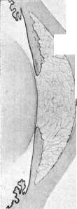
IRITIS, inflammation of the iris, ciliary body, or rather, the anterior segment of the vascular tract. Although not particularly common among other eye diseases, this condition is classified as very serious, as according to Trousseau's statistics, involvement of the vascular tract is the cause of 13% of all cases of bilateral blindness and 15% of unilateral blindness. These percentages almost exactly correspond to the frequency of unilateral and bilateral iridocyclitis: 49.5% for the former and 50.5% for the latter. Occurring at all periods of life, iridocyclites predominantly affect the most productive age group—21-40 years. According to their course, irites are divided into acute and chronic. Etiologically, iridocyclites can be divided into several groups that are by no means of equal importance in clinical practice; the most common are infectious, which are divided into two subgroups: exogenous and endogenous, followed by toxic and traumatic. Exogenous forms result from the penetration of infection due to violation of the integrity of the eyeball or due to the spread of a purulent process from adjacent foci (e.g., in orbital phlegmon). Endogenous (the most common form) arise from the carriage by the bloodstream of various pathogenic microorganisms. Toxic forms occur under the influence of toxins circulating in the blood or developing in foci of infection in the eyeball itself and its membranes or entering the anterior chamber from the conjunctival sac. Traumatic forms are due to various types of injuries. In general, most iridocyclites are metastatic diseases indicating the presence of an infectious focus somewhere in the body, not always easily diagnosable. According to Terrien, out of 100 iridocyclites and iridochoroiditis, 42 are due to general infectious diseases, 22 to postpartum conditions, 21 to surgical infections, and 14 to cerebrospinal meningitis. Gilbert, based on 500 cases of iridocyclites, obtained the following data regarding their etiology. Diseases: Syphilis without tuberculosis—Syphilis + tuberculosis—Rheumatism—Gonorrhea—Influenza—Angina—Dental diseases—Herpes zoster. Number: 173—1—55—79—15—15—15. Percent: 45.6—16.6—1.6—1.2—1.2. Mainly on the basis of clinical data, iridocyclites are divided into simple and parenchymatous, and the former, depending on changes in the composition of the aqueous humor, into serous, sero-fibrinous, and hemorrhagic; parenchymatous forms in turn are divided into simple and purulent. Much of this division is not so essential, since for therapy the etiology is more important than the form. True, there is some connection between the cause and the picture of the disease, but it is far from constant. Pathologically and anatomically, exogenous and then endogenous iridocyclites were first studied by means of experiments. In this way it was established that in any inflammation of the anterior segment of the vascular tract, the aqueous humor becomes richer in protein, which, when the preparations are hardened, coagulates to form a homogeneous or fine-granular mass, while fibrin, when present, covers all surfaces of the anterior and posterior chambers with a delicate network, sometimes connecting them with thin, interlacing threads (see separate table, fig. 3), in the loops of which lie detached and dead cells and pigment grains. Thin threads or clumps of fibrin can also be found in the parenchyma of the iris, which shows a picture of fine-cellular infiltration and edema. The same phenomena, but in a more intense form, are also found in the ciliary body and posterior chamber, with the ciliary processes appearing swollen, and the exudate exuding from them collecting not only on their surface but also between the covering layers of pigment and cylindrical cells, dissecting them and forming cavities. The exudate initially consists only of leukocytes that have come here both through the posterior pigmentous layer of the iris and from the ciliary body, but later the cells of the ciliary body covering begin to proliferate, participating in the exudation into the vitreous body, with the pigment cells gradually becoming poorer in pigment and in the future no longer differing from the others. The infiltration that occurs in the iris and ciliary body in acute inflammation is usually diffuse, but it is most densely located in the area of the sphincter as well as in the posterior layers of the iris. Its cellular composition is initially predominantly represented by multinucleated leukocytes, and later mononuclear cells, lymphocytes are added. Besides cellular elements of hematogenous origin, elements of the iris stroma and in particular chromatophores participate in the inflammation. In chronic forms of iridocyclitis having specific causes such as tuberculosis, syphilis, etc., the infiltration of the iris tissue consists mainly of small lymphocytes, plasma cells, histiocytes, and the infiltration may be diffuse or form separate foci. In composition, the exudate may be cellular, predominantly leukocytic, or fibrinous, or sero-fibrinous, with transitional forms being histologically very common. The most frequent complication of iridocyclitis is the adhesion of the pupillary margin to the anterior capsule of the lens, effected by means of the exuding fibrin. Such an adhesion is initially very fragile and can be broken with the help of strong mydriatics, leaving a grayish deposit on the lens capsule. When observing such cases during pupil dilation, one can notice that at the site of the adhesion as the pupillary margin begins to retract, a dark brown tooth appears—the pigmentous layer of the iris, which sometimes has a kind of grayish border—a layer of adhering fibrin. In parenchymatous or in more severe simple iridocyclites, the parenchyma of the iris may also be attached because the infiltrate may spread to the posterior surface of the iris or, conversely, fibrinous deposits pass from its anterior surface directly to the pupillary area in the form of a strand or membrane, which in the future may not only organize but even become vascularized. The symptomatology of iridocyclites includes subjective and objective signs. Subjective signs. The patient's complaints first note discomfort in the affected eye, which quickly turns into pain, localized not so much in the eyeball itself as in the orbit, or even in the corresponding half of the head. At the height of the process, the pains are so severe that they poison the patient's existence. Pressure on the area of the ciliary body gives a sharp painful effect, and even movement of the eyelid during blinking can sometimes be sharply painful. Photophobia varies considerably in intensity from the most moderate, appearing only in bright light, to complete inability to tolerate light at all. Visual acuity is initially unchanged, but later may fall to simple light perception. Lacrimation is usually insignificant. Objective symptoms. Pericorneal injection is sometimes accompanied by mild conjunctivitis and chemosis. The anterior chamber in size in most cases shows no deviation from the norm, except in very rarely occurring serous I., in which there is an increase in its depth. Changes in the transparency of the aqueous humor are the result of exudate entering it. With the help of a microscope and a slit lamp, abnormal impurities in the anterior chamber fluid are clearly visible, sometimes in the form of suspended particles, and sometimes attached to thin fibrinous threads filling the entire chamber (see separate table, fig. 3). Besides suspended particles, with this method of investigation, it is easy to notice deposits and precipitates on the posterior surface of the cornea—precipitates consisting of conglomerates as well as individual pigment or non-pigment cells, clumps of fibrin, grains of pigment, fragments of cells, and other products of inflammation. They are located mainly in the pupillary area and below it in the form of a triangle with its apex toward the center of the cornea. This area must be examined with particular care, at least with an ordinary magnifying glass and focal illumination, because often only the presence of small grayish, fat-like droplets gives the first indication of a chronically running iridocyclitis. The predominant precipitation of precipitates in this area occurs because it always occupies the most sloping position (see separate table, fig. 7). It should be noted that in the formation of precipitates, the endothelium, if it participates at all, does so only in very advanced cases. The aqueous humor sometimes contains so much protein that it precipitates, forming a spherical transparent body resembling a lens in shape. More often, however, fibrin threads, pigment grains, and cellular elements settle at the bottom of the anterior chamber in the form of a semicircle or segment of grayish-white or yellow color, occupying a larger or smaller part of the chamber depending on the intensity of the process (hypopyon). Depending on various etiological factors, hypopyon can also be sterile.
A change in the color of the iris is a consequence of the filling of its numerous blood vessels, due to which gray and blue irises acquire a greenish tint, while brown ones become chestnut-rust-colored. The surface of the iris loses its characteristic luster, and its relief pattern appears blurred, erased. Under a magnifying glass, blood-filled vessels are visible, and under a microscope (especially in chronic cases), small, initially semi-transparent, round grayish nodules appear on the pupillary margin, as if emerging from under the iris. Subsequently, they merge into larger, non-transparent formations [see separate table (pp. 791-792), Fig. 7]. These formations, similar to precipitates on the Descemet's membrane, are a product of deposition on the iris stroma, not a proliferation of its cellular elements. With significant stromal infiltration, the iris appears thickened, which is particularly pronounced on the pupillary margin. The infiltration itself can be either diffuse or focal, superficial and deep. The size of foci varies widely: from visible only under a magnifying glass to occupying almost the entire surface of the iris and filling the entire anterior chamber. The pupil is usually constricted, reacting sluggishly or not at all to light. Expansion under the influence of atropin occurs very slowly, initially the pupil takes on an irregular shape, and then expands, but less than normal, sometimes taking on a very capricious outline due to posterior synechiae-adhesions of its edge or the posterior pigment layer of the iris to the anterior lens capsule [see separate table (pp. 791-792), Fig. 8]. Due to the appearance of exudate in the pupillary area, the pupil acquires a grayish or even purely gray tint. With sufficient intensity of the process, a more or less significant decrease in visual acuity is observed. Intraocular pressure is usually slightly elevated at the very beginning, but later it becomes normal or even decreased; only in the serous form of iridocyclitis does intraocular pressure remain elevated. Complications occurring during iridocyclites make the prognosis serious. Thus, initially single synechiae, increasing in number, can lead to annular adhesion around the entire circumference of the pupil-seclusio pupil-lae, which leads to the separation of the anterior chamber from the posterior chamber. This circumstance leads to a violation of the circulation of aqueous humor, which, accumulating in the posterior part, bulges the iris forward (see separate table, Fig. 4), so that it surrounds the pupil lying in the depth in the form of a valva. This condition usually leads to an increase in intraocular pressure-secondary glaucoma (see). Adhesion of the pupillary margin is often accompanied by the formation of a membrane covering the pupil (occlusio pupillae), which at this time is sometimes so constricted that it is barely visible (atresia pupillae). With abundant exudation on the posterior surface of the iris and its organization, a complete adhesion of the iris to the lens and ciliary body occurs, as a result of which the chamber in the center will have normal depth, while on the periphery it will be larger, and in such cases the lens is often surrounded by exudate, which subsequently organizes into connective tissue membranes, which inevitably leads to a violation of its nutrition-inflammatory cataract. Relatively rarely, only in chronically occurring iridocyclites, a pulling of the newly formed membrane of the pigment epithium onto the anterior surface of the iris is observed-ectropion uveae. In all the above cases of complications of iridocyclites, the appearance of the iris itself also changes sharply: it becomes poorer in pigment - it discolors, its surface becomes smooth and at the same time matte, while the parenchyma becomes more flaccid and thins. Sometimes the own tissue of the iris seems to be absent in places, as a result of which the pigment layer is visible, which in turn becomes lighter, and in places may almost completely disappear, which is particularly clearly manifested during diaphanoscopy as well as during ophthalmoscopy. This entire process is designated as atrophy of the iris and is usually accompanied by the same process in the ciliary body, as a result of which the eyeball becomes softer, its configuration changes, flattening occurs corresponding to the four rectus muscles-phthisis bulbi incipiens (shrinking of the eyeball). It is necessary to mention the effect of iridocyclites on the optic nerve, in the membranes of which in the nearest segment to the eyeball, small-cell infiltrations were observed. Iridocyclites caused by exogenous infection almost always extend to the posterior part of the vascular tract, causing either the purulent inflammation of all the membranes of the eyeball-panophthalmitis, or the plastic inflammation of the vascular coat of the eye (uveitis plastica); the latter is generally often observed in severe iridocyclites, whatever their cause. The diagnosis based on the data presented is not difficult, and it is quite possible to avoid the most dangerous error of confusing iridocyclitis with an acute attack of glaucoma, if one remembers that in acute glaucoma intraocular pressure is sharply elevated, the anterior chamber is shallow, the pupil is wide, vision is sharply reduced from the very beginning, the cornea is cloudy, whereas in iridocyclitis the tension is either normal or reduced, the anterior chamber is normal, and in serous I. even deepened, the pupil is narrow, the cornea is unchanged". Depending on the etiological factor, the course and clinical picture of iridocyclites may differ in some features, sometimes so characteristic that the etiological diagnosis can be made based on them alone. Not so long ago, syphilis was considered the most common cause of iridocyclites; at present, Western European researchers put tuberculosis in first place, which, according to Gilbert's statistics (see above), underlies almost half of the diseases of these parts of the eye. Clinically, tuberculous lesions of the anterior part of the vascular tract occur in two forms: 1) in the form of serous or parenchymatous iridocyclitis and 2) tuberculosis of the iris and ciliary body. The first variety is much more common than the second and occurs either in the form of parenchymatous, characterized by blurring of the relief of the iris and easily ruptured synechiae, or serous iridocyclitis, characterized mainly by greish precipitates on the Descemet's membrane and almost complete absence of macroscopic changes in the iris; even the pupil, which expands very moderately with strong atropinization, retains its round shape for a long time. The disease proceeds chronically, differing in a very capricious course: long periods of
s \
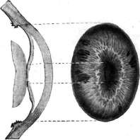
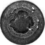
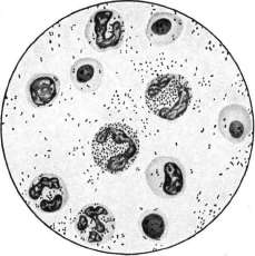
Figure 1. Brucella abortus (Bang). Figure 2. Bact. abortus equi. (Figures 1 and 2 according to Kolle-Wassermann).
Figure 3-7. Iridocyclitis. Periods of calm alternate with acute flashes, which again pass into a sluggish subacute state. At the height of the inflammatory process, hemorrhages are observed more frequently than in other iridocyclites. Extremely characteristic of these forms is early focal and diffuse depigmentation. These forms are most commonly observed in adults and are benign in nature, however, a transition to a more severe form - tuberculosis - is possible, characterized by the appearance of small grayish nodules - in serous iridocyclitis and yellowish ones - in parenchymatous (see separate table, fig. 5).-Iritis of the iris and ciliary body in childhood more often occurs in the form of a solitary tubercle (a more malignant form). The process usually begins at the periphery, at the root of the iris, with the appearance of a coarse-grained yellowish-pink formation, around the base of which there are always small tubercles, although initially distinguishable only in the corneal microscope. Gradually increasing and merging with the main central granuloma, they fill the entire anterior chamber and lead to the destruction of the cornea or sclera. A more benign form is characterized by the appearance of small nodules, grayish when superficial and yellowish when deep, scattered over the entire surface of the iris and having no particular tendency, in contrast to syphilitic ones, to be located in a specific area. Usually these tubercles develop on the basis of a pre-existing chronic iridocyclitis in youth and more mature age, however, very rarely appearing after thirty years. Syphilis gives no less diverse forms of iridocyclitis, and the latter among generally syphilitic eye lesions occupy first place in frequency - 44.7%. This does not include changes in the iris occurring in the roseola stage without any subjective sensations, expressing sharp hyperemia of its superficial vessels - first in the ciliary part and then passing to the pupillary area. The most common clinical form is the fibrinous iridocyclitis, which usually arises three months after infection. Serous forms are rare exceptions. In the former, due to the abundant fibrinous exudate, posterior synechiae develop quickly, which are distinguished by their special strength because in the formation of the adhesion not only the fibrinous effusion but also the infiltration of the iris parenchyma participates, the pupillary edge of which sharply thickens. Particularly characteristic of this form of I. is the lesion of the vascular walls, on which thickenings develop, as a result of the accumulation of epithelioid cells, to which endarteritis is subsequently added, leading to narrowing of the vessel lumen. In the parenchyma, round-cell accumulations are also found, which are not characteristic of the luetic process. It should be noted that in a luetic, iridocyclitis can develop on any other basis, and rheumatic iridocyclites are often accompanied by hypopyon, which is not encountered in the purely syphilitic form. Significantly less frequently than the form described above, papular I.'s are encountered in the early stages of syphilis, characterized by the appearance of reddish nodules - papules, both single and multiple, developing mainly in the pupillary area, which is explained by the peculiar structure of the arterial system here. The appearance of papules is usually accompanied by fever. The size of papules varies widely - from microscopic to wheat grain and larger. They rarely break down, but after their resorption, a trace in the form of thinning and atrophy of the iris parenchyma remains (see separate table, fig. 6).-As for later manifestations - gummas, they are rarely found in the iris, and more often under this name are described gummas of the ciliary body that have grown through its root into the anterior chamber. Regarding the latter, it should be noted that their appearance does not always coincide with the tertiary period of syphilis; on the contrary, they are much more often observed in the secondary, which is why it is more correct to call them syphilomas. For a long time they can remain unrecognized in the picture of a sluggish or rather acutely proceeding iridocyclitis, until finally in the angle of the anterior chamber a yellow-orange tumor appears or on the sclera in the area of the ciliary body a brown nodule appears - a syphiloma breaking through outward, simulating a sarcoma. The differential diagnosis in this case is complicated by the fact that a syphiloma can develop in the form of a single node and multiple ones, as well as a ring-shaped formation. It is built from lymphocytes, as well as accumulations of epithelioid cells with not infrequent admixture of giant cells; in places, nests of polynuclears are also found. The prognosis is always very serious, because even with a favorable course - resorption and organization of the infiltrate - blindness occurs in 62%. The given percentage refers to the pre-salvarsan period, and it must be assumed that at present the prognosis with early recognition should be more favorable. Rheumatic iridocyclitis can be considered only a disease developing on the basis of existing acute or chronic inflammation of the joints and muscles. Acute rheumatic iridocyclitis of moderate severity often begins with hyperemia of the conjunctiva, without bacteriologically confirmed etiology. Gradually, hyperemia of individual large, radially running vessels of the iris and their superficially lying branches is revealed. With the gradual intensification of this hyperemia, a sharply expressed pericorneal injection and pupil narrowing appear. On the surface of the iris, delicate fibrinous precipitates fall out in separate nests, and on the lower half of the cornea - precipitates. The iris is usually not thickened; its adhesions with the lens are located in one place or another depending on the position of the body in which the patient spends the greater part of the day. It is possible that sometimes the cause of such iridocyclites are generally metastases from not particularly virulent microorganisms from some unrecognized foci. Gonococcal iridocyclitis usually appears at the height of the development of urethritis and predominantly in men, and the moment of metastasis to the eye is accompanied by a sharp decrease in the discharge from the urethra, which resumes after a few days with the same force. Often, the eye disease is preceded by the lesion of one or many joints. The disease proceeds in the form of fibrinous iridocyclitis, and it is precisely with this form that the above-described lentiform clots in the anterior chamber are most often encountered. Almost as a rule, iridocyclitis is accompanied or even begins with a metastatic conjunctivitis with chemosis and mucopurulent discharge. In subjects with advanced or chronic gonococcal lesions of the genitourinary apparatus, recurrences are especially often observed, due to which in the pupillary area pictures corresponding to figure 8 of the separate table (pp. 791-792) can be obtained. Patho-anatomically, this form has not been studied for quite understandable reasons. The experiment on animals does not correspond to the pathogenesis in humans.-Leprous iridocyclitis is often encountered in anesthetic leprosy, and in tuberous leprosy it is observed in 74%, developing as a result of the transition of the process from the cornea or sclera or independently, and proceeds in a chronic form with the formation of small nodules in the pupillary and large ones in the ciliary area. The descriptions given of the characteristic forms of metastatic iridocyclites by no means exhaust all their types, but since the etiology of the others does not affect their clinical picture, it will be sufficient to dwell only on the typhoid iridocyclite, observed in such large numbers during the epidemic of the intervention and civil war period. According to data from the city of Rostov, eye complications were observed in 5.3% of typhoid patients. The disease usually began with slight clouding of the vitreous body, then ciliary injection appeared, followed by the development of a typical picture of iridocyclitis, with flakes in the vitreous body, synechiae and often hypopyon. Pathogenetically, the disease is not yet clear. Among the iridocyclites arising under the influence of toxins of local origin, the greatest clinical significance is attached to those in purulent processes of the cornea, when the toxin, penetrating into the anterior chamber, acts directly on the iris and ciliary body. The same is observed in severe trachomatous pannuses, then in deep extensive burns of the cornea and sclera - iritis putrida. Toxin can also form in the posterior half of the eyeball and from there influence the anterior part - iridocyclitis with a limited abscess in the vitreous body, with parasites in it or in the retina, with the disintegration of intraocular tumors. As for I. in retinal detachment, it is more probable that the cause here lies in some unrecognized inflammatory focus in the flat part of the ciliary body.-Some chemical substances (e.g. Podophyllin, juice of some species of Euphorbia used for removing warts, juice of Primula obconica flowers, etc.), getting into the conjunctival sac, cause iridocyclitis. Toxins can also be carried by the general blood flow and cause the disease.
Such iridocyclitis can include gouty, for which a particularly characteristic feature is the discrepancy between weakly expressed inflammatory phenomena from the iris and ciliary body and sharp sclero-conjunctival ones, often having a focal nature. An attack of I. is often a precursor of a gouty seizure and is distinguished by a prolonged course with frequent exacerbations and relapses. In women, iritis menstrualis is sometimes observed in connection with menstrual disorders, which would perhaps more correctly be classified as metastatic. There is a special form of iridocyclitis known as sympathetic inflammation. The treatment of iridocyclitis with established etiology should first and foremost be etiological, and in this respect, specific therapy for syphilitic iridocyclitis is accompanied by the greatest success—energetic use of mercury (intravenous administration); Erlich preparations are especially indicated here. Tuberculin therapy for tuberculosis of the iris and ciliary body is less reliable. Climatotherapy and general strengthening treatment are of great importance in the latter processes; in recent years, radiant energy has been successfully used locally. As symptomatic treatment for all forms of iritis, local treatment is of enormous importance, consisting in the use of atropine and its analogues in normal, and even more so slightly lowered, pressure. 1% solutions are used 2-3-4 times a day until maximum dilation of the pupil is achieved. By paralyzing the sphincter of the pupil and the accommodative muscle, atropine gives rest to the affected organ, and by reducing the surface of the iris, it simultaneously reduces the capacity of the vascular bed, decreases hyperemia, and finally prevents the formation of synechiae by drawing the pupillary edge to the periphery, where contact between it and the lens surface will no longer be so close. If the pupil does not dilate despite prolonged atropinization, leeches are applied to the temple: natural ones in the amount of 2-3 or artificial ones—Herteloup. Such bloodletting gives relief from pain and, by reducing stagnant phenomena in the eye, facilitates atropine's pupil-dilating effect. Furthermore, warm compresses or hot fomentations are applied; dark glasses or bandages on the affected eye. In recent years, radiant energy has been acquiring ever greater importance in the therapy of iridocyclitis in general. With increased intraocular pressure, atropine is contraindicated, but frequent bloodlettings and cautious pilocarpin are absolutely indicated. Sometimes it is necessary to resort to puncturing the cornea and releasing aqueous humor. In purulent exogenous iridocyclitis, such release of fluid is recommended to be done repeatedly with washing of the chamber. General methods of treatment consist of mild laxatives, hot foot and general dry-air baths. In general, for iridocyclitis of the most diverse origins, intravenous injections of 25% grape sugar every 1-3 days in 40 cm3 doses are very beneficial. To this solution, one can add collargol, cyanide of mercury, sodium salicylate, urotropin, etc., as desired. Lactotherapy also deserves attention (its excellent effect in post-typhoid iridocyclitis). Chronic, stubbornly relapsing iridocyclitis often yields only to surgical intervention—iridectomy, which is also indicated in pupil occlusion, whereas in seclusio with protrusion of the iris into the anterior chamber, transfixion is limited. With complete posterior synechia, iridectomy is not applicable, and the operation of Wenzel is performed.
In the period of complete and prolonged failures, it seems especially pleasant. Thus, I. is a special case of the integrity of our perceptions. The question of the physiological basis of the phenomena indicated cannot yet be considered sufficiently developed. Lit.: Dejerine J., Semiologie des affections du systeme nerveux, P., 1926; Goldschneider A., Schmerzproblem, B., 1920.
Related articles
Mentioned in
Cite this page
“Iritis.” Soviet Medical Encyclopedia. English translation of Bolshaya Meditsinskaya Entsiklopediya, 1st ed. (Moscow, 1928–1936), ed. N. A. Semashko. https://sovietmedicalencyclopedia.pages.dev/article/iritis/