Meningitis
Historical document, translated for reference. It reflects medical knowledge of the 1920s–30s and is not medical advice.
Summary
This article from the 1928-1936 Soviet medical encyclopedia discusses epidemic cerebrospinal meningitis, covering its causes, symptoms, diagnosis, and treatment approaches during that period.
Encyclopedia article (1928–1936)
811 Epidemic cerebrospinal meningitis
841 Meningitis (syn. leptomeningitis), inflammation of the soft meninges (see). Etiology. Any pathogenic microorganism can be the cause of acute M., but some of them (tubercle bacilli, Friedländer's diplococci, streptococci, typhoid bacilli, etc.) especially frequently, and the meningococcus of Wexelbaum, the pneumococcus even predominantly affect the soft meninges. Besides infection, M. can be caused by soluble poisons (lead, CO, alcohol) penetrating into the subarachnoid spaces. Intralumbar injections of various medicinal drugs sometimes cause aseptic meningeal irritations, in some cases with polynuclear cytosis. Uremia is sometimes accompanied by a meningeal reaction. Infection of the soft meninges occurs in three ways. 1. Directly - in complicated open fractures of the skull bones with exposure of the soft meninges. Skull fractures without damage to the outer coverings can spread to the pyramid of the temporal bone, ethmoid, sphenoid bones and frontal sinuses, whereby communication is established between the natural accessory cavities of the skull and the subarachnoid spaces, leading to infection of the soft meninges. Trauma even without a skull fracture can activate a latent infection existing in the body, promoting its fixation in the soft meninges. Early and late traumatic M. are distinguished. 2. Infection from parts nearest to the brain (skin, bones, accessory cavities) can spread to the soft meninges. Especially often the source of infection are purulent inflammations of the middle ear and carious processes in the pyramid of the temporal bone, and in such cases the process directly passes to the soft meninges, more often by local lymphatic paths, perineural paths or venous sinuses. Suppurations not only of the eyeball but also of the orbit itself, osteoperiostitis, simple conjunctival infections with chemosis can infect the soft meninges through the orbital fissure or through the bony walls of the orbit. Frontal, ethmoid, sphenoid sinuses are direct sources of infection of the soft meninges. Inflammation of the maxillary cavity usually first causes inflammation of the orbit, and with further spread the soft meninges also become infected. Lesions of the spine, more often tuberculous, less often fractures or exposure of the spine in deep penetrating bedsores, can be complicated by infection of the soft meninges. 3. Metastatic M. develop when infection is transferred by the hematogenous route. Puerperal, bronchogenic, intestinal sepses, endocarditis, infection of the gallbladder and urinary bladder can result in metastatic M. Infection of certain organs especially often causes M. (pneumococcal M. in inflammations of the lungs, bronchiectases (less often), suppurations in the pleura and lungs). Complications with metastatic M. also occur in intestinal infections, mainly of the large intestine, in infections of the skin cover (furuncles, erysipelas, phlegmons of the face) and more distant parts of the body (anthrax, infectious processes in serous cavities, bones). Endocarditis causes M. by mycotic embolism of the vessels of the soft meninges, although in endocarditis embolisms of the cerebral vessels are more common. General infectious diseases are also complicated by M. (secondary); in such cases infection also spreads to the soft meninges by the hematogenous route. According to the course, acute and chronic inflammations of the soft meninges are distinguished; acute M. in turn are divided into serous and purulent forms according to the nature of the histopathological process and exudate. An etiological classification of acute M. cannot always be carried out, since often the causative agent of the disease cannot be established and moreover different causative agents sometimes give a uniform picture. From the group of purulent M., epidemic meningococcal M. has been isolated; other forms of purulent M., although they represent some peculiarities, are nevertheless difficult to distinguish in practice. From the serous forms, tuberculous and syphilitic M. have been isolated. The classification of acute M. is complicated by the fact that along with the peculiarities of the causative agent, the properties of the macroorganism and the external living conditions of the patient often come to the fore. The form of the local inflammatory reaction of the soft meninges, the character and course of the entire process are determined by the immunobiological state or resistance of the organism to infection. Thus, the same causative agent, e.g. the meningococcus, with different individual susceptibility and different external conditions causes different tissue reactions in the form of serous or purulent M., having different clinical pictures and course - acute in some cases and more chronic in others. Meningeal symptom complex. The individual etiological forms constituting the group of acute M. are united by a common core - the meningeal symptom complex, which consists of general cerebral and local symptoms of irritation of the soft meninges and phenomena of loss from the spinal nerve roots and cranial nerves. Headache is one of the most constant symptoms of the meningeal symptom complex. Noise, light, movement intensify the headache, which is often accompanied by vomiting without nausea, usually without tension, in the form of a stream. From the intestine, persistent constipation are often observed, sometimes the abdomen is drawn in. The second characteristic sign of M. are contractures: 1) rigidity of the occipital and back muscles causes extensor resistance during passive flexion of the head; contracture of the long back muscles causes limitation of mobility of the spine; 2) contractures in the lower extremities are expressed by the Kernig symptom (see Kernig's symptom), the contralateral Brudzinski symptom and the occipital symptom (see Brudzinski's symptoms). The Lesage symptom (suspension symptom) also belongs to the group of contractures, which is expressed in that if a sick child is lifted by the armpits, the meningitic child holds the legs bent and fixed at the knee and hip joints, while a healthy child constantly bends and straightens the legs. Contractures are distributed in the form of mono-, hemi-, paraplegia. On the face, individual groups of muscles - ocular, masticatory, lingual, pharyngeal, etc. - can be affected by contractures. Contractures can be both temporary and permanent. Less constant in the picture of M. are convulsions, more common in children; partial and generalized convulsions are distinguished. Sometimes acute M. begins with a seizure of general epileptiform convulsions. More common are partial convulsions in mono-, hemiplegic form or with the character of tics. Movements in the form of chewing, teeth grinding, sucking, trismus also belong here. In acute M., pseudospontaneous movements of a stereotyped character - rubbing some part of the body, patting oneself, etc. - rarely occur. Phenomena of loss in acute M. are expressed by paralyses, which are of two kinds. In some cases they are incomplete, last for a short time, often occur after convulsions or are established gradually. These paralyses usually develop in the first days of the disease. In other cases, paralyses appear in later stages of the disease, develop slowly, and once established, do not regress; the spread of these paralyses occurs in the order of the anatomical arrangement of cortical centers. From the side of the sensory sphere, in the initial stages of M., phenomena of irritation (hyperesthesias, paresthesias) are observed, in later stages of the process - anesthesias. Hyperesthesias can be generalized and partial. Their intensity varies from slight increases in sensitivity to the most severe pains from the slightest touch; they are usually observed in the stage of pronounced M., very rarely in the prodromal period (tuberculous M.); having lasted several days, hyperesthesias usually disappear. Anesthesias are also generalized and partial, sometimes unilateral; they are constant in the later and residual stages of M., but it is very difficult to detect them in this stage of the disease due to the clouding of the patient's consciousness. Usually, hyperesthesias of the sense organs - hearing, vision, less often smell - sharply stand out. Hyperesthesia of vision is expressed in marked photophobia, pains in the eye muscles, reflex narrowing of the pupils. From the side of hearing, hyperacusia is noted. From the side of the eyes, inequality of pupils is observed, narrowing in the initial and paralytic dilation of the pupils in later stages of the disease. In acute stages of M., sometimes a decrease in visual acuity, neuritis, stagnant phenomena in the papilla of the optic nerve are observed. In the residual stages of acute M., various degrees of weakening of vision, hearing, smell are detected on the basis of lesions of the peripheral apparatus. Tendon reflexes in acute M. are normal, sometimes increased to clonus, but can also disappear; vasomotor phenomena are caused by central disturbance of the regulation of the tone of skin vessels; the latter are often greatly dilated, which is objectively confirmed upon examination with a capillaroscope. Vasomotor disorders in acute M. are expressed in marked dermographism (Trousseau's meningeal sign), frequent changes in redness and paleness of the face (Trousseau's spots).
From the pulse side, arrhythmias, dissociation between the pulse and te (slowing of the pulse with elevated temperature and increased pulse with normal temperature), fluctuations in blood pressure, irregularities in rhythm, and increased cardiac activity are observed. From the respiratory side, irregularity in rhythm, respiratory arrests, discrepancy between thoracic and abdominal breathing, Cheyne-Stokes breathing, and asthmatic attacks in the terminal stages of M. are observed; the latter is sometimes preceded by cessation of cardiac activity. In the terminal stages of acute M., disturbances in swallowing are also observed, which are partly caused by the unconscious state of the patient (coma). Secretory disorders are expressed by increased sweating and salivation; these phenomena are particularly pronounced in the initial stages of acute M. Temperature is usually elevated; in certain forms it remains very high, in others, for example, in tuberculous and certain forms of serous M., t° does not reach high degrees. The type of the temperature curve is irregular, remittent, often with large fluctuations. There is no parallelism between the axillary and rectal t°. In some cases, before death, high elevations of t° are observed. In acute M., polymorphous skin rashes sometimes occur, which appear quickly and also disappear quickly. Sphincters are not affected in acute M., but due to suppression of the psyche or transition of the inflammatory process to the roots and spinal cord, retention or incontinence of urine, less frequently of feces, may develop. The cerebrospinal fluid has great importance in the clinic of acute M., both in symptomatic and differential-diagnostic relations. The cerebrospinal fluid in some cases is turbid, of a purulent nature, in others it is transparent, sometimes colored yellowish or yellowish-green (xanthochromia), in some cases it has a hemorrhagic character. In certain forms of acute M., a clot forms in the expelled fluid (tub. M., some cases of serous M.). The pressure of the fluid in acute M. is elevated, so that during puncture the fluid is discharged in a stream or very frequent drops. In some cases, when inflammatory adhesions exist, the pressure of the overlying column of fluid is decreased or eliminated, so that the pressure of the discharging fluid is minimal. The cerebrospinal fluid contains an increased amount of albumins and globulins—more than 0.25% (positive Nonne-Appelt reaction, phase 1, Pandi, etc.). In acute M., especially in tub. M., the sugar content is significantly reduced, and later it completely disappears from the fluid (normal sugar content in the fluid ranges from 0.5 to 0.75 mg%). The content of NaCl and generally of chlorides is reduced. The cryoscopic point is lowered, except in purulent meningitis, in which an increase in A is observed (average cryoscopic point—0.56 according to Ashar). The permeability of the soft meninges in acute M. is significantly increased. The number of cellular elements in the cerebrospinal fluid in acute M. is very increased (in normal fluid their number does not exceed 3-6 cells per 1 mm3). In some forms of acute meningitis, lymphocytes predominate, in others—polynuclear cells; sometimes pathogenic microorganisms are found in the fluid. Mental phenomena are constant in the clinical picture of acute M. In the prodromal period (tub. M.) in children, changes in character appear. Sometimes psychomotor excitation is sharply expressed, in other cases, on the contrary, indifference, depression, negativism, refusal of food, hallucinations predominate; consciousness in some forms of acute M. remains clear until the end (this is often observed in epidemic M.), sometimes it is lost quite quickly, coma develops. In such cases, patients often emit inarticulate sounds, cries (cri hydrencephalique), especially when changing position, perform a series of uncoordinated, aimless movements with their hands, bite their lips, pick their nose, scratch their body. The meningeal symptom complex under the influence of various conditions is modified. Thus, the picture of acute M. reflects the predominant localization of the anatomical process, the age of the patient, the presence of general infection. M., localized mainly on the convex surface, are characterized by the predominance of psychomotor excitation. M. of the base (e.g., tuberculous) are characterized by early and predominant involvement of the nerves of the base. Most often in this affection, the oculomotor nerve is affected (ptosis, paralysis of individual eye muscles), due to which double vision occurs; paralysis of the internal eye muscles—sluggish pupillary reaction to light, change in their size, inequality of pupils. Almost as often as the oculomotor nerve, the facial and abducens nerves are affected. In addition, in M. of the base, early appearance of respiratory and vascular disorders is observed. Involvement of the brain ventricles in the meningeal process with obliteration of the outflow pathways from the ventricles (foramina of Magendie and Luschka) is expressed by the picture of acute hydrocephalus (acute acquired hydrocephalus): increased intracranial pressure, headaches, often in attacks, vomiting, stagnant phenomena in the papillae of the optic nerves. In infants, bulging of the fontanelles, progressive increase in skull volume, and convulsions are particularly pronounced. Secondary M. in acute infections arise during the main disease, therefore their onset may pass unnoticed, as the general infection can also cause vomiting, headache, delirium, convulsions, etc. Sometimes in general infections, the exhausted organism weakly or does not react at all with meningeal symptoms to the secondary infection of the soft meninges; in such cases, latent M. is sometimes spoken of. The picture of the meningeal symptom complex changes extremely depending on the age of the patient. In infancy, the central nervous system is not yet fully differentiated, therefore in the picture of acute M., general reactions in the form of general and partial convulsions, increased intracranial pressure (bulging of fontanelles, drowsiness) predominate. In old people, the meningeal symptom complex is mostly reduced: headache is often weakly expressed, and the patient does not complain of it, psychomotor excitation is almost absent, the content of delirium and all the behavior of the patient have the character of dementia. At the beginning of the disease, insomnia, restlessness are usually observed, which later pass into indifference, a stuporous state and even coma. Vomiting is observed very rarely; meningeal contractures are often absent; trembling in the limbs, in the head is usually sharply expressed. The disease often develops gradually, slowly, but proceeds rapidly and sometimes ends fatally in a few days. Pathophysiology of the meningeal symptom complex. Headache in M. is caused by affection of the soft meninges, as a number of researchers established the fact that with direct irritation of both the dura mater and the brain substance, no sensation of headache occurs. The infection, acting directly on the nerve endings in the soft meninges of the brain, causes their irritation. Due to affection of the vegetative apparatus of the soft meninges and the vascular plexus, active hypersecretion of cerebrospinal fluid and increased intracranial pressure develop, which in turn irritates the endings of the vegetative nerves of the soft meninges, thereby intensifying the headache. The patient's condition significantly improves when the fluid is released, which lowers the intracranial pressure. The Kernig symptom represents a normally existing synkinesia, intensified by topical tension of muscles due to irritation by the process of the vegetative apparatus of the soft meninges, spinal roots, and the accompanying peripheral irritation in connection with passive flexion of the leg. Fixation of the limb in a certain position in the Kernig symptom is due to contracture of antagonists and predominance of physiologically stronger flexor muscles. The contralateral Brudzinski symptom also represents a phylogenetically old synkinesia, returning due to increased muscle tone as a result of irritation of the spinal roots and simultaneous suppression of the activity of the brain by the meningeal process. The upper Brudzinski symptom belongs to the group of cervical and labyrinthine reflexes of Magnus-Kleijn (see Magnus-Kleijn reflexes), which has been established experimentally. Cerebral and bulbospinal focal phenomena of loss and irritation in acute M. are caused by encephalitic and meningo-radiculo-myelitic processes on the basis of the same infection. Generalized convulsions, in the picture of which one can note the successive irritation of individual centers of the cortex, are caused by widespread meningoencephalitis in the cortex; partial epilepsy attacks are caused by limited meningoencephalitis in the cortex, their clinical picture entirely depends on the localization of these foci. Paresthesias and hyperesthesias are caused by irritation of the posterior roots and spinal nerves, less frequently they have a central origin. The state of tendon reflexes depends on the state of the reflex arc.
On the basis of compression of the spinal cord by exudate or increased intracranial pressure, pathological reflexes and reflex-spastic symptoms sometimes appear. Serous M. Acute serous M. can occur with almost all infections, but is most frequently observed in typhoid fevers, bronchopneumonias, and influenza. Epidemic parotitis, colibacillary infection, general sepsis, rubella, and angina are also sometimes accompanied by meningeal symptoms, and the cerebrospinal fluid is secreted under increased pressure and sometimes shows inflammatory changes. Diseases of the middle and inner ear, nasal cavity, conjunctiva, orbit, and other parts adjacent to the meninges, as well as inflammatory processes located far from the cranial cavity (lymphangitis, typhlitis), can cause serous inflammations of the meninges. In a number of cases, serous M. represents the initial stage of purulent M., into various forms of which it subsequently passes. In all the cases listed above, acute serous M. is secondary. There are also primary forms of acute serous M., occurring without preceding infection. These forms are characterized by an acute onset in complete health and a relatively favorable course. The causative agents in primary serous M. are often not found; apparently there is no specific causative agent for this form of M., and acute primary serous M. develops as a result of infection with various ordinary and filterable bacteria, the virulence of which is altered or weakened due to certain physiological conditions. It has been experimentally proven that weakened cultures of typhoid bacilli cause serous M. The latter is also caused by meningococcal infection, and the etiological identity of these cases with meningococcal M. is established only serologically (agglutination), and partly epidemiologically. Besides infection, serous M. can also be caused by dissolved exogenous and endogenous poisons circulating in the blood. Besides infection and toxins, acute serous M. is also caused by head trauma. One must distinguish cases of acute serous M. that accompany infected skull wounds, as these cases belong rather to secondary infectious serous M., and cases of aseptic serous M. arising from superficial head injuries, even without visible violations of the skin cover. Positive findings of bacteria in these cases are extremely rare, so the cerebrospinal fluid is considered sterile; the transition of these cases to purulent forms of M. is almost excluded. Some authors even express the opinion that trauma in traumatic serous M. is only a predisposing, but not a causative factor, while infection is the causative factor. Among other predisposing factors, one must mention sunstroke (insolation), chronic alcoholism, and chronic nephritis. - Symptomatically, one must distinguish two forms of acute serous M.: 1) diffuse and 2) localized. Diffuse serous M. is not accompanied by high elevations in temperature, consciousness is not clouded, sometimes mild mental disorders are observed, rarely a soporose state or delirium. Neuritis or stagnant phenomena in the optic nerve papilla are often found. In the initial stages of serous M., pareses of the cranial nerves of cerebral or bulbar type are already found, whereas in other forms of M., paralysis of the cranial nerves has a peripheral character and usually develops in later stages of the disease. The protein and globulin content in the cerebrospinal fluid is elevated; cellular elements in the fluid, mainly lymphocytes, are sometimes present in very large numbers. The fluid, usually clear, often has a hemorrhagic character, sometimes takes on a greenish-yellow color. A small number or absence of polymorphonuclear cells in the fluid, rare clot formation, and a large number of lymphocytes in connection with the clinical picture, course, and outcome are characteristic of serous M. and speak against tuberculous M. Against acute syphilitic M. speak the absence of an etiological factor and negative serological reactions of the fluid. Besides the composition of the fluid, for serous M. the constant and early appearance of cerebral symptoms and the presence in some cases of infectious diseases (typhoid, pneumonia, etc.), sometimes complicated by serous M., are characteristic. A favorable outcome of the disease confirms the diagnosis of serous M. The diffuse form of serous M. must be distinguished from 'meningism,' which is a rapidly developing and equally rapidly disappearing meningeal symptom complex observed in various infectious diseases; in such cases, cerebrospinal fluid is secreted under increased pressure, is sterile, and shows no other changes. The phenomena of meningism are caused by toxic-infectious irritation of the vegetative apparatus of the meninges of the brain and spinal cord; with the elimination of toxic-infectious products, the phenomena of irritation quickly pass, and meningism disappears. - Patho-anatomical changes in diffuse serous M. are expressed by hyperemia, edema of the meninges with exudation of serous fluid into the subarachnoid spaces; microscopically, inflammatory exudative-proliferative changes are found in the meninges, desquamation of the endothelium of the lymph spaces. In the cortical substance of the brain, softening is found, more rarely hemorrhages in connection with disorders of meningeal circulation. The nerve cells of the cortex are in a state of fibrinolysis and tigrolysis. The localized form of serous M. (syn. localized arachnoiditis, serous meningeal cysts, cystic meningitis, arachnoidal cyst) can be localized in any part of the brain and in the spinal cord. The etiology of primary localized serous M. (arachnoidal cysts) is not yet sufficiently clarified, although it is undoubtedly that the main etiological factor is infection, trauma, intoxication (alcohol, lead, arsenic), especially in people who have previously suffered meningo-cerebral lesions; among chronic infections - syphilis, tuberculosis. In patients with tuberculosis, the meninges often react to the presence of tuberculous bacilli in them with hyperemia, serous M., on the basis of which serous arachnoidal cysts develop. The latter are found on autopsies of these individuals who die from other causes or from pulmonary tuberculosis. Localized forms of serous basal M. and internal hydrocephalus are caused by an inflammatory process in the meninges, due to which the subarachnoid cisterns of the base are isolated into closed cavities-cysts, which press on the brain substance and nerves of the base. The pathological process localized in the area of the circle of Willis and the central cistern affects the sympathetic plexuses of the carotid artery, branching from the superior sympathetic ganglion on both sides, as a result of which the regulation of blood and fluid pressure is disturbed, hypersecretion develops, and fluid accumulates in the ventricles. During operations on opening the skull, the dura mater of the brain and spinal cord does not pulsate and is tense; it is thickened and fused with the meninges and the bones of the skull. On the surface of the brain, cysts leave depressions, in the area of which the cerebral cortex is thinned. When the process is localized in one of the basal cisterns, the cerebellum under the influence of the pressure of the cyst can be forcefully displaced upward and pressed against the tentorium cerebelli, which causes a number of cerebellar symptoms. Localized serous M. of the brain base is clinically expressed mainly by paralyses of the nerves of the base (most often of the oculomotor nerve), symptoms of lesion of the brain appendage and internal hydrocephalus. Meningeal contractures, inflammatory changes of the cerebrospinal fluid are absent or weakly expressed. Increased intracranial pressure predominates in these cases; patients complain of headache, mostly in attacks, of decreased memory, working capacity, disturbance of statics, dizziness; stagnant papillae, changes in the rhythm and regularity of the pulse are observed; in some cases, mental phenomena predominate. These cases are considered as acute acquired internal hydrocephalus. In clinical terms, abortive forms of acute acquired internal hydrocephalus are very important, which were described by Quincke. In the anamnesis of such patients, one can establish frequent attacks of headache, nausea, constipation, and noise in the ears. On ophthalmoscopy, mild stagnant phenomena are found from the side of the optic nerve; the mentioned attacks are significantly relieved after lumbar puncture. Quincke described an abortive form of internal hydrocephalus expressed by migraine. In other cases, mental phenomena in the form of periodically occurring attacks of excitement, depression, or neurasthenic phenomena predominate. The asthenic form of abortive hydrocephalus is expressed by headaches, muscle pains, dizziness, adynamia with signs of increased intracranial pressure. Due to the different localization of arachnoidal cysts, the clinical picture of the latter is polymorphic.
The localization of arachnoid cysts on the convex surface of the cerebral hemispheres is comparatively rare; they are much more frequently found on the base, especially in the posterior cranial fossa in the area of the cisterna cerebello-medullaris and cisterna acustico-facialis of the cerebellar-pontine angle. Even in the diffuse form of serous M., marked cerebellar symptoms are often found due to compression and even indentation of the cerebellum into the foramen occipitale magnum. The clinical picture of localized serous M. in the posterior cranial fossa resembles the picture in cerebellar tumors, so that all the characteristic symptoms of the latter are present also in cysts of the posterior cranial fossa: localized tenderness of the skull on percussion, pain in the occiput, rigidity of the occipital muscles, attacks of dizziness, nystagmus, early choked disk (in rare cases the latter is absent), static ataxia with falling to one side, cerebellar asynergy, adiadochokinesia, the Barany symptom complex, disturbance of muscle sense and sense of weight. Sometimes localized serous M. manifests as a picture of a tumor of the cerebellar-pontine angle. The localization of localized serous M. in the perigypophyseal area manifests as a symptom complex of a tumor of the brain appendage. In these cases, the bulging floor of the third ventricle compresses the optic nerves and the brain appendage, although Stumpf considers that in this case there is not direct compression of the brain appendage, but a disturbance of the relationship between it and the brain due to interruption of the nerve pathways connecting them, or insufficient transition of secretory products from the glandular to the nervous part of the hypophysis. In such cases, concentric narrowing of the visual field, phenomena of dystrophia adiposo-genitalis and other secondary endocrine disturbances are observed. The clinical picture of localized serous M. on the convex surface of the cerebral hemispheres presents certain characteristic features: comparatively slight increase in pressure of the cerebrospinal fluid on lumbar puncture, which may increase in the further course of the process, absence or late appearance of choked disks, localized attacks of partial epilepsy, rhythmic myoclonias of varying frequency, spreading in the order of arrangement of cortical centers. The myoclonias in these cases are very close to the myoclonias observed in epidemic encephalitis (Claude). The clinical picture of spinal localized serous arachnoiditis resembles an extramedullary tumor, from which it is very difficult, sometimes impossible, to differentiate an arachnoid cyst. Cystic arachnoiditis is indicated by fluctuations in the intensity and distribution of sensory disturbances (hyperesthesia and anesthesia), the upper boundary of which never changes, while the lower is movable. Arachnoid cysts in the spinal cord are single and multiple; they are located at different levels of the spinal cord, more often in the cauda, in the thoracic and lumbar regions. The cysts are located between the pia and arachnoid membranes, almost always on the posterior surface of the spinal cord. On patho-anatomical examination, the walls of arachnoid cysts present either fresh inflammatory changes or residual changes after a completed inflammatory process. In the first case, the newly formed tissue of the cyst is rich in cellular elements, penetrated by numerous capillary vessels; in traumatic cases, blood pigment is found intra- and extracellularly. In the residual stages, acute inflammatory changes give way to the formation of fibrous scar tissue; in most cases, the contents of such cysts are sterile. The development and course of arachnoid cysts are acute, subacute, and chronic, often with characteristic remissions. Arachnoid cysts must first be differentiated from brain tumors, and the following points speaking in favor of cysts must be taken into account. 1. The presence in the patient's past of recurrent attacks of increased intracranial pressure. 2. The existence in the anamnesis of head trauma, ear or eye disease, although in this case the possibility of a brain abscess is not excluded. This possibility decreases with the increase in the interval between trauma and the onset of cerebral phenomena and is excluded in the presence of remissions and attacks of exacerbation of the process. 3. Acute onset of the disease, sometimes with elevated temperature. 4. Paralysis of the nerves of the base, especially of the eye movements, early appearance of choked disk. 5. Favorable effect on the choked disk, headache, and general well-being of lumbar and suboccipital puncture. 6. Prolonged (sometimes for years) benign course of the symptom complex. Nevertheless, even with tumors, especially with lipomas, psammomas, and cholesteatomas, remissions of general and focal symptoms are also often observed, but in these cases the remissions are less stable and complete than in cystic arachnoiditis. Solitary tubercles, gummas, cysticerci, echinococci can shrink and calcify; the transudate of cystic cavities inside tumors can be absorbed, and some of the phenomena disappear in such cases. Secondary hydrocephalus, complicating brain tumors, gives attacks of worsening and remissions in connection with changing conditions of outflow of fluid from the brain ventricles. Arachnoid cysts with localization in the cortex must be differentiated from solitary tubercles, tumors, and meningo-cerebral hemorrhages. In the presence of cystic arachnoiditis, it is necessary to keep in mind localized tuberculous M. in the form of separate foci (localized tuberculous pachi- or leptomeningitis). Their clinical pictures are similar with the only difference that in tuberculous M. slight increases in temperature, positive Pirquet skin reaction, clonic convulsions or paralysis, increase in protein (mainly globulins) in the cerebrospinal fluid, often lymphocytes are observed; Hasselt found tuberculous bacilli in such cases. Tuberculous localized leptomeningitis constitutes an intermediate stage to cystic arachnoiditis in tuberculosis and to diffuse tuberculous leptomeningitis. The prognosis for life and recovery in the diffuse form of serous M. is in most cases favorable. The latter depends partly on early recognition. Complications leading to defects and death are also observed. The prognosis in localized serous M. depends on the localization of the cystic process, its spread, size, and accessibility of the cyst for surgical intervention. In making the prognosis, the property of serous M. to recur must be kept in mind; the age of the patient and the condition of the organism are also of great importance. In elderly and weakened patients, serous M. proceeds much worse, remissions are shorter, and the disease takes a protracted course. In spinal cystic arachnoiditis, the prognosis depends on early diagnosis of the condition and the operation undertaken on this basis. It is undoubtedly true that in serous M., both diffuse and localized, spontaneous recoveries occur. Treatment. In the diffuse form of serous M., frequent drainage of large amounts of fluid with the introduction of a 1% solution of urotropin endolumbally is used. When serous M. is a variety of meningococcal epidemic M., it is necessary to use antimeningococcal serum endolumbally. In localized M. of the base, lumbar and suboccipital punctures are used; the latter are mandatory in case of stagnant phenomena in the optic nerve. In acute hydrocephalus of the ventricles, repeated lumbar punctures, ventricular punctures, puncture of the fontanelles in infants, and ruptures of the corpus callosum often have a favorable effect, and sometimes even are accompanied by cessation of the process and recovery. In cystic arachnoiditis of the base and convex surface, lumbar and suboccipital punctures in most cases do not give special benefit; the most rational in these cases is direct surgical intervention in the form of decompressive trepanation, if the localization of cystic arachnoiditis is not quite clear or there is a picture of acute internal hydrocephalus. Opening of the posterior cranial fossa is indicated in cases when the cyst gives a symptom complex of a tumor of the cerebellar-pontine angle; opening of the middle cranial fossa is indicated in the infundibular form. The success of the operation depends on its early application. In spinal arachnoiditis, when the clinical picture corresponds to an extradural tumor, laminectomy is necessary. If localized serous M. complicates some chronic disease of the spinal cord, the operation can even cause an aggravation of the patient's condition due to exacerbation of the basic disease. Purulent M. Etiology. Purulent M. are not an etiological form, since under certain conditions various pathogenic microorganisms can cause suppuration in the soft cerebral meninges. More often than others, pneumococcal, streptococcal, colibacillary, influenza, typhoid, etc. M. are encountered; in purulent M., mixed infections are also observed; from the general infections, each can cause purulent M. Patho anatomy. Purulent M. are localized mainly on the convex surface of the cerebral hemispheres, although they are also found on the base and in the spinal cord.
Pus-thick, more or less rich in fibrin, often greenish-yellow in color, covers the surface of the brain completely in the form of a cap or in separate plaques; the latter is often observed when the purulent process spreads from adjacent foci of suppuration (arachnoid abscesses). In the initial stages, only hyperemia of the soft meninges and a large accumulation of serous exudate in the arachnoid spaces are found; subsequently, the exudate turns into seropurulent and purely purulent, sometimes with an admixture of blood. The exudate accumulates in the basal cisterns and in the convolutions of the cortex; its composition is mainly polymorphonuclear leukocytes. In the tissue of the soft meninges, perivascular accumulations of leukocytes, infiltration of the vessel walls and arachnoid tissue by round-cell elements, proliferation, desquamation, and necrosis of the endothelium of the subarachnoid spaces are found. A similar purulent inflammatory process occurs in the region of the choroid plexus and ependyma of the ventricles, which are purulently melted and necrotized. Often, the purulent inflammation spreads to the brain substance, and the spread of the process occurs along the adventitial lymph spaces, which together with the vessels penetrate from the soft meninges into the brain substance. In the brain tissue, in addition to diffuse infiltration, limited foci of purulent melting of tissue (abscesses) and local thrombophlebitis are formed. In the central nervous system, the pathogens spread through the cerebrospinal fluid and along the subarachnoid spaces. From a pathogenetic standpoint, two groups of purulent meningitis are distinguished: 1) Meningitis spreading per continuitatem, and 2) metastatic, or hematogenous purulent meningitis. The first form originates from adjacent foci of suppuration near the soft meninges, the pathogens of suppuration correspond to those present in the main purulent focus. In terms of frequency, otogenic forms are in the first place, which are associated with chronic inflammation of the middle ear. The most common sources and routes of infection in purulent meningitis are as follows: 1) direct contact of the affected skull bone with the soft meninges, 2) spread of infection through the lymph spaces of the labyrinth, which widely communicate with the subarachnoid spaces, 3) spread through the petro-squamous fissure and the facial nerve canal, 4) spread through the carotid canal, 5) suppuration in the paranasal sinuses (they spread directly through the bones to the dura mater, and then to the soft meninges through venous anastomoses or through lymph vessels), 6) orbital suppurations, 7) sinus thrombophlebitis, 8) rhinogenic general septicemia (rare), 9) purulent processes in the skin of the head (e.g., erysipelas, furunculosis, especially with nutritional decline), 10) complex fractures of the skull bones, as well as trauma without violation of the integrity of the skull. In the latter case, infection penetrates through the injury to the soft parts of the skull and may be detected already after several hours or later when the wound is contaminated or when osteophlebitis develops. With fractures of the skull base, infection of the soft meninges can originate from the external auditory canal or nasal cavity. The microorganisms causing traumatic purulent inflammation of the soft meninges are the same as those causing suppuration of the soft tissues in infections (most often Staphylococcus aureus is found, less often Streptococcus and Pneumococcus). The metastatic form of purulent meningitis occurs much less frequently; in some cases it is caused by septic emboli, in others it is one of the symptoms of general sepsis. Purulent leptomeningitis in infectious diseases are of hematogenous origin; they develop particularly often in typhoid fever, paratyphoid fever, influenza, pneumonia, pleurisy. In some of these cases, purulent meningitis originates from local foci of suppuration: e.g., purulent rhinitis in influenza, purulent otitis in other infections, etc. In purulent pleurisy, infection spreads along the intercostal nerves and roots, thereby penetrating the soft meninges. In a small number of cases, primary purulent meningitis is spoken of, since visible foci of suppuration are not found in these cases; apparently the portals of entry of infection in such cases are the tonsils, bronchi, intestine. Individual bacteriological forms of purulent meningitis. Pneumococcal meningitis very often complicates various infectious diseases (typhoid, influenza, endocarditis, etc.) due to the presence of pneumococcus in the paranasal sinuses. Otogenic, rhinogenic meningitis and meningitis after angina are often of pneumococcal etiology. Pneumococcal meningitis are purulent, although in some cases they have the character of serous meningitis. Pus-greenish-yellow, very thick, located mainly between the pia and the brain substance, often accompanied by cellular-protein dissociation of the cerebrospinal fluid; leukocytosis may be absent.-Streptococcal meningitis are purulent, sometimes serous in nature, mostly of secondary origin (bronchopneumonia, erysipelas, puerperal infection, etc.), occur
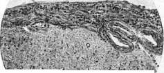

», .'; ' "-,.-; ... if-- '- ''Л,-' :-4^J'Л-Ь.> l : - :$<!if'* ?' :x$:<:-s § . 'A .-v^Ts -':- i-'-v :...: 'Xfh \ ': ijiv-: ;:-. Figure 1. Chronic fibrous leptomeningitis. Thickened, scar-like soft meninges; thickened vessel walls; significant lymphoid infiltration. Fig. 2. Purulent leptomeningitis. Soft meninges of the brain infiltrated with leukocytes and lymphoid elements. Fig. 3. Tuberculous meningitis with caseous tubercles. Fig. 4. Purulent cisternal leptomeningitis. Infiltrated soft meninges with altered nerve roots. Fig. 5. Purulent meningitis: a-purulent soft meninges; b-cerebellum. Figure 6. Tuberculous meningitis: a-tuberculous tubercles in the soft meninges; b-infiltrated soft meninges; c-pia mater; d-medulla oblongata; e-cerebellum. Pneumococcal M. The disease is very severe, but recovery is possible. Staphylococcal M. occurs very rarely; white staphylococcus is more frequently found in otogenic M. Pneumobacillary M. (Friedländer's pneumobacilli) rarely occurs as a primary condition, more often as one of the localizations of generalized infection. Recovery is not excluded. Cases have been observed after trauma. Colibacillary M. occurs rarely, almost exclusively in children; sometimes in colitis, atonic stages. The cerebrospinal fluid is clear or purulent; recovery is observed. Gonococcal M. is caused by the gonococcus, which from a bacteriological standpoint must be differentiated from the meningococcus. Actinomycosis also causes purulent M., most often as a complication of chronic actinomycotic process. Symptomatology of purulent M. The onset is sudden, less often with prodromal symptoms: headache, fatigue, general malaise, etc., which often go unnoticed against the background of the main infectious or septic disease. Simultaneously with severe headache, there is a high temperature spike, often with chills; the typical temperature curve is constant, later it may take a hectic character, normal or subfebrile temperature is very rarely encountered. The pulse is weak, irregular, rapid. Breathing is also rapid, nausea, vomiting; general hyperesthesia, consciousness is depressed (stuporous, later comatose state); sometimes delirium with psychomotor excitement and hallucinations. General and partial convulsions (especially frequent in children). Meningeal contractures are sharply expressed, later paralysis of the limbs. In the initial stages of the disease-bulbar phenomena. The cerebrospinal fluid is purulent, the cryoscopic point is lowered, the percentage of sugar and chlorides is reduced, but not to the same extent as in tuberculous M. Protein in large quantities. The permeability of the soft meninges is significantly increased. Cellular elements are present in large numbers in the fluid, mainly polymorphonuclear, in most cases degenerated. Sometimes bacteria are found in the fluid. The course of purulent M. is rapid, sometimes lightning-fast, death in such cases can occur within 24-48 hours or less, usually the disease lasts from 6 to 8 days and ends in death from paralysis of respiration in a comatose state or during convulsive seizures. In purulent M., there are no characteristic epidemic M. outbreaks and remissions of the process. The literature describes individual cases of recovery after purulent M., with defects remaining in the form of paralysis of cranial nerves and limbs, internal hydrocephalus and mental lesions; less often-complete recovery. Diagnosis of purulent M. is based mainly on the properties of the fluid (large number of polymorphonuclear cells, sometimes microorganisms) and on the presence of a source of suppuration or infectious disease. Epidemic meningococcal M. differs from purulent M. in that it is a primary disease; in some cases, the presence of an epidemic or sporadic cases of meningococcal M. can be established, the meningococcus is often found in the fluid; other bacteria are absent. The course of meningococcal M. is remitting, which does not occur in purulent forms; herpes, arthralgia and skin rashes are often observed, which usually do not occur in purulent forms. Consciousness in meningococcal M. usually remains until the last moments and, conversely, is quickly lost in purulent forms. Serous forms of M. differ in the nature of the fluid (large number of lymphocytes), often hemorrhagic fluid, absence of microorganisms. The course of serous M. is much milder than purulent ones, consciousness is clear. Meningitides in infectious diseases differ in their transient course, absence of changes in the fluid, increased pressure of the latter. M. in infectious diseases is often obscured by the main disease; the lesion of the soft meninges in these cases is expressed by paralysis of cranial nerves, meningeal contractures and changes in the fluid. Treatment: repeated punctures with the release of large amounts of fluid and endolumbar administration of polyvalent streptococcal and staphylococcal sera, endolumbar administration of 5-10 cm³ of 20-40% solution of urotropine. In purulent foci in the bones of the skull due to traumatic injuries of the latter or in suppurations in the paranasal sinuses, surgical treatment is used with the aim of removing the source of suppuration.
m.
Margulis. Epidemic cerebro-spinal meningitis (meningitis cerebro-spinalis epidemica), an acute infectious disease with inflammation of the soft meninges as the most important symptom of the disease. Epidemic cerebro-spinal M. is distributed throughout the entire globe and is observed in all countries year after year as an endemic disease. Etiology. Epidemic cerebro-spinal M. is caused by the meningococcus of Weichselbaum (1887), which is usually found in the cerebrospinal fluid, often in the blood, in secretions from the pharynx, and in pus in complications (see Meningococcus). Epidemiology. Infection with meningococcus occurs mainly through "droplet" infection and contact. As with other infections, bacillus carriage has great importance for the occurrence and spread of epidemic meningitis. In this regard, the research of the English is interesting: thorough examinations of troops free from epidemic M. revealed from 2% to 5% carriers of meningococcus. From further research, it was found that an increase in the number of bacillus carriers to 10% already signaled impending trouble in terms of M. disease, and when the percentage of bacillus carriers reached 20, there was an immediate danger of an epidemic developing, as individual cases of M. were already appearing. An epidemic of M. usually develops slowly and gradually. Epidemic M. is more common where overcrowding predominates, where elementary sanitary-hygienic practices are lacking, and where material conditions are poor. The youngest age group is most predisposed to epidemic M., mainly children under 3 years, with the highest numbers occurring in children under 1½ years. After 3 years and up to 5 years, this predisposition decreases noticeably, yet still yields high numbers in these age groups, and only after 5 years does the number of cases of epidemic M. decrease more significantly. As for the constitutional peculiarities of the child, it is hardly possible to speak of any particular constitution as a factor especially predisposing to epidemic M. The influence of the external environment as a predisposing factor and one that often has primary importance is very great. This includes climate, local peculiarities, certain seasons of the year with different relationships between the number of sunny days and cloudiness, the amount of precipitation, the magnitude of barometric pressure, etc. Furthermore, the socio-economic conditions of the environment, its sanitary-hygienic appearance, and finally, diseases that are currently prevalent among the population. For example, it can be assumed that influenza infection activates the virulence of meningococcus and thereby increases the number of cases of epidemic M. Similarly, any catarrhs of the nose and throat can serve as a predisposing factor for M., as they decrease the resistance of the mucous membranes of the upper respiratory tract and facilitate the penetration of meningococcus. - Routes of infection penetration. The usual place of penetration of meningococcus into the meninges, according to the opinion expressed by Westenhofer, is considered to be the lymphoid pharyngeal ring, from which it penetrates the blood via lymphatic pathways, and then to the soft meninges. However, considering that epidemic cerebro-spinal M. is often preceded by catarrhal phenomena of the upper, and often lower, respiratory tract, it must be thought that meningococcus can penetrate the blood at any point in the respiratory tract, starting from the nasal mucosa and ending with the alveoli. Some authors believe that it can penetrate, bypassing the bloodstream, directly to the soft meninges, namely: 1) through the ethmoid bone along the fibers of the olfactory nerve, 2) through the sinus and body of the sphenoid bone (Mackenzie), and 3) through the pharyngeal opening of the Eustachian tube into the middle ear, and from there to the soft meninges. The relative frequency of involvement of the anterior parts of the brain speaks in favor of the first route. Finally, the possibility cannot be excluded that meningococcus, when reaching the intestinal tract from the mucous membranes of the upper respiratory tract, can penetrate the blood through the intestinal wall, especially in young children. Pathological anatomy. Pathological-anatomical changes in epidemic cerebro-spinal M. are not always identical and vary depending on the severity of the case, duration of the disease, etc. In cases of ultra-acute, fulminating forms, the soft meninges are swollen, hyperemic, slightly cloudy in places, but there is no exudate yet or it begins to form in small quantities in the sulci. However, when it comes to cases of epidemic M. in which the process lasted longer, in typical cases the entire convex surface of the brain is as if enveloped in a dense, purulent, greenish-yellow exudate, forming the most abundant deposits along the sulci, both large and small. Subsequently, the purulent exudate spreads to the base of the brain, giving especially abundant accumulation in the region of the optic chiasm, pons, medulla oblongata, and extending to the cerebellum. However, it is more often observed that the base of the brain is free of exudate or it is found in small quantities at the optic chiasm, and the soft meninges of the base are hyperemic, edematous, and cloudy. Often the purulent exudate selectively impregnates the frontal lobes of the brain, forming a kind of green cap. Sometimes the purulent exudate is absent both on the surface and at the base of the brain, and then the soft meninges appear cloudy, markedly edematous, with the edema also extending to the brain tissue itself, due to which the brain convolutions are compressed and flattened. On the surface of the brain, hemorrhages of various sizes are observed, and in places small inflammatory foci extending into the depth of the cortex, sometimes necroses. The brain tissue on section is soft, edematous, hyperemic, with markedly dilated vessels. The ventricles are usually dilated. In case the process extends to the ependyma, the ventricles are filled with cloudy, serofibrinous or purulent fluid, sometimes with an admixture of blood. The vessels of the plexus choroidei are markedly dilated, infiltrated, and abundantly filled with blood; the ependyma of the ventricles is swollen with marked inflammatory phenomena. As for the spinal cord, inflammatory changes of the same character are found there, and the purulent or seropurulent exudate is not equally abundant in all parts: the thoracic and lumbar parts are more often affected and covered with exudate, while the cervical part is more or less free of it. As a rule, the posterior surface of the spinal cord is more affected than the anterior. - In prolonged cases of the disease, from the brain's side, symptoms of its hydrocephalus come to the fore, which is why this chronic form is also called hydrocephalic. The fluid in the ventricles loses its purulent properties, becomes clear and only slightly cloudy; the causative agent is not always found in such exudate. In these cases, the soft meninges thicken, become cloudy, and acquire a whitish appearance. - From the internal organs in epidemic cerebro-spinal M., significant changes are also often noted. Thus, from the heart side, myocarditis (leukocytic and lymphocytic infiltrates of the myocardium) are observed, accompanied by various degenerative changes. Meningococcal endocarditis is a rare phenomenon. Usually, catarrh of the upper respiratory tract, pharynx, and nasopharyngeal space is noted. In most cases, catarrhal-purulent bronchitis, bronchiolitis, and small catarrhal-pneumonic foci are also found, and in the corresponding exudates, the causative agent of the disease can sometimes be found, usually however together with other cocci, especially the diplococcus of Fränkel. Degenerative changes in the parenchymal organs (liver, kidneys, etc.) rarely fail to occur; inflammatory infiltrates along the stroma are less frequently observed in them. Relatively often in acute cases, Zenker's degeneration phenomena are found in the striated muscles of the skeleton, especially the rectus abdominis muscles. From the epidermis side, necrotic changes are noted, especially in cases with hemorrhagic skin rashes (see below). Sometimes in the clinical picture of the disease, septic phenomena are most prominent, and the processes from the soft meninges may seem to recede to a second place or even be absent (acute meningococcal sepsis). The possibility of skin rashes and metastatic foci in various organs increases in this case. Clinical picture. The clinical picture of epidemic cerebro-spinal M. or meningococcal infection is extremely varied, and this variety is explained by the fact that this M. is not exclusively a local disease, but a general, septic disease in which the entire organism is often involved in the pathological process. The difference is only that in one case, meningococcus, having penetrated by one route or another into the blood and causing general blood infection, secondarily localizes on the soft meninges, while in the other case, primarily arising M. gives rise to secondary sepsis.
It should be noted, however, that not all agree with this point of view, and some believe that meningococcal infection can in one case proceed as a pure, localized M., and in another as M., accompanied by sepsis. Epidemic M. was obtained experimentally in rabbits by Zdrodovsky. - Symptomatology. After a very short incubation period lasting 1-3 days, the disease begins suddenly and abruptly: temperature quickly rises to 39-40°, severe chills appear; simultaneously the patient develops severe headaches, vomiting, and stiffness of the neck appears. This violent, sudden onset is extremely characteristic of epidemic M., in contrast to M. of other etiology, especially tuberculous. Sometimes from the very beginning, clonic and tonic convulsions are observed. In a very limited number of cases, prodromal phenomena of an indefinite nature are observed, expressed as slight chills, general malaise and weakness, pains and aches in the joints, headaches, etc. In the future, in typical cases, more or less pronounced meningeal symptoms appear, and first place must be given to the most severe headaches, which are located either throughout the head, or in the temporal lobes, or in the forehead area; headaches are so severe that in many patients, including even one-year-old children, the face takes on a characteristic suffering expression, with eyebrows drawn together, contracted frontal muscles, with open suffering eyes; if the patient is sleeping, then due to increasing headaches, he cries out in his sleep, and young children often automatically bring one hand or the other to their head. Headaches are accompanied by vomiting, sometimes frequent, uncontrollable, sometimes more rare, 1-3 times a day - depending on the severity of the patient's condition and the duration of the disease; vomiting is more common at the beginning of the disease and in the first week of the illness and less often in the second, third week and later, however it almost never completely disappears, only changing in its intensity and increasing during relapses of the disease or when developing hydrocephalus. The neck stiffness that appears in typical cases from the very beginning of the pathological process passes into persistent neck retraction, and the simultaneous rigidity of almost the entire musculature of the body, especially the rigidity of the lower extremities, give a whole series of phenomena characteristic of the disease. These include: Kernig's symptom (see Kernig's symptom), Brudzinski's symptom (see Brudzinski's symptoms), Marbe's symptoms (Marbe): a) the neck-plantar reflex, which consists in the fact that if in a patient lying horizontally, the head is quickly bent to the chest with the left hand, while at the same time supporting the extended lower extremities with the right hand, then dorsal flexion of the big toes of both feet occurs; b) the abdominal-femoral reflex (Bauchbeinreflex) is also obtained in a patient lying horizontally in the case of passive, sufficiently strong pressure on the abdomen; then flexion of the lower extremities in the hip joint occurs; with unilateral pressure, flexion occurs on the same side, but in severe cases it can also be observed on both sides. The presence of each of these symptoms is not constant, but depends on the severity of the disease, the nature and localization of the pathological process itself. Neck retraction, associated with spastic contraction of the neck muscles, and simultaneous spastic contraction of the muscles of the whole body, and especially the muscles of the back, in pronounced cases of M. causes the patient to have a very characteristic position - opisthotonus, which is observed significantly more often in early childhood children than in older children and adult subjects. As a result of inflammatory changes in the soft meninges, on the one hand, and on the other hand, inflammatory changes in the sheaths of individual nerves and especially the spinal cord, hyperesthesia and as a separate manifestation of it - hyperalgesia are often revealed very early, even from the first day of the disease. This increased sensitivity extends not only to the skin but also to deeper located parts, especially the musculature. In some cases, instead of hyperesthesia, there is a decrease in sensitivity or complete anesthesia; the latter phenomenon is most often observed in cases that begin severely from the very start and are usually accompanied by complete loss of consciousness, prostration, etc. The same spread of the inflammatory process to individual nerves, as well as to individual parts of the brain in the form of focal, encephalitic foci, also explains the focal symptoms, such as hemiplegia, paraplegia, monoplegia, paralysis of individual nerves (optic, auditory, oculomotor, facial, etc.) and convulsions of various kinds: either of individual muscle groups, or of half the body, or of the whole body. In some cases, along with other symptoms, the disease from the very beginning gives convulsions, in others they join during the course of the disease, and in thirds they occur only at the end of the disease shortly before or just before death. - Symptoms of a general cerebral nature include various disorders of skin and tendon reflexes (tendon reflexes are normal or absent; large fluctuations in the state of reflexes within a short time; clonus of the feet, rare cups; Babinski's phenomenon is relatively common), as well as disorders of mental activity: in mild cases - restlessness, agitation, insomnia, in severe cases - drowsiness, often passing into a deep, sound sleep, then clouding or complete loss of consciousness, delirium, rage, hallucinations, etc. - From the side of the sense organs, mainly from the eyes and ears, hyperesthesia is also observed: the patient is disturbed by strong sounds, noise, he cannot tolerate bright light, turns away from the light source; the pupillary reaction is not always constant and the same: the pupils are sometimes dilated, sometimes narrowed, sometimes uneven, and they react differently to light (up to complete lack of reaction). In addition to cerebral symptoms, in epidemic cerebrospinal M., increased temperature and symptoms from individual organs are almost always observed. - Temperature is extremely varied and irregular; M. does not give a characteristic temperature curve: t°c is either high and irregular, or subfebrile, or falls before death, or gives a high preterminal rise, which occurs much more often. It should be noted that subfebrile and normal temperatures, appearing from the very beginning of the disease, indicate a severe, often hopeless process. - Pulse in epidemic M. in most cases is sharply increased, regular and of good filling and often does not cause special concern until death; however, during the disease, irregular pulse and bradycardia can also be observed. The heart in this disease usually does not give deviations, and only in very severe cases, and then only at the end of the disease, signs of heart weakness appear. - The respiratory organs from the very beginning of the disease show one degree or another of catarrhal phenomena; this mainly applies to the upper respiratory tract: rhinitis, almost constant catarral angina, bronchitis. Breathing is rapid; depending on the severity of the pathological process and especially its localization, at the beginning, during, and more often at the end of the disease, if the process proceeds unfavorably, all kinds of irregular breathing are observed - Cheyne-Stokes, Biot's, Kussmaul's. - The gastrointestinal tract in children over 1-2 years old as a rule does not show significant deviations from the norm, however in the vast majority of cases constipation is always present. The situation is different in infants, where gastrointestinal dyspepsia may occupy the main place, masking the underlying disease. - The liver and spleen in epidemic M. give clinically noticeable enlargement in a very limited number of cases, and even then not from the first days of the disease. Enlargement and tenderness of the liver are observed especially in severe cases accompanied by jaundice, which is based on general, septic nature damage to the body. - In the kidneys in the vast majority of cases no pathological phenomena are found, in individual cases nephritis. In the urine, occasionally protein, sugar; diazo reaction is always negative. Polyuria (from 2 to 4 l) is a constant phenomenon; at the beginning of the disease urine is sometimes scarce. Glycosuria, acetonuria in severe cases with damage to the vegetative centers, indicanuria, sometimes urobilinuria. Urinary incontinence or retention are observed in later stages of the disease and are caused by damage to the roots, more rarely the spinal cord, or suppression of the psyche. In some patients, urinary disorders (retention or incontinence of urine, which is the result of an inflammatory process in the roots or spinal cord) may be observed. - Blood in epidemic cerebrospinal M. has all the general features characteristic of a severe infectious disease: namely, hyperleukocytosis reaching 30-40 thousand.
This high leukocytosis at the beginning of the disease is due to an increased number of neutrophils, while lymphocytes decrease in number, and the initial stages of the disease are characterized by lymphopenia. Subsequently, in cases with a favorable course, the number of neutrophils decreases, giving way to an increase in the number of lymphocytes. From the morphological side, it is extremely important to note that the increase in band and young forms of leukocytes runs parallel to the severity of the pathological process. As for eosinophils, at the beginning of the disease their disappearance is observed (in 2/3 of cases); as a rule, eosinophils are completely absent in severe cases; in the further course of the disease, especially if the disease is on the path to recovery, eosinophils gradually begin to appear, giving in some cases eosinophilia (up to 11%). Monocytosis has no particular practical significance: if an increased monocytosis is observed at the beginning of the disease, it is present in far from all patients and is not constant. -Manifestations from the skin in epidemic M. are very frequent. Being in their essence an expression of the general infection, they show no signs of regularity: they can be in the form of an erythematous rash covering either the entire body or individual parts of it with spots of various sizes and shapes, or in the form of a measles-like or scarlet fever-like rash, or a urticarial or miliarial rash, or in the form of papules, diverse in size and position; in rarer cases, skin lesions are observed having the character of nodular erythema. The most prognostically significant are rashes of a hemorrhagic and purulent nature. Finally, a fleeting, transient erythema is sometimes observed, which is the result of impaired vasomotor activity. This same impaired vasomotor activity also explains the spots of Trousseau. Skin lesions also include herpes, which is observed with varying frequency during different epidemics. Herpes appears on the 2nd-3rd day in the corner of the mouth, at the nasolabial fold, on the auricle, in the area of distribution of individual nerve roots, nerves, spinal ganglia; herpes usually lasts for several days; in the herpes vesicles, meningococcus can sometimes be found. The examination of cerebrospinal fluid in epidemic cerebrospinal M. is very important. Cerebrospinal fluid as a rule is turbid, sometimes purulent, but can only be opalescent; it usually flows out under increased pressure, in some cases the pressure reaches high figures, especially in the presence of hydrocephalic phenomena. Sometimes at first the fluid is of a purulent nature, and then becomes turbid; in the puncture, separate small clots can form immediately, while in rarer cases the entire puncture quickly clots into one compact clot (Froin's phenomenon). Both the turbid CSF and especially the purulent one have a slightly yellowish or yellow or yellowish-green color. On chemical examination, a sharp increase in protein, especially its globulin fraction, and a decrease in the amount of chlorides are found; the amount of sugar in the CSF is also changed, and its increase or decrease has no particular practical significance, only the ratio of the amount of sugar in the fluid to the sugar in the blood is important: while normally this ratio is 50%, in M. it is lowered (Joschor), and this relative hypoglycorrhia depends, on the one hand, on the direct action of microorganisms, and on the other, on the glycolytic function of the inflamed meninges. Globulin tests are positive. The number of formed elements (cytosis) is sharply increased, and in the case of purulent CSF it is impossible to count them; in most cases their number reaches 3-5-10 thousand in 1 mm³, and the very nature differs depending on the stage of the course of M.: while in the acute period polymorphonuclear cells predominate or are almost exclusively present, in the recovery period pleocytosis is caused by lymphocytes, which can also predominate in cases of chronic M. The most important for the diagnosis of epidemic M. is the detection of meningococci in the CSF, which are located intra- and extracellularly (see Meningococcus). The blood-brain barrier in M. is sharply affected: its permeability to foreign substances circulating in the blood is significantly increased and their reabsorption from the cerebrospinal fluid is difficult, due to which they remain in the CSF for a long time. Forms of epidemic cerebrospinal M. and its course. According to Heppert, the following forms of epidemic M. can be distinguished: Group I: acutely proceeding cases: 1) fulminating forms of M.; 2) cases lasting up to 4-6 weeks. Group II: prolonged cases: 1) with long-lasting inflammatory phenomena and intermittent course; 2) with the formation of hydrocephalus. Group III: epidemic M. in infants. 1) Fulminating, ultra-acute form-meningitis fulminans, siderans-is characterized by a sudden, abrupt onset, rapid development of M. phenomena, with sharply expressed septic-toxic phenomena: consciousness is almost immediately lost, the patient is in complete prostration, hardly reacts to any manipulations with him. Temperature is high, and in more severe cases it is normal or even below normal; pulse is frequent, small, sometimes arrhythmic; breathing is irregular. The meningeal syndrome is not always equally expressed, and sometimes gives only individual symptoms-Kernig, Trousso; even neck stiffness may be absent; skin and tendon reflexes are absent; pupils react sluggishly to light or do not react at all. Lumbar puncture gives a turbid or purulent CSF, in which meningococci are found. The patient, without regaining consciousness, dies, and the entire disease lasts from several hours to 3 days. M. fulminans is observed in each epidemic, but not with the same frequency. 2) The acute form lasts from one to three weeks, and here already various variants in the course of the disease itself are observed. The onset is also usually sudden, with high temperature, chills, vomiting, but the meningeal syndrome is not always detected early, sometimes it takes two to three days before it is possible to clearly detect the symptoms of M. In very rare cases, the disease does not start so quickly, with a certain gradualness, and subsequently the symptoms of M. also appear slowly. Depending on severity, mild, severe, and moderate forms can be distinguished. The course in many cases is wavy: after improvement has occurred, the pathological process flares up again, causing a worsening of all clinical manifestations. In cases with an atypical course, phenomena from other organs (e.g. gastrointestinal) predominate, or the picture of general sepsis comes to the fore, while the meningeal syndrome is weakly expressed, and only the nature of the CSF speaks of the true nature of the disease. The abortive form also belongs to the acute forms, observed not infrequently but the most favorable in terms of outcome; the pathological phenomena arise quickly, quickly increase in intensity, but just as quickly disappear, giving complete recovery. 3) Prolonged forms can be divided into subacute with a duration of up to 6 weeks and chronic-over 6 weeks and up to several months. These are all cases where inflammatory phenomena, maintained by the simultaneously existing sepsis, last unusually long, giving the most diverse pictures of the course of M. The disease is either stationary or sharply intermittent with a malaria-like temperature, with 2-3 day improvements, expressed not only by weakening of the symptoms of M., but also by improvement in the CSF (often mistaken for malaria), and finally the disease proceeds with relapses. There can be from 3 to 8 such relapses during the course of the disease, and as in general with a prolonged course, the patient reaches extreme degrees of exhaustion. A third of these patients die either from the disease itself or from complications that have joined; another third recovers, but recovery is usually not complete, and there always remain some defects from the nervous system, sensory organs, or hydrocephalus develops; finally, the last third slowly and gradually recovers, giving complete restitutio ad integrum. 4) Epidemic M. in infants is rightly singled out as a separate group, because in its course and in the variety of atypical forms it sharply differs from M. in older children.
Representing all gradations in terms of severity and duration of course (by severity—severe, moderate, and mild, and by duration—fulminating, acute, subacute, and chronic cases), it can be classified according to the nature of its clinical manifestations into: a) the typical form, where the onset of the disease, the development of the meningeal symptom complex, and the further course all bear the characteristic features of M. and where the entire clinical picture fits within the framework of a similarly typical course in older children; b) the gastrointestinal form, when the meningeal syndrome is present but often incomplete and weakly expressed, while phenomena from the gastrointestinal tract predominate, starting with dyspepsia and ending with signs of intoxication; in the further course, the meningeal symptom complex may become more pronounced and definite; c) the catarrhal form, in which phenomena from the upper and lower respiratory tract occupy the first place, while the meningeal phenomena, being not always sharply expressed, are limited to 'meningismus,' thereby diverting attention from the true nature of the disease; only lumbar puncture decides the issue in such cases; d) the encephalitic form, where along with the meningeal symptom complex, encephalitic phenomena either predominate or are equally brightly expressed; this form is less favorable in terms of restitutio ad integrum, as it gives the greatest number of complications and subsequent phenomena from the central nervous system; e) the hydrocephalic form, in which from the very beginning of the disease, symptoms of increased intracranial pressure predominate, and in the further course, hydrocephalus develops; f) the septic form, in which meningococcal sepsis occupies the main place: characteristic temperature, skin lesions, often of a hemorrhagic nature, sometimes jaundice; meningeal phenomena are weakly expressed or there are isolated symptoms. As for the frequency of one form or another, it should be said that the typical form should be placed first, followed in decreasing order by the gastrointestinal, catarrhal, septic, hydrocephalic, and finally the encephalitic form. Complications in epidemic M., both from the nervous system and from other organs, are extremely diverse. The most severe complication is internal hydrocephalus, especially in early childhood (in 5% of all cases), and the main factor creating favorable conditions for the development of this complication should be considered the duration of the disease process. Another fairly frequent complication can be various types of paralysis and pareses: monoplegia, diplegia, hemiplegia, and finally general spastic paralysis; they may be accompanied by disturbances and disorders of speech and defects in intellect. The frequency of involvement of the external eye muscles varies: thus, the abducens muscle (VI pair) is affected more often than the muscles of the III pair; muscle involvement is usually asymmetric, unilateral, causing diplopia, ptosis, strabismus. Paralysis of muscles usually regress with the regression of other symptoms, sometimes they persist. Next come lesions of vision and hearing. In this case, central lesions are combined with peripheral ones, which are a particular manifestation of general meningococcal sepsis. From the side of the visual organs, the following are noted: neuritis of the optic nerves with subsequent atrophy of them, loss of vision of central origin, keratitis, clouding of the vitreous body, septic uveitis, etc. In the presence of changes in visual acuity, the fundus of the eye may be unchanged. Decreased vision or even blindness may regress, and vision may be restored. As a result of changes in the optic nerve, partial or complete atrophy of it may occur. More rarely, optic nerve damage develops during the recovery period and is caused by processes of scarring in the membranes, which result in secondary degeneration of its fibers. From the side of the auditory organs—central deafness, internal otitis, purulent media otitis (0.6%). As a consequence of general meningococcal sepsis, polyarthritis, pyelitis, nephro-nephritis, lymphadenitis, jaundice, etc., are observed. Finally, a large group of complications affects the respiratory organs; here it can be emphasized as a rule that the more severe the course of M., the more reason there is to expect complications from the respiratory organs; bronchitis is noted in 24%, and bronchopneumonia in 12.5%, and the causative agent is not only the meningococcus but also other types of bacteria. Thrombophlebitis of the sinuses can sometimes be mistaken for epidemic cerebrospinal M., but local pains in the ear, mastoid process, and other cavities, stagnant phenomena in the peripheral veins of the face and head, facial edema, eyelid edema, clear, unchanged fluid help establish the correct diagnosis. A brain abscess can cause a meningeal symptom complex when localized near the soft meninges, but along with meningeal symptoms in such cases there are also focal phenomena of loss, in addition the cerebrospinal fluid is usually sterile and unchanged; the course and outcome of the abscess are different from M. The diagnosis of epidemic M. should be based not only on the clinical picture, the presence of a meningeal syndrome, the nature of the course of the disease, but also on the detection of the meningococcus in the cerebrospinal fluid bacteriologically and bacteriologically, and in old, chronic cases where the meningococcus may be absent in the liquor—on serological studies (agglutination reaction). As for differential diagnosis, one must first think of tuberculous M. Among the purulent non-specific M., in differentiation one must first consider pneumococcal M. It also begins acutely and violently and immediately takes a severe course, giving a high constant type temperature, unlike epidemic M., where it is irregular; in the blood with it, such a high leukocytosis is not observed as in epidemic M.; the cerebrospinal fluid is cloudy or purulent, and in the smear almost a pure culture of Gram-positive diplococcus—pneumococcus is found, located extracellularly. Other non-specific purulent M.—streptococcal, staphylococcal—are rarely primary, and more often secondary, mainly of otogenic origin. In these cases, the corresponding bacteriological findings in the cerebrospinal fluid and the presence of a purulent focus establish the nature of the disease. M., occurring in various infectious diseases, such as typhoid fever, besides the presence of the main infectious process, has the distinction that it usually occurs at the end of the disease, and the typhoid bacillus is found in the liquor. Purulent M., caused by the influenza bacillus, are not uncommon, especially in infants. Without analysis of the cerebrospinal fluid, it is often very difficult to decide whether a given patient has M. or only a meningeal syndrome (meningismus), which occurs, for example, in helminthiases. Clear cerebrospinal fluid flowing under sharply increased pressure, absence of pleocytosis, and most importantly, negative colloid reactions decide the diagnosis. The same applies to the phenomena of meningismus accompanying various other diseases—pneumonia, pyelitis, etc. The prognosis in epidemic cerebrospinal M. often presents a very difficult task: the disease is too diverse in its course, sometimes it changes unexpectedly in one direction or another. In any case, the following points must be taken into account: the age of the patient, the severity and duration of the disease itself, the morphological changes in the blood, the nature of the cerebrospinal fluid, the accompanying phenomena of meningococcal sepsis, the complications that have joined, and finally the nature and time of onset of treatment. It is quite obvious that the younger the child, the more severe M. proceeds, the greater mortality it gives, the more complications and subsequent phenomena it leaves behind. In this respect, infants occupy first place—mortality among them is especially great, reaching 90-95% in children under 6 months. On the contrary, the older the child, the more chances of recovery, all other things being equal. The duration of the disease, the severity of its course worsen the prognosis, and if to this are added one or another complications, especially from the respiratory apparatus, then the prognosis becomes very doubtful. It is further worsened when the phenomena of meningococcal sepsis begin to predominate in the picture of the disease: most such patients die. The appearance of eosinophils in the blood with a simultaneous lymphocytosis serves as a favorable sign in terms of prognosis. It is quite understandable that a rapid change in the liquor for the better is a good sign. Finally, therapy, its nature and time of onset are of great importance for the prognosis. It can be definitely stated that the earlier treatment is started, the greater the chances of recovery. Treatment. Among the methods of drug therapy, silver preparations were proposed—2-5% collargol intravenously, rubbing in Credé's ointment; 1% urotropin into the cerebrospinal canal, 5-10 cm3, daily or every other day, as well as intravenously (40%); washing the canal with physiological NaCl solution, and more recently—washing the canal with a 1:5,000 solution of optochin (Optochinum hydrochloricum).
In adults, intralumbar administration of a 1% solution of urotropin from 5.0 to 10.0 is recommended, on the following day - intravenous infusion of 5.0 of a 40% solution of urotropin with electargol (5.0) or 1.0 of an 8% solution of colargol. This sequence is designed to weaken the blood-brain barrier. There are advocates of therapy for M. using only lumbar punctures and baths, with the puncture being performed every other day or daily and a significant amount of cerebrospinal fluid being released, while the baths are hot - daily (temperature 37.5-40°, duration 10-15 minutes, depending on the patient's condition). However, the main place in therapy is occupied by specific methods - treatment with specific serum and vaccine, each separately or in combination, as well as a vaccine prepared together with urotropin or optochin (Vrubel). Serum is administered intralumbally, suboccipitally, into the ventricular cavity (in infants), intravenously and intramuscularly. Most authors recommend combined methods of administration - intralumbally and intramuscularly, because it is not possible to introduce significant amounts of serum into the canal, especially in small children. Literary data on treatment results are very varied - there are authors who did not obtain an effect from serum treatment or obtained a weak effect, while others are great advocates of this method. The question is complicated by the fact that it is completely impossible to compare treatment results applied in different epidemics by different authors (at different doses) and especially with differently prepared sera. In experimental M. in rabbits, when using monovalent typed serum, Zdrodovsky achieved 100% success. The question requires new very careful work on parallel material under the control of all clinical and laboratory methods and careful study of the 'natural course of the disease'. The latter circumstance is especially important, because in many cases, with symptomatic treatment, one can see rapid improvement in the disease in cases that started very severely. The best results are obtained by the method of using large doses. Recently, a number of authors conduct therapy exclusively with large doses, administering serum intramuscularly, but part of the serum (10-15-25 cm3) can be administered intralumbally, and the remaining amount - intramuscularly. Sometimes a large obstacle( for further treatment, as well as an unpleasant complicating factor during treatment with large doses, is serum disease, which is observed in 57.4% with this therapy. But daily use of brewer's yeast (vitamin B) apparently weakens serum reactions. The scheme of therapy with large doses is as follows (Leonov): 1) daily systematic administration of serum; 2) daily lumbar puncture with removal of as much cerebrospinal fluid as possible and systematic observation of the number of formed elements in it; 3) systematic, from the very first day of serum use, administration of brewer's yeast, 1-2 tablets 3-4 times a day; 4) the single dose of serum should be based on 3 points - the age of the patient, the duration of his illness before the start of specific treatment and the severity of the pathological process itself; on average, schematically, the single dose of serum can be as follows: 30-40 cm3 daily in children up to 6 months; 60-80 cm3 daily in children over 6 months and up to 1½ years; 80-100-120 cm3 in children from 1½ to 8 years and finally in children 10 years and older - 150-200 cm3; 5) all treatment should be coordinated with the general condition of the patient under the mandatory control of clinical manifestations and the nature of the cerebrospinal fluid. As for vaccine therapy, it is proposed to be applied only in cases where treatment starts for one reason or another late, mainly after the third week of the disease and in cases with chronic course. Both autovaccine and polyvalent vaccine can be used, starting with 25-50 million microbial bodies and gradually increasing to 500 million, injecting under the skin every other day or every two days. Finally, symptomatic treatment in one form or another should be applied to meningitis patients. This includes painkillers - opium, morphine, bromine - and anticonvulsants - chloral hydrate, etc. Prevention. First of all, registration of M. diseases and identification of epidemics must be carefully organized. Patients with epidemic M. must be hospitalized immediately, and in the treatment facility they must be strictly isolated, not in general wards with other patients; patients should be discharged from the hospital only after establishing bacteriological recovery. In view of the fact that the main evil in the spread of this disease is carriers, it is necessary to direct attention to their identification. For quite understandable reasons, carriers can be isolated only under special conditions (barracks, boarding schools, schools), but it is necessary to identify them, count their '27 825' number and, if possible, carry out sanitation of the nasal mucosa, oral cavity and pharynx (systematic rinsing with H2O2 solution or smearing the nasal and pharyngeal mucosa with pyocyanase three times a day). Although the meningococcus is not stable, all items that have come into contact with the patient should be thoroughly disinfected; this especially applies to both underwear and bedding, handkerchiefs, etc. The question of the possibility of preventive vaccination in foci unfavorable in terms of epidemic M. has not been studied at all; finally, sanitary education in the fight against this disease should occupy the first place: informing the population about the disease itself, the ways of infection spread, the dangers of being a carrier, the necessity of observing the condition of the nasal and pharyngeal mucosa, the methods of their sanitation, the necessity of urgent hospitalization of patients, etc.
V. Leonov. Tuberculous meningitis, a very serious disease, which, with extremely rare exceptions, ends fatally. To this day, no more than 60 cases of confirmed tuberculous meningitis ending in recovery have been described in the literature. Tuberculous meningitis is primarily a disease of childhood. The most affected age is from 2 to 6 years. Diseases in the first months of life occur very rarely. In adults, tuberculous meningitis is observed much less frequently than in children. Etiology and pathogenesis. Tuberculous meningitis in children is most often one of the manifestations of the generalization of tuberculosis. Usually, at autopsy, miliary tubercles are also found scattered in other organs. This combination is especially characteristic of early childhood, whereas in older ages, when there is not such a pronounced tendency to generalization of the process, it is sometimes possible to observe lesions of the meninges without generalization. Given such a frequent combination of tuberculous meningitis with miliary tuberculosis, the question of the etiology of tuberculous meningitis essentially comes down to the question of the reasons for the more frequent susceptibility of children to general contamination of the body, ending in lesions of the meninges. According to Stefko, predisposition to tuberculous meningitis is caused by hypoplasia of the vascular apparatus in general and of the arteries of the base of the brain in particular. As a factor predisposing to the generalization of tuberculosis and tuberculous meningitis, non-family infection of children is also indicated: with such an infection, the process in the child supposedly proceeds especially violently, more often ending in tuberculous meningitis. Finally, the time of year is also of importance. Tuberculous meningitis is most often observed in children during the spring months. It is possible that the meteorological shifts characteristic of each season also lead to vegetative restructuring of the body, and the latter, in connection with endocrine factors, is decisive for the disruption of the immunobiological equilibrium of the child. Engel believes that the decrease in vitamins in food in the spring is also of significance. However, all these questions require further study. Measles, whooping cough, and influenza most often lead to the generalization of the process, which often ends in tuberculous meningitis. That head injuries or falls in a child can awaken a latently proceeding tuberculous process and lead to its generalization and tuberculous meningitis is theoretically permissible, but with careful study of the anamnesis, it is almost impossible to establish a causal connection between these phenomena. Pathological anatomy. The tuberculous infection, usually spreading hematogenously from the primary tuberculous complex (see Focus of infection, Tuberculosis), mainly affects the soft meninges. More than half of all cases of miliary tuberculosis in children are accompanied by tuberculous meningitis, which sometimes joins the general dissemination after a few weeks. In only a few cases does tuberculous meningitis develop on the basis of tuberculous lesions of the ear, nose, or bones of the skull. The almost exclusive localization of hematogenous tuberculous meningitis is the base of the brain. Tuberculous bacilli mainly make their way to the choroid plexus, from where they enter the cerebrospinal fluid, and with it to the base of the brain. Two main forms of tuberculous meningitis are noted, connected by countless transitional forms. The first form is often not easily recognizable macroscopically. Along with moderate hyperemia, only upon more detailed examination are sparse miliary-sized tubercles noted, mainly in the densely fused Sylvian fissure and between the frontal lobes in the area of the corpus callosum. No exudate is noted. Some pathologists interpret this form as miliary tuberculosis of the soft meninges (tbc miliaris meningum). The exudative form is noted much more frequently, i.e., proper tuberculous meningitis. The areas mentioned above are also studded with small tubercles in this form. They are even more clearly visible on the choroid plexus, and sometimes also in the form of tiny dewdrops on the ependyma of the ventricles. At the base of the brain, an abundant gelatinous greenish exudate (gelatinous infiltration) is noted, and in the ventricles, slightly cloudy watery fluid, with the ventricles appearing dilated. The brain, especially the wall of the ventricles, is edematous, and the cerebral vessels are engorged with blood. Histologically, the exudate contains an abundance of lymphoid cells; the vessels are also clogged with these cells, and sometimes tuberculous endarteritis is noted. In some places, hyaline-fibrinoid swelling and caseous necrosis are noted. Along with this diffuse form of tuberculous meningitis, a limited form is much less frequently noted; it rarely combines with the first. In these cases, somewhere near the cerebral fissure, a plaque of fused yellow tubercles is noted. This plaque extends inward, sometimes involving other gyri, giving a picture of tuberculous meningoencephalitis. Under the microscope, typical tuberculous, partly caseously degenerated granulation tissue, penetrated by tuberculous bacilli, is noted. Clinically, the process proceeds unnoticed or with fever, meningeal phenomena, and an abundant amount of cerebrospinal fluid. It should be borne in mind that tuberculous meningitis, especially its exudative forms, very often combines with pictures of encephalitis (see Encephalitis), especially of the third ventricle. The question of the causes of death in tuberculous meningitis has not yet been finally resolved. It is not fully understood why, with relatively minor changes in the meninges and a small exudate, death occurs in 100% of cases in tuberculous meningitis, whereas with larger lesions, cerebrospinal meningitis gives recovery in 50-70% of cases. Czerny explains the inevitability of a fatal outcome in tuberculous meningitis by the already developed during life gastromalacia, while Szontagh explains it by a catastrophic disturbance of metabolism. Ambrus, considering the constant presence of cerebral edema, thinks he can explain the meningeal symptom complex by this edema in connection with increased local acidosis. It is possible that the predominant lesion of the base of the brain by an exudative process, which is most characteristic of tuberculous meningitis, especially sharply disrupts the function of the most important subcortical areas and nerve trunks (vagus), to a lesser extent occurring in other forms of meningitis (Medovikov). Obviously, the specific action of the tuberculous toxin on the nerve cell is also of importance. These circumstances can explain the fatal outcome of lesions of the base of the brain. Indeed, we know that similar tuberculous lesions of other serous membranes (pleura, pericardium, peritoneum, joints) not only do not give a fatal outcome but in most cases allow for a favorable prognosis. Moreover, the biological reactions in tuberculous meningitis usually give a positive result, so that anergy cannot be spoken of here. Symptomatology. The first symptoms of meningitis sometimes appear quite unexpectedly in a strong, well-nourished, apparently healthy child (this mainly applies to early childhood). In these cases, the child does not yet have time to lose his rosy complexion. However, in a very large number of cases in older children, tuberculous meningitis develops against a background of prior weight loss. The disease develops in these cases either as a separate lesion of the meninges or is preceded by some intercurrent disease (measles, whooping cough, influenza). In other cases, any local tuberculous process (glands, bones, joints, intrathoracic) can end in tuberculous meningitis. Finally, tuberculous meningitis is the last phase of the disseminated miliary process. The child loses his cheerfulness, begins to lose weight, loses his appetite, sometimes coughs slightly, and temperature shows greater or lesser fluctuations. Detailed examination, especially with the help of X-rays, makes it possible to establish that the child is developing miliary tuberculosis. This picture lasts 3-4 weeks, until typical meningeal symptoms appear against this background. The first symptom of meningitis is severe headache, sometimes so persistent that the child stops playing and seeks rest. Infants show general restlessness, rub their face with their hand, eyes. At the same time, vomiting appears, and at first it occurs after meals, arousing suspicion of gastric origin, and later on an empty stomach. Vomiting is either single or repeated, sometimes lasting for several days. The headache meanwhile intensifies; the child prefers to lie in bed with widely open, rarely blinking eyes, taking a comfortable position on his side with thighs drawn to the abdomen. At the same time, the child remains fully conscious, answers questions, complaining only of headache. In a number of cases, the child is excited, tosses in bed, jumps up, but most often he asks to be left alone. Appetite is reduced, but the child still eats the food offered. In the second week, the lethargy noted at the beginning, which could be explained by the headache, gradually intensifies, reaching a state of indifference to the surroundings. The child answers questions sluggishly and reluctantly, reluctantly drinks the offered liquid food (he can no longer chew), preferring to lie on his side with his head thrown back. Left to himself, the child lies quietly, only occasionally emitting a piercing cry. He lies either with his eyes closed, dozing, or with his eyes open, staring into the distance, in a state of complete apathy.
By the end of the second and beginning of the third week, the child completely loses consciousness, twitching of the limbs is observed, which sometimes takes on a rhythmic character. Sharp weight loss. The tendency toward constipation noted at the beginning gradually progresses to complete retention of stool. The abdomen is boat-shaped. Swallowing is severely impaired, and in the last days the child cannot swallow at all. Breathing is not entirely even at the beginning of the illness, and its rhythm becomes further disturbed, and at the end it takes on a Cheyne-Stokes character. The temperature in meningitis, as in miliary tuberculosis, presents nothing characteristic. Usually it rises to 38°, only reaching 39-40° at the end. Objective examination of the child at first gives little data, as the symptoms of preceding miliary tuberculosis are intertwined with the already beginning manifestations of the rash on the meninges, but with time the so-called basal symptoms increasingly come to the fore, i.e., symptoms of compression of the nerves and centers of the diencephalon emerging at the base of the brain (paralysis of the facial nerve, ptosis, paralysis of the eye muscles, anisocoria). In the very first days, rigidity of the neck muscles can be determined, although not sharply expressed; later it becomes more pronounced. Sometimes older children allow their head to be bent, but complain of pain during this manipulation, and the pupils dilate. Quite early in infancy, tension of the fontanelle is also noted, which increases later as the exudate accumulates. By the end of the first week, paresis of the eye muscles (mainly the abducens nerve) develop, leading to strabismus, although initially transient but distinct, as well as facial nerve paresis, which is clearly revealed by the unevenness of facial mimicry during the child's crying. From the side of the eyes, other symptoms are also noted, namely unevenness of the pupils - their dilation or narrowing, mainly the sluggishness of the pupillary reaction to light or its paradoxical dilation. In the terminal stage, marked injection of the scleral vessels, conjunctivitis, and clouding of the cornea are noted. In the very first days, a noticeable slowing of cardiac activity is observed, indicating an increase in the tone of the vagus nerve. The pulse drops to 50-60 beats per minute, and at first irregularity of the rhythm is noted - alternation of acceleration with slowing, and then clear extrasystoles. 1-3 days before death, paralysis of the vagus nerve causes a sharp increase in the pulse. Along with neck rigidity, in the first week phenomena of limb rigidity with increased tendon reflexes are noted, and such over-irritability is observed in one limb or even a group of muscles, and not everywhere, depending on the greater or lesser pressure on the nerve trunks and centers. At the same time, in the first week, the symptoms of Kernig, Babinski, and three symptoms of Brudzinski (see above - meningeal symptom complex) can be found on the lower extremities. In the second week, rigidity in individual limbs gives way to rhythmic convulsive movements, while in other groups there is paralysis of the muscles, and a day or two before death, in many cases, attacks of general convulsions occur with the child in a completely unconscious state, with the face turning red, the pupils dilating ad maximum, and breathing becoming intermittent. Already in the first week, so-called Trousseau's spots are noted on the skin. Redness, as if without cause, of one cheek is observed, which changes to redness of the other or the appearance of spots on the trunk and extremities. When a stroke is drawn on the skin, a wide red stripe that appears quickly and lasts a long time is observed due to dilation of the skin vessels. Ashner and Dresdel give a strongly vagotropic reaction. In a number of cases, when miliary tuberculosis is simultaneously present, scattered tuberculides are noted on the skin. On examination of the lungs, in most cases nothing is found except for symptoms of enlargement of the bronchial glands. With greater spread of the primary focus, some assumption (shortening of sound, diminished breathing, occasionally rales) can sometimes be made regarding its localization. The presence of miliary rash on the x-ray allows for clarification of the diagnosis. From the side of the blood, symptoms of its thickening are noted. The erythrocyte sedimentation reaction is sharply slowed. Urine is normal. The disease lasts 2-3 weeks depending on age. The older the child, the longer the duration of the disease. Diagnosis. In the initial stage, tuberculous meningitis is most often taken for a gastric disease, especially if headache is not sharply expressed. In cases where the appearance of cerebral symptoms is preceded by an increase in temperature for a longer or shorter time, the disease is usually taken for typhoid fever, all the more since nothing is found on the part of the internal organs. The presence of an enlarged spleen (which is also noted in miliary tuberculosis) seems to confirm this diagnosis, but the progression of the meningeal complex symptoms allows one to think of the possibility of tuberculous meningitis after only 2-3 days. In this period, along with neck rigidity and the Kernig symptom, drowsiness, slow and irregular pulse, and red dermographism are particularly suspicious in relation to tuberculous meningitis. For recognition, it is desirable to perform a tuberculin test. The Pirke reaction is positive in 60-70% of all cases, and the Mantoux reaction by Engel in 94.4%. The diagnosis is confirmed by the presence of miliary rash in the lungs on x-ray. In infants, the diagnosis of a tuberculous process is confirmed by the presence of tuberculides. But complete certainty in the diagnosis is given only by lumbar puncture. In tuberculous meningitis, fluid is released under great pressure, is transparent, so-called 'dust particles' are visible in transmitted light (slightly opalescent), specific gravity 1.010-1.020, contains protein in the amount of 0.1-0.7%, there is no sugar in it. The amount of globulin is increased at the expense of albumin, which is why various precipitation reactions (Pandy, Nonne-Appelt, etc.) can be obtained. When standing for several hours, a web-like clot of fibrin forms in the cerebrospinal fluid, whereas normally no film is obtained. In the sediment on centrifugation (and in the film), a large number of lymphocytes and a few polymorphonuclears are noted; in the film, Koch's bacilli can be found in 50-70% of cases. In the first days, with unclear symptoms of tuberculous meningitis, one might think of various other non-specific diseases (e.g., acute gastritis in older children), and when meningeal symptoms are already present, it is necessary to exclude other lesions accompanied by a meningeal reaction. With the gradual progression of meningitis symptoms, one can first think of its tuberculous nature, since all other forms of meningitis begin abruptly, but it must be kept in mind that sometimes tuberculous meningitis also begins acutely (in early childhood). The matter is clarified again by puncture. Purulent meningitis gives a cloudy punctate immediately, while cerebrospinal meningitis, although in some cases giving initially a transparent and relatively poor in cells fluid, still has polymorphonuclears predominating. It remains to exclude cases of meningeal reaction accompanied by transparent fluid, and serous meningitis accompanying some infectious diseases; in these cases, no film is usually obtained in the punctate and sugar is present. In the overwhelming majority of cases, in a clinical setting where puncture and other examinations can be performed, the diagnosis of tuberculous meningitis can still be made relatively early. Prognosis - absolutely unfavorable. Cases of recovery are extremely rare. Treatment. Attempts at specific treatment (0.5-1.0 mg of tuberculin endolumbally) did not give a positive result, although individual cases of recovery have been described. The idea of surgical intervention (Sokolov) has been abandoned. Diverting treatment is not recommended. Symptomatic treatment aims to alleviate the severe manifestations of the disease. Reduction of intracranial pressure is best achieved by lumbar punctures, which sometimes eliminate clouding of consciousness and headaches. For the latter, pyramidon, phenacetin with caffeine are indicated. For convulsions, agents that eliminate them are used: enemas with chloral hydrate (0.5-1.0 per dose), luminal sodium in doses of 0.05-0.2 intramuscularly. Given the sharply expressed symptoms of vagotonia, recently attempts have been made (Medovikov) to use antagonistically acting adrenaline, calcium chloride, caffeine, ephedrine. Deep radiotherapy was also used, thinking that by such an effect on the choroid plexus, the accumulation of cerebrospinal fluid could be limited. These attempts also ended in failure, as did the insufflation of oxygen or air into the spinal canal.
P. Medovikov. Tuberculous M. in adults is observed significantly less frequently than in children. -Symptomatology. The onset of tuberculous M. is gradual, preceded by a prodromal stage characterized mainly by psychic symptoms—lassitude, apathy, change in character. Then headache appears, which is often localized in the occipital region, dizziness, pains in different parts of the body; loss of appetite, frequent constipation, slight elevation of temperature, loss of body weight. The prodromal stage may last for weeks, after which vomiting suddenly appears, which is often repeated, apathy increases, sometimes general convulsive seizures occur, temperature rises. In the initial period remissions are observed; often at this stage of the disease paralysis of the nerves of the base are already found. Clouding of consciousness is established early. The pulse rate fluctuates greatly, slowing down and great lability of the pulse on movement, pain, emotions, etc., predominating. Slowing of the pulse in the terminal stages usually passes into acceleration. Breathing is often irregular. The temperature elevation is of remittent type, in the evenings from 38° to 39.5°, although in some cases the temperature gives very slight fluctuations; in rare cases subnormal temperature or a sharp elevation of temperature is observed several days before death. In the internal organs usually no changes are found, enlargement of the spleen is often noted, in other cases local changes in the lungs or phenomena of miliary tuberculosis (X-ray) can be found. On examination of the fundus of the eye—stagnant papillae or tubercles in the chorioidea. Changes in the blood are not characteristic, usually moderate leukocytosis with predominance of lymphocytes. On lumbar puncture even in the initial stage elevation of pressure, lymphocytosis, increase of protein and globulins are found; at this time sugar has usually not yet disappeared; on standing of the fluid a delicate clot of fibrin forms. At this time stiffness of the neck and the Kernig symptom are already noted. Simultaneously or somewhat later hyperesthesia—general and of the sense organs—develops. Motor phenomena of irritation are varied—automatic pseudospontaneous, sometimes choreic movements in the limbs and trunk, mixed tonic and clonic convulsions. Tendon reflexes are increased, sometimes decreased. The initial stage of the disease without sharp boundaries passes into the next, in which meningeal contractures, paralysis of the cranial nerves, particularly of the ocular ones—strabismus, ptosis; paralysis of the facial nerve; lagophthalmus, asymmetry of the facial muscles—predominate. In the lower extremities paresis is masked by contractures and rigidity, sometimes hemipareses and parapareses are found. The soporous state becomes deeper. Crying out and restlessness occur less frequently; involuntary urination and defecation. The pupils in the beginning of tuberculous M. are usually wide, later they narrow. In the next few days the paralysis increase, become general, voluntary movements disappear, tendon and skin reflexes disappear, a comatose state develops, sometimes convulsive seizures; breathing is superficial, sometimes Cheyne-Stokes; extreme exhaustion; death. The last stage rarely lasts longer than 2-3 days. The average duration of tuberculous M. is 22-3-4 weeks, although there are cases of shorter duration; rarely does tuberculous M. last longer than 4 weeks; there are known exceptional cases in which remissions lasted for months and even years, so that the appearance of complete recovery was obtained. In exceptional cases actual recovery was observed. In some cases an atypical acute onset of the disease is observed, sometimes in the form of epileptiform convulsions, often in the form of Jacksonian epilepsy with pronounced paresis of one half of the body. In these cases there is an unusual localization of the process on the inner surface of the brain, mainly in the motor area. -Prognosis is unquestionably unfavorable. Diagnosis is made on the basis of the character of the clinical picture and course of the disease, lymphoid pleocytosis of the cerebrospinal fluid with the characteristic formation of a membrane; usually on examination of the membrane in a large percentage (50-80 and more) tubercle bacilli are found. Inoculation of the fluid into an animal gives a positive result (miliary tuberculosis). Syphilitic M. Syphilitic M. develop in all stages of syphilis. Symptoms from the side of the soft meningeal membranes appear before the appearance of the specific exanthema (meningite preroseolique). M. are more often found in the secondary and tertiary stages of syphilis. Usually the process predominates at the base of the brain, simultaneously affecting the spinal cord, i.e. taking the form of cerebrospinal M. The duration of the initial period of early neurosyphilis should on average be considered from 2 to 3 years. M. arising during this period of syphilis are classified as early; M. appearing in the later stages of early syphilis are classified as late M. Syphilitic leptomeningitis most often develops subacutely and chronically, but acute forms are also observed; the latter are more often found in the initial period of early syphilis. The path-anatomical picture of acute and subacute leptomeningitis in the initial period of early syphilis is macroscopically expressed to a greater or lesser extent by clouding and thickening of the soft meningeal membranes, sometimes by accumulation along the course of vessels of a grayish-yellow exudate, mainly at the base of the brain (region of the chiasm). Microscopically lymphocytes are found, more rarely—plasma cells, still more rarely leukocytes are encountered. The vessel walls are infiltrated and surrounded by lymphoid accumulations. The inflammatory process spreads along the vessels and partitions to the substance of the brain, spinal cord, to the roots and cranial nerves, causing inflammation and secondary degeneration of the latter. In the morphological picture of late M. the proliferative moment predominates in the form of a chronic hyperplastic scar or granulomatous process with the formation of limited accumulations of polymorphonuclear cells—multiple gummatous nodes (gummatous M.). The predominant localization of gummatous M.—the base of the brain, and usually the nerves of the base are also involved in the process. Gummatous M. can also be localized on the convex surface of the hemispheres, especially in the frontal and occipital lobes. In the spinal cord diffuse syphilitic and gummatous leptomeningitis spread along its entire length, but predominate in the thoracic section of the spinal cord. 1. Early syphilitic M. The clinical picture of early M. is expressed by the meningeal symptom complex, which in less intense reaction is limited only to changes in the cerebrospinal fluid—pleocytosis, which is sometimes accompanied by positive protein and globulin reactions of the fluid. The cerebrospinal fluid is already changed in very early stages of syphilis; at the same time increased pressure of the fluid is usually also noted. The changes in the fluid are the first meningeal symptom and are already noted in the seronegative primary period. With the development of the syphilitic exanthema the changes in the fluid intensify: first lymphocytosis appears, then globulin reactions (phase 1), increase in the total amount of protein, W.R. Such an order of appearance of individual changes in the fluid is not constant and can change. Usually the changes in the fluid increase slowly and fluctuate considerably in intensity, so that one can always find partial changes in the composition of the fluid and cannot expect the presence of all 4 reactions in the initial stages of early syphilis. Lymphocytosis, increase in the amount of protein and globulins in the cerebrospinal fluid are caused by the meningeal inflammatory process, which is confirmed by path-anatomical studies (Volville, Jacob, Königstein and Spiegel), which found in the secondary stage of syphilis lymphoid infiltration of the soft meningeal membranes, the adventitia of the vessels of the membranes and in individual cases—of the brain substance. These anatomical changes, as well as the changes in the cerebrospinal fluid and blood indicating the specific nature of the disease, can also be found without any clinical symptoms of damage to the nervous system. The frequency of changes in the fluid in the initial stages of early syphilis fluctuates within wide limits. Thus, in untreated cases of the initial period changes in the fluid are found in 17-80% of cases, in untreated generalized syphilis—in 48-85%. Steiner considers that with the exception of cured abortive cases in the seronegative primary stage of syphilis pathological changes in the fluid are found in all cases; they are especially expressed at the beginning of the secondary period, and these changes can be temporary and transient. The number of cases with positive changes in the cerebrospinal fluid gradually increases from the beginning of the infection; during or immediately before the exanthema the changes in the cerebrospinal fluid intensify, and the change progresses up to 10 months after infection; from this period—a gradual decrease in the number of cases with positive changes in the fluid (Königstein and Goldberg). Two moments are of great importance in the pathogenesis of the changes in the fluid: timely started treatment and the nature of the latter. Almost similar data was also obtained by Shu in his detailed study of the cerebrospinal fluid in the early periods of syphilis.
Shu believes that inflammation of the cerebral meninges occurs in one out of every five patients and is most often detected in the second half of the year following infection. A certain parallelism is observed between changes in the fluid and certain forms of skin lesions, for example, papular syphilids, specific alopecias, leukoderma, in which changes in the cerebrospinal fluid are observed in 70-100% of cases. Simultaneously and almost as frequently as changes in composition, an increase in fluid pressure is encountered. Symptoms from the nervous system are observed some time before, during, or after the appearance of the syphilitic exanthema; they manifest as a series of subjective sensations—headaches, a feeling of pressure in the eyeballs, dizziness, ringing in the ears, vomiting, a feeling of internal restlessness, pains in the extremities, various sensations in the heart area, insomnia. The most frequent and widespread symptom is headache; according to Shu, it was observed in 62 out of the 202 cases of primary and secondary syphilis he described, and in patients suffering from headaches, Shu found in all cases an increase in cerebrospinal fluid pressure. The aforementioned nervous phenomena are caused by meningeal irritation, which also extends to the nerve roots and cranial nerves, manifesting as neuralgias, root pains, hyperesthesias with root distribution, papular rashes following the course of the root nerves. As with the meningeal symptoms, neuralgic pains arise during the primary or secondary stage of syphilis before or during the eruption in the form of occipital, intercostal, sacral-lumbar neuralgias (root sciatica). The localization of pains is very varied; the most frequent localization of pains is the head, occiput, area of the trigeminal nerve, posterior roots. Neuralgic pains and other phenomena of irritation and loss from cranial nerves and roots in the early stages of early neurosyphilis constitute a transition to the symptom complex of recurrent paresis (see), or rather meningeal recurrences (or meningoneuritis) (Steinor); the latter also occur in untreated cases, which refutes the opinion of their medicinal origin. Often, lesions of cranial nerves are preceded by general phenomena of meningeal irritation—headache, dizziness, ringing in the ears, vomiting, etc. These prodromal or accompanying symptoms may also be absent, so that the clinical picture of the disease begins suddenly with paralysis of cranial nerves. According to the statistics of E. Zimmerman, neurorecurrences occur in 1.6% of cases; most often the auditory nerve is affected, then the optic nerve, less frequently the facial, oculomotor, abducens, trochlear, and trigeminal nerves. The nature of the paralysis is peripheral; most often the nerves of the base are affected—from the exit of the nerve to the place where it leaves the cerebral meninges; the optic nerve is an exception in this regard and is affected in the most distal parts, which is caused by its anatomical structure (the existence of special sheaths). The degree of involvement of individual nerves varies from mild paresis to complete paralysis. When the auditory nerve is affected, sometimes the vestibular and cochlear apparatuses are affected separately; sometimes the affection is bilateral. The optic nerve is affected in the form of papillitis, hyperemia of the papillae, optic neuritis, neuritis retrobulbaris. Lesions of the optic nerve are usually bilateral. Paralysis of the oculomotor nerve occurs in isolation or is combined with a lesion of the optic nerve. Paralysis of the facial nerve, more often unilateral, manifests as paralysis of all its branches. Neurorecurrences are usually accompanied by more or less pronounced changes in the fluid, so this circumstance is a connecting link between other forms of early syphilitic meningeal lesions and meningeal recurrences. Acute syphilitic cerebrospinal M. also belong to early syphilitic meningeal lesions; symptomatically they are close to common infectious M. Path.-anat. changes in acute diffuse syphilitic cerebrospinal M. consist of widespread lymphoid and plasmacytic infiltration of the cerebral and spinal meninges; in some places the process also extends into the brain substance. In the cerebral meninges, mainly in the perivascular lymph spaces, spirochetes have been found in some cases. The onset is acute, but in some cases a gradual onset with a prodromal period is observed, in which headache, insomnia, remittent fever, which more often persists at moderate levels (rarely it is completely absent), come to the fore. The meningeal symptom complex is fully expressed: paralysis of the basal nerves, epileptiform seizures (rarely), changes in consciousness—from drowsiness to coma. Psychiatric symptoms: strong excitement or depression; sometimes both conditions alternate, but more often depression is observed, sometimes brief losses of consciousness. Changes in the fluid are found in all cases and consist of an increase in the number of cellular elements, but their quantity and character vary; lymphocytes are most commonly encountered, but in some cases polynuclears as well. The total amount of protein and globulins is increased; sometimes a cellular-globulin dissociation is observed. The fluid is clear; sometimes cloudy; a clot forms on standing, as in tuberculous M. Serological reactions in the blood and fluid are positive, although it should be kept in mind that in the presence of syphilis, a Wassermann reaction in the fluid can be found in M. of another etiology. The course of syphilitic cerebrospinal M. is variable: in some cases very rapid, in others slow or recurrent. Flare-ups of the process are characteristic. In some cases a fatal outcome is observed; in others the disease improves, and recovery is even possible under the influence of specific treatment. In terms of treatment, combined intralumbar and intravenous administration of neosalvarsan is preferable.
2. Late syphilitic M. Meningitis of the late stages of early neurosyphilis (tertiary period) manifests with almost the same clinical picture as M. of the initial period of early syphilis with the difference that in the tertiary stage meningeal contractures are absent or very weakly expressed, hyperesthesias are absent, a retracted abdomen and increased temperature are not observed; the development and course of the pathological process are as a rule chronic, rarely subacute. Late syphilitic M. have diffuse spread with predominance of the process in certain places—the base, convex surface of the brain. Patients almost always suffer from headaches, often intensifying at night; dizziness, clouding of consciousness and more pronounced psychiatric phenomena in the form of confusion, disorientation, mild delirious states are often observed. In the symptomatology of syphilitic M. of the base, lesions of the brain's nerves dominate. Of the motor nerves, the oculomotor is affected more often than others; out of 167 cases of cerebral syphilis, Uthgo found paralysis of the third pair in 66 cases, of the abducens nerve in 29 cases, and of the trochlear nerve in 6 cases. According to Uthgo, bilateral paralysis of the third pair occurs almost as often as unilateral. Complete paralysis of the internal and external branches of the oculomotor nerve is less frequent than partial paralysis of individual branches of this nerve. Thus, Alexander found complete paralysis of the oculomotor nerve in 19 cases, par
paralysis of individual branches - in 145 cases. Paralysis of the oculomotor nerve, as well as other nerves of the base, are caused by compression of the nerve by newly formed granulation and gummatous tissue in the soft meninges of the base, or the inflammatory process in the meninges directly extends to the nerves in the form of neuritis or perineuritis. When the trunk of the oculomotor nerve is affected by syphilitic process, usually only part of its fibers is affected. Clinically, several combinations of paralysis of the III pair are observed (see Ophthalmoplegia). Argyll-Robertson symptom in cerebral syphilis, according to Utgoff, occurs in 10%, and in 4% without paralysis of the oculomotor nerves. Complete areflexia of the pupils is most frequently observed. For paralysis of the oculomotor nerves in basal syphilis, fluctuations in the intensity of paralysis and combination with paralysis of other nerves of the base are characteristic, especially with neuritis of the optic nerves. - Of other nerves of the base, the abducens nerve is affected more frequently, but half as often as the oculomotor nerve, usually together with other nerves of the base. Bilateral paralysis of the abducens nerve in basal M. is encountered as an exception: in 270 cases, Utgoff noted bilateral paralysis of the abducens nerve in 6 cases and unilateral in 21 cases; in 17 autopsy cases of abducens nerve paralysis, Utgoff found it in 3 cases. - The facial nerve in syphilitic basal M. is not affected in isolation, bilateral affection is very rare, affection of this nerve in basal syphilitic M. is observed in 5% of cases (Utgoff). - The facial nerve and the motor portion of the trigeminal nerve are usually affected in combination with other basal nerves (III, V, VIII). The motor portions of X, XI, XII nerves are extremely rarely affected, usually in combination with affection of other nerves of the base. Affection of the optic nerve in basal syphilitic M. is expressed by stagnant phenomena, hemianopic disorders, and often combines with paralysis of the oculomotor nerve. Stagnant papilla of the optic nerve is encountered in basal M., according to Utgoff, in 10% of cases, more often bilateral, less often unilateral. It is characteristic for the disappearance of the stagnant papilla, often complete, after specific treatment, and recurrences and early appearance of the stagnant papilla in the absence of other symptoms of basal syphilitic M., although the latter is an exception; as a rule, paralysis of other basal nerves is observed simultaneously. The pathogenesis of the stagnant papilla partly coincides with that in brain tumors, and partly is explained by local constrictions of the optic nerve by newly formed gummatous tissue. Neuritis of the optic nerve in syphilitic basal M. occurs in 12% of cases (Utgoff). Clinically, neuritis of the optic nerve is expressed by central scotoma, especially for colors, and hemianopic defects; often bitemporal hemianopia is encountered; nasal hemianopia has not been observed. In mild cases, it is a matter of concentric narrowing of the visual field; the fundus of the eye, even with a sharp decrease in visual acuity, may remain normal (retrobulbar neuritis), but usually symptoms of neuritis or neuritic atrophy of the optic nerve are observed; less frequently, but still cases of simple atrophy are encountered. Sometimes in slowly developing cases, blindness suddenly appears. Simple atrophy of the optic nerve in basal syphilitic M. has a different origin than in late syphilis. In basal M., atrophy of the optic nerve is caused by secondary descending degeneration of nerve fibers due to affection of the cross-section of the nerve in some part of its length. In descending atrophy, visual impairment precedes objective changes in the fundus of the eye, whereas in cases of simple atrophy in late syphilis, loss of visual functions and changes in the fundus of the eye usually proceed in parallel. Moreover, simple atrophy of the optic nerve in basal M. develops acutely or subacutely, often in spurts, often remains unilateral, does not progress relentlessly and does not necessarily end in blindness, as in late forms; on the contrary, final amaurosis in basal syphilitic M. should be considered rare. - The prognosis of simple atrophy of the optic nerve in basal M. is variable; secondary atrophies, which are more frequently observed, give a better prognosis than primary ones. The sensitive portion of the trigeminal nerve is affected in basal syphilitic M. relatively rarely: in 17 autopsy cases of Utgoff, affection of the trigeminal nerve was found in 3 cases, constantly unilateral. Of 150 cases collected by Utgoff from the literature, the trigeminal nerve was affected in 22 cases, and only once was the paralysis bilateral; sometimes the nerve is affected in isolation, in other cases its individual branches are affected. Clinically, affection of the trigeminal nerve in basal syphilitic M. is expressed by neuralgic pains, anesthesias, and trophic disorders. Willbrand and Saenger from 22 cases of trigeminal nerve affection in 11 cases observed neuroparalytic keratitis. - Affection of the cochlear and vestibular nerves occurs less frequently than affection of the optic nerve. Nystagmus is noted rarely, dizziness is more frequently encountered. Sometimes basal meningitis primarily localizes in the region of the cerebellopontine angle; in such cases, a symptom complex characteristic of a tumor of the same localization develops (deafness, dizziness, ataxia, nystagmus, cerebellar phenomena). The prognosis regarding restoration of hearing in basal lesions of the auditory apparatus is relatively favorable, somewhat less favorable regarding dizziness, which persists for a long time and may even remain forever. - Isolated affection of the glossopharyngeal nerve in basal syphilitic M. has not been observed; usually the aforementioned nerve is affected simultaneously with other nerves of the base. Clinically, affection of the IX pair is characterized by disorder of taste. - Affection of the vagus nerve is expressed by paralysis of the soft palate, pharyngeal and laryngeal muscles, and phenomena from the pulse, respiration, vocal cords, hoarseness of voice. Paralysis of the recurrent laryngeal nerve, paralysis of the vagus nerve are observed in connection with paralysis of other bulbar nerves. - The hypoglossal nerve in basal M. is affected simultaneously with other cranial nerves. - Affection of the olfactory nerve in basal M. manifests in the form of decreased or heightened sense of smell - olfactory paresthesias (irritation of the nerve). In anatomical examination of Westphal's case, adhesions and penetration of gummatous masses from pia mater into bulbus olfactorius were found, compression of the nerve by gummatous masses with subsequent secondary degeneration of nerve fibers. - In basal syphilitic M., a number of symptoms are observed which are caused by affection of the hypophysis and infundibular part of the III ventricle (interthalamic-hypophyseal system). Clinically, affection of this system is expressed by phenomena of dystrophia adiposo-genitalis, hemianopia, sometimes there are unilateral or bilateral stagnant papilla, polyuria, polydipsia, diabetes, partial paralysis of the oculomotor nerve. In some cases, the acromegalic syndrome appears. In the cerebrospinal fluid, a large lymphocytosis is found (from several tens to 1,000 or more cellular elements in 1 mm3). The total protein and globulin content is elevated, positive serological reactions in the blood and cerebrospinal fluid. For basal syphilitic M., extreme lability of general cerebral and focal symptoms, improvement and worsening of the clinical picture of the disease, appearance of new and disappearance of old symptoms without special external causes are characteristic. Course. Late M. most often develop after a prodromal period, although sudden development of paralysis of basal nerves is also observed. Thus, affection of the oculomotor nerves often manifests as acutely developing diplopia, complete immobility of the pupils, less frequently reflex immobility of the pupils to light. In affection of the chiasma, amblyopia, hemianopia, amaurosis develop; often neuritis of the optic nerve develops acutely. When the brain substance is affected simultaneously, focal phenomena are observed: aphasia, hemiplegias, hemianesthesias, paralysis of all four limbs, bulbar phenomena. In the diagnosis of late basal syphilitic M., besides the characteristic development, affection of cranial nerves, absence of meningeal contractures, the lymphoid composition of the fluid is of great importance, which distinguishes these M. from purulent ones, including cerebrospinal epidemic M. They differ from tuberculous M. not only in course but also by the presence of Wassermann reaction in the fluid, although it should be borne in mind that any meningeal process, even non-specific, in a syphilitic patient can cause Wassermann reaction in the fluid due to change in permeability of the soft meninges by the accompanying non-specific process.
With less frequent localization of the process on the convex surface of the cerebral hemispheres, the general phenomena usually come to the forefront immediately or after a prodromal period: headaches, dizziness, vomiting; subsequently, limited sensitivity of the skull, partial cortical epilepsy seizures, sometimes general epileptiform seizures; psychic phenomena in the form of psychomotor agitation are very common; focal phenomena in the form of monoplegias, aphasia, apraxias, hemiplegias, cortical sensory disorders, etc., also often appear early. Optic neuritis is less common in this form. With localization in the spinal cord, the phenomena correspond to the level of the most pronounced specific process. Isolated lesion of the soft meninges of one spinal cord is very rare. Prodromal phenomena most often manifest as pains in the occiput, between the shoulder blades, shooting pains in the limbs, especially the lower ones, usually with nocturnal exacerbations. Rigidity of the spine, its pain on percussion, girdle pains, paresthesias are observed. With further development of the root processes, symptoms of sensory loss, herpes, paralysis and muscle atrophy join. To these root phenomena, symptoms of spinal cord substance damage gradually join, expressed by Babinski's symptom (spastic phenomena, clonus, etc.). Disorders of sphincters are often observed. The course of the disease is very variable, usually alternating periods of worsening and improvement of the process, with frequent remissions. Later the process becomes more chronic and often even with appropriate treatment becomes stationary. The prognosis compared to cerebral forms is less favorable; recovery usually occurs with defects, complications (cystitis, pyelonephritis) are more common, which can lead to a fatal outcome. Treatment for late syphilitic cerebrospinal M. is specific; undertaken in the early period of the process and applied in a rational form, it usually gives good results in terms of delay and partial regression of the process. Chronic M. I. Chronic cerebral leptomeningitis is localized mainly on the convex surface of the hemispheres; it is expressed by clouding, thickening and adhesions of the soft meninges with each other, and in some cases by the presence of meningeal exudate. The localization of the meningeal process may partly indicate etiology; thus, basal chronic leptomeningitis in the vast majority of cases is of syphilitic origin, in fewer cases - remnants of transferred acute epidemic M. Of other chronic infections, tuberculous intoxication should be mentioned. Dyscratic states and auto-intoxication (scurvy, nephritis) cause thickening of the vessel walls and the tissue of the soft meninges themselves; among exogenous intoxications, chronic leptomeningitis is particularly often noted in alcoholism (chronic), sometimes in pellagra. Chronic leptomeningitis often complicates various chronic cerebral diseases - mental illnesses, epilepsy, progressive paralysis. There undoubtedly exist primary chronic serous and fibrous leptomeningitis, most often of the base, apparently also of infectious origin. Chronic leptomeningitis occurs at any age, but for each period of life a certain etiological cause is characteristic or predominates; thus, in newborns and infants, tuberculous chronic leptomeningitis is rarely observed, while in later age they predominate. In men, this condition occurs much more often than in women. Mental and physical fatigue, emotional shocks are predisposing factors. The symptomatology of cerebral diffuse chronic leptomeningitis can only be artificially constructed by combining various symptoms noted in individual confirmed autopsy cases of diffuse chronic leptomeningitis. The most frequent symptoms are as follows: headache, spastic or flaccid paralyses, more often in the form of hemiplegia, muscle contractures, convulsions, speech disorders, choked discs, delirium, psychomotor agitation, soporous and comatose states. The long-term isolated existence of individual symptoms is most characteristic of cerebral diffuse M. Differences in the gradual appearance, duration and combinations of the above symptoms determine the polymorphism and explain the absence of a characteristic clinical picture in cerebral diffuse leptomeningitis. Depending on the greater or lesser involvement of the soft meninges, brain substance, and the character and degree of spread of chronic leptomeningitis, several clinical forms of the latter are distinguished. In the torpid mental form, psychic phenomena predominate and retardation of the patient comes to the forefront; focal phenomena, paralyses and contractures are absent or only outlined. Another form is represented by scattered limited chronic leptomeningitis in the form of separate plaques, which can be localized on the convex surface and base, involving the brain substance and cranial nerves in the process. The clinical picture of such cases is polymorphic and is determined by the localization of the plaques. In most cases, the etiology of this form of chronic leptomeningitis is syphilis and tuberculosis. In most cases, the symptoms of chronic leptomeningitis are overshadowed by the main disease of the central nervous system. Patho-anatomical changes in chronic diffuse leptomeningitis are as diverse as their clinical picture. A hypertrophic form is distinguished, in which there is thickening and clouding of the soft meninges, their scar transformation, sclerosis. In such cases, phenomena of vascular inflammation are absent. In other cases, edema predominates, and vascular-inflammatory phenomena may be weakly expressed. Specific chronic leptomeningitis morphologically presents a picture of gummatous leptomeningitis or tuberculous process. Finally, in some cases there are remnants of a transferred acute M. that has passed into a chronic state. The limited form of serous chronic leptomeningitis, which can be localized on the convex surface and base of the brain (see above), has great practical significance. Recognition of chronic diffuse leptomeningitis in view of the absence of a definite symptomatic picture is extremely difficult and should be based mainly on etiological data, course, and data from cerebrospinal fluid examination. In terms of therapy, syphilitic cases are mainly accessible. II. Chronic spinal leptomeningitis. Changes in the soft meninges are similar to those in chronic cerebral leptomeningitis, ossified plates are often found. According to spread, diffuse and limited forms are distinguished. The first occurs with various etiological influences of endogenous and exogenous origin (infection, alcoholism and other intoxications, dyscratic diseases, prolonged exposure to cold, old age). Chronic spinal leptomeningitis can occur without pronounced clinical symptoms. It can complicate a number of chronic diseases of the spine and spinal cord, but can also develop primarily, in some cases being a spinal localization of serous cerebral M. The chronic limited spinal leptomeningitis (cystic arachnoiditis, see above) is of great importance.
MENINGITIS. They distinguish diffuse and limited forms. The first occurs with various etiological influences of endo- and exogenous origin (infection, alcoholism and other intoxications, dyscratic diseases, prolonged exposure to cold, old age). Chronic spinal leptomeningitis can occur without pronounced clinical symptoms. It can complicate a number of chronic diseases of the spine and spinal cord, but can also develop primarily, in some cases being a spinal localization of serous cerebral M. The chronic limited spinal leptomeningitis (cystic arachnoiditis, see above) is of great importance.
Related articles
Mentioned in
Cite this page
“Meningitis.” Soviet Medical Encyclopedia. English translation of Bolshaya Meditsinskaya Entsiklopediya, 1st ed. (Moscow, 1928–1936), ed. N. A. Semashko. https://sovietmedicalencyclopedia.pages.dev/article/meningitis/