Microorganisms
Historical document, translated for reference. It reflects medical knowledge of the 1920s–30s and is not medical advice.
Summary
This article provides a comprehensive overview of the classification and biological characteristics of microorganisms as understood in the early 20th century. It details the various groups, including bacteria, fungi, spirochetes, protozoa, and ultramicroscopic viruses, while discussing the historical development of bacterial taxonomy.
Encyclopedia article (1928–1936)
MICROORGANISMS. Contents: Classification.................107 Tables of microbes: I. Pathogenic bacteria..........115 II. Spirochetes................179 III. Lactic acid bacteria........183 IV. Principal species of bacteria causing acetic acid fermentation.....187 V. Protozoa found in humans .... 191 Cultivation of microbes .........199 Cultivation of protozoa.........208 Microorganisms are single-celled living beings belonging partly to the plant and partly to the animal kingdom. (For the history of the subject, see Bacteria, Bacteriology.) Classification. Pathogenic microorganisms are an artificial group united by a common feature: the ability to cause a disease process in humans or animals. The following groups belong to microorganisms: 1. Bacteria (see). 2. Trichobacteria (Chlamydobacteria according to Migula's terminology) are long, unbranched filaments (sometimes forming false branches). Often the filament has a sheath surrounding the bacteria on all sides; inside the sheath in the body there are numerous granules. Reproduction occurs by the separation of filaments or individual cells equipped with cilia (gonidia). Trichobacteria include Leptothrix buccalis, which is a permanent inhabitant of the oral cavity (see Human Microflora), as well as many aquatic species (Cladothrix, etc.). 3. Trichomycetes constitute an intermediate group between bacteria and fungi. These are thin filaments (hyphae) having a diameter of no more than 1 µ; there are no transverse septa in them; the filaments are branching, the branching is true. The body structure is simple, like that of bacteria; the existence of a nucleus has not been proven with certainty. Reproduction occurs in such a way that the terminal part of the hyphae forms aerial spores, arising as a result of the fragmentation of the protoplasm into a whole series of segments. In other cases, spores are formed differently: the protoplasm of the hyphae entirely or partially passes into the spore, and the latter reaches significant sizes as a result. Trichomycetes include streptothrix and actinomycetes. The difference between them lies in the fact that in the organism, streptothrix are found in the form of individual filaments, often breaking down into short segments in the form of rods or even cocci, or in the form of small mycelium-like clusters; as for actinomycetes, in the animal organism they are formations consisting of densely interwoven hyphae (filaments) like felt, with radial free filaments extending from this plexus to the periphery, often ending in club-shaped swellings at the ends. Lehmann and Neumann classify diphtheria and pseudodiphtheria bacilli, as well as tubercle bacilli and the entire acid-fast group (Coryne- and Mycobacteria), as trichomycetes. The basis for this is that these microbes, under certain conditions, can produce true branching and sometimes form flask-shaped swellings. Furthermore, under certain conditions, tubercle bacilli transform into streptothrix (Karwacki) or into actinomycetes (V. I. Kedrovsky). Other researchers isolate Coryne- and Mycobacteria into an independent group. 4. Filamentous (true) fungi consist of one or many cells and are devoid of chlorophyll. Their vegetative body is a plexus of thin filaments (hyphae), forming a mycelium. The thickness of individual filaments is not less than 3-5 µ; the filaments form true branches. A hypha consists of a membrane (wall), protoplasm, and one or, most often, several nuclei. With age, the membrane thickens and forms two layers: inner and outer. The protoplasm is dense-liquid and contains many highly light-refracting granules, as well as inclusions in the form of vacuoles, fat droplets, etc. Nuclei can usually be differentiated only with the help of special staining methods; both direct and indirect division have been proven in them. The mycelium of fungi consists of hyphae, among which there is a certain degree of specialization; there are hyphae serving for the extraction of food, others are suckers for attachment; so-called sclerotia are formations consisting of densely interwoven hyphae; reserves of nutrient material are concentrated in them, etc. A characteristic feature of fungi is the variety of spores they form (see Fungi); in some species, a sexual cycle of development also takes place. Being devoid of chlorophyll, fungi are not capable of an autotrophic lifestyle, i.e., they cannot create organic matter from an inorganic nutrient substrate. They are most often parasites living in (or on) animal or plant organisms, or they lead a saprophytic lifestyle. Among them, as among bacteria, there are obligate and facultative parasites, but their parasitism does not go as far as that of bacteria. More often than in bacteria, phenomena of symbiosis are encountered in fungi. 5. A special group is constituted by blastomycetes (see), or yeast fungi (saccharomycetes) (see Yeast). 6. Spirochetes are long filaments, in the form of a spiral; they are single-celled microorganisms. The body consists of protoplasm wrapped around an elastic, perfectly straight line, which is a formation of a skeletal order. A nucleus has not been detected in spirochetes. Flagella and spores are absent. There is no membrane. Spirochetes owe the active contraction of their body to their protoplasm. Morphologically, spirochetes are similar to spirilla, which belong to bacteria, but differ from them by the pointed ends of the body, elasticity, and the lack of a membrane. Spirochetes apparently represent an intermediate group between bacteria and protozoa, and they are significantly closer to the latter than to the former. Circumstances speaking in favor of this are the cyclical course of infections caused by some of them, as well as the complex cycle of development of certain spirochetes (development in an intermediate host). 7. Protozoa. 8. Ultramicroscopic viruses (ultraviruses) are characterized by the ability to pass through a filter; due to this, they also bear the name of filterable viruses. Charles Nicolle calls them inframicrobes. Ultraviruses lead only a parasitic lifestyle; no free-living forms are known among them. The infections caused by them are most often limited to humans or a specific species of animals, without passing to other species. In general, if we leave aside algae, among which no pathogenic forms are known, all microorganisms fall into the following groups: bacteria, fungi, spirochetes, protozoa, and ultramicroscopic viruses. Bacteria stand at the lowest level of the plant world, fungi belong to plants and fall into true and lower, spirochetes form an intermediate group between bacteria and protozoa. Protozoa belong to animals; the position of ultraviruses in the system of living beings has not been clarified, but apparently there are relationships between them on the one hand and spirochetes and protozoa on the other. Such a grouping is not free from objections, however. Recently, facts have been accumulating indicating that it is not entirely correct to view bacteria as living beings standing at the lowest level of development. The doctrine of the cyclogeny of bacteria (Almquist, Lohnis, Enderlein) proves that bacteria occupy a higher place in the system of living beings and are direct descendants of fungi, having changed their properties due to prolonged existence in saprophytic or parasitic conditions. For the role of microbes in nature, see Nitrogen-fixing bacteria, Fermentation, Putrefaction, Cycle of substances. Systematics of bacteria. The classification of bacteria has been developed by a whole series of researchers. Over 25 classifications are known: Müller (1785), Ehrenberg (1838), Dujardin (1841), Naegeli (1851), Davaine (1868), Hoffmann (1869), Cohn (1875), Magnin (1878), Winter (1881), Burrill (1882), Zopf (1880-82), Flügge (1886), Hüppe (1886), Baumgarten (1890), Sternberg (1892), Lehmann and Neumann (1896), A. Fischer (1897), Migula (1900), Chester (1901), Smith (1905), Winslow (1908), Benecke (1912), Buchanan (1915-18), Orla-Jensen (1919), Castellani and Chalmers (1919), Lieske (1922), Pringsheim (1923), and the Society of American Bacteriologists (1920). - (The first attempts to classify bacteria should be attributed to 1786, when the Danish scientist O. Müller published his work: "Animalcula infusoria fluviatilia et terrestra". The next researcher who classified bacteria was Ehrenberg (1838), who classified bacteria as animals, divided the entire class of infusoria into 22 families, three of which united the group of bacteria. In Ehrenberg, we already encounter the generally accepted generic names: Bacterium, Vibrio, Spirillum, Spirochaetae, and Spirodiscus, and the double botanical nomenclature of Linnaeus applied to bacteria for the first time, for example Bacillus subtilis. Cohn (1875) established that organisms currently belonging to bacteria and previously considered infusoria belong to the plant world, and gave a purely botanical classification for bacteria, dividing them as follows: 1. Sphaerobacteria-spherical bacteria: gen. 1. Micrococcus. 2.
Microbacteria—rod-shaped bacteria: genus 2. Bacterium. 3. Desmobacteria—filamentous bacteria: genus 3. Bacillus; genus 4. Vibrio. 4. Spirobacteria—spiral bacteria: genus 5. Spirillum; genus 6. Spirochaeta. At approximately the same time, an opponent of Cohn's classification, the botanist Nägeli, gave bacteria the name Schizomycetes (fission fungi), which has survived to the present time. In his classification, Cohn proceeded from the constancy of forms (monomorphism), which met with many objections from proponents of pleomorphism, led by Nägeli, Hallier, Billroth, and others. Pleomorphists found that there were no definite, strictly morphologically delimited genera and species, that forms depended on external conditions, and that different forms could transition into one another. For example, Billroth believed that all pathogenic bacteria belonged to one and the same genus, Coccobacteria septica. In connection with the improvement of bacteriological technique and the obtaining of pure cultures, the doctrine of pleomorphism died out, but at the same time, the classification based on monomorphism had to give way to the new classification of Zopf, which was based not on morphological characteristics, but on a division into natural-historical genera and species that pass through various forms in their development. The classifications of Cohn and Zopf, which recognized the constancy of genera and species to an equal degree and differed only in their definition, encountered great difficulties in view of the uniformity of the forms of bacterial bodies. There arose a necessity to have some constant morphological characteristics for classification. In 1884, de Bary divided all bacteria into Endosporeae and Arthrosporeae. Another morphological characteristic that can be used for classification purposes is the presence of flagella in bacteria, and finally, one more criterion is used, consisting of determining the shape and structure of individual colonies on solid nutrient media. The lack of morphological characteristics forced a turn also to physiological characteristics; these include saprophytism, parasitism, aerobic, anaerobic, zymogenic, chromogenic, and pathogenic properties. Among the newest classifications, the following can be named. Classification of A. Fischer (1897). Order I: Haplobacterinae (unicellular vegetative bodies of various shapes in the form of individual cells or combinations thereof). Family 1: Coccaceae. Subfamily 1: Allococcaceae (with irregular division in all directions); genus 1: Micrococcus (non-motile cells). Genus 2: Planococcus (motile cells). Subfamily 2: Homococcaceae (with a definite order of cell division). Genus 3: Sarcina (non-motile cells, dividing in 3 mutually perpendicular planes). Genus 4: Planosarcina (the same, but motile cells, monotrichous). Genus 5: Pediococcus (cells with alternating division in 2 perpendicular planes—groups of 4 or more). Genus 6: Streptococcus (division always in one and the same direction—chains). Family 2: Bacillaceae (cylindrical cells). Subfamily 1: Bacilleae (rods that do not change their regular shape during spore formation); genus 7: Bacillus (non-motile rods); genus 8: Bactrinium (monotrichous); genus 9: Bactrillum (lophotrichous); genus 10: Bactridium (peritrichous). Subfamily 2: Clostridieae (spore-bearing cells of spindle shape with a spore in the middle part of the cell); genus 11: Paracloster (non-motile rods); genus 12: Clostridium (peritrichous). Subfamily 3: Plectridieae (spore-bearing cells in the form of a drumstick with a spore at the end); genus 13: Paraplectrum (non-motile rods); genus 14: Plectridium (peritrichous). Family 3: Spirillaceae (spirally curved bacteria); genus 15: Vibrio (comma-shaped, monotrichous); genus 16: Spirillum (with several coils, lophotrichous); genus 17: Spirochaete (with numerous small coils, flagella not known). Order II: Trichobacterinae (bacteria connecting into branching and non-branching filaments; reproduction by gonidia or hormogonia). Family 1: Trichobacteriaceae (filamentous bacteria): a) dense non-motile filaments enclosed in a sheath, aa) non-branching; genus 18: Chlamydothrix (free filaments with motile cylindrical gonidia); genus 19: Thiothrix (attached filaments with sulfur inclusions; motile cylindrical gonidia); genus 20: Crenothrix (attached filaments not containing sulfur; round non-motile gonidia). - bb) Filaments with pseudodichotomous branches; genus 21: Cladothrix (including Sphaerotilus) (false-branching filaments, lophotrichous cylindrical gonidia). - b) Pendulum-like moving filaments without a sheath; genus 22: Beggiatoa (containing sulfur). Classification of Migula (1900). Order I: Eubacteria (colorless or weakly colored cells without a nucleus, not containing sulfur, bacteriopurpurin, and chlorophyll). Family 1: Coccaceae (round cells, assuming an ellipsoidal shape before division); genus 1: Streptococcus (non-motile, division in one direction; individual paired cells and chains); genus 2: Micrococcus (non-motile, division in 2 directions, sometimes forming plates of 4 cells); genus 3: Sarcina (non-motile, division in perpendicular directions; forming packets of cells); genus 4: Planococcus (like Micrococcus, but motile); genus 5: Planosarcina (like Sarcina, but motile). Family 2: Bacteriaceae (straight cylindrical cells; division perpendicular to the long axis): genus 1: Bacterium (non-motile rods, sometimes with an endogenous spore); genus 2: Bacillus (peritrichous, often with an endogenous spore); genus 3: Pseudomonas (with polar flagella, spore formation rare). Family 3: Spirillaceae (spirally twisted cells, division perpendicular to the long axis); genus 1: Spirosoma (without flagella); genus 2: Microspira (with 1-3 polar flagella); genus 3: Spirillum (with many polar flagella); genus 4: Spirochaeta (organs of movement not known, cells are snake-like twisted). Family 4: Chlamydobacteriaceae (cylindrical cells connected into filaments and surrounded by a sheath; reproduction by gonidia); genus 1: Chlamydothrix (cylindrical, non-motile cells connected into non-branching filaments, in which the apex cannot be distinguished from the base); genus 2: Crenothrix (filamentous, non-branching bacteria, in which the apex and base are distinguished, attached filaments with a dense sheath, sometimes impregnated with iron hydroxide, reproduction by non-motile gonidia); genus 3: Phragmidiothrix (cells connected at first into non-branching filaments and then dividing in three directions, forming non-motile gonidia; individual cells can branch upon germination); genus 4: Sphaerotilus (including Cladothrix) (cylindrical cells enclosed in a sheath; dichotomously branching filaments, without distinction between apex and base, reproduction by gonidia, with a subpolar tuft of cilia). Order II: Thiobacteria (cells containing sulfur, colorless or colored in shades of red and violet). Family 1: Beggiatoaceae (filamentous bacteria without bacteriopurpurin); genus 1: Thiothrix (non-motile, non-branching, attached filaments enclosed in a delicate sheath; at the end of the filament are motile cylindrical gonidia); genus 2: Beggiatoa (pendulum-like moving, non-branching free filaments; gonidia not known). Family 2: Rhodobacteriaceae (cells contain bacteriopurpurin). Subfamily 1: Thiocapsaceae (cells connected in groups, division in 3 directions); genus 1: Thiocystis (groups of cells small, motile, sometimes surrounded by a gelatinous mass); genus 2: Thiocapsa (flat groups on the surface of the substrate, consisting of round non-motile cells); genus 3: Thiosarcina (sarcinae containing sulfur). Subfamily 2: Lamprocystaceae (groups of cells dividing first in 3, and then in 2 directions); genus 1: Lamprocystis (groups at first compact, then forming something like loops, eventually breaking up into small motile clusters of cells). Subfamily 3: Thiopediaceae (groups of cells dividing in 2 directions); genus 1: Thiopedia (groups of motile cells of 4 individuals). Subfamily 4: Amoebobacteriaceae (groups of cells dividing in one direction); genus 1: Amoebobacter (groups of cells, amoeboidly moving, connected by cytoplasmic filaments); genus 2: Thiothece (groups with thick gelatinous cysts; motile cells enclosed in a common gelatinous mass); genus 3: Thiodictyon (groups of cylindrical cells connected in the form of a network); genus 4: Thiopolycoccus (groups of small, tightly connected non-motile cells). Subfamily 5: Chromatiaceae (free cells, sometimes motile). Genus 1: Chromatium (comparatively large cylindrical or ellipsoidal cells); genus 2: Rhabdochromatium (cells rod-shaped or spindle-shaped); genus 3: Thiospirillum (cells spirally twisted). Classification of Lehmann and Neumann (1896). A. Class: Schizomycetales. Family 1: Coccaceae—spherical bacteria. Etc.
Gram-positive, do not form spores: a) genus: Streptococcus; cells divide in one direction, forming chains; under certain conditions, instead of chains, diplococci are formed; b) genus: Sarcina; cells divide in three directions; c) genus: Micrococcus; cells divide irregularly in equal directions, arranging themselves in the form of separate cocci, in the form of short chains of 2-4 cocci, and in groups in the form of clusters. 2nd family: Bacteriaceae—straight rods and filaments; do not form spores; predominantly Gram-negative; with or without flagella: a) genus: Bacterium—Gram-negative, aerobic, common 113
MICROORGANISMS
114 form rod; b) genus: Fusobacterium—Gram-negative, anaerobic, pointed at the ends rod; c) genus: Erysipelothrix—Gram-positive, thin, delicate rod; d) genus: Plocamobacterium—Gram-positive, aerobic, large rod. 3rd family: Desmobacteriaceae—filamentous higher fungi. The filaments are often firmly attached at one end, so that it is possible to distinguish the anterior and posterior ends; Gram-negative; often form thick sheaths. 4th family: Vibrionaceae—Spirillaceae—rods curved in the form of a screw or spiral; Gram-negative; motile due to flagella: a) genus: Vibrio—short, mostly thin cells, slightly curved, in the form of a comma, with one terminal flagellum; b) genus: Spirillum—long cells, curved spirally or corkscrew-like with one, mostly polar, tuft of flagella, consisting of many long main and short additional flagella. 5th family: Spirochaetaceae—flexible spiral-shaped cells without flagella, but motile due to serpentine movements of the entire body; the inclusion of this family in the system is not yet fully established; the similarity to Spirillaceae is mostly only external. 6th family: Bacillaceae—straight rods containing endospores; mostly Gram-positive; with or without flagella. B. Actinomycetales—a group not yet fully defined, which includes the causative agents of diphtheria, tuberculosis, leprosy, and bacteria standing close to them, as well as the branching typical genus Actinomyces. The group includes thin filamentous cells, not containing chlorophyll, forming true branching; some species have a clearly expressed mycelium, others form conidia. In young cultures, unbranched rods are often encountered, indistinguishable from ordinary bacteria. In many species, there is a tendency to form bulb- and club-shaped thickenings at the ends of the filaments. Pleomorphism is sharply expressed. This group is divided into a) Proactinomycetaceae, including Corynebacteriae and Mycobacteriae, and b) Actinomycetaceae, containing one genus, Actinomyces. A precise classification of non-spore-forming bacillary forms into suborders and genera is given by Castellani and Chalmers (1918–19). These authors, just like Lehmann and Neumann, retain the name Bacillus for spore-forming species and create many new genera, each of which approximately corresponds to one of the old large species. A new American classification, developed by the Society of American Bacteriologists (Bergey, Harrison, Breed, Hammer, Huntoon), was worked out by a special committee. In this classification, bacteria are divided into 6 orders, each of which is divided into families, suborders, and genera. All bacteria are described according to one specific scheme: size of bacteria, character of growth on agar, gelatin, litmus milk, potato, blood serum, biochemical reactions, etc. In addition, literature is provided—where to find the described bacteria. The subdivision by biological properties is generally close to the scheme of Lehmann and Neumann. Classification of the Society of American Bacteriologists. Class Schizomycetes; I order: Eubacteriales. 1st family: Nitrobacteriaceae. 1st genus: Nitrobacterieae; 1st species: Hydrogenomonas; 2nd species: Methanomonas; 3rd species: Carboxydomonas; 4th species: Nitrosomonas; 5th species: Nitrosococcus; 6th species: Nitrobacter; 7th species: Acetobacter; 8th species: Thiobacillus. 2nd genus: Azotobacterieae; 9th species: Azotobacter; 10th species: Rhizobium. 2nd family: Coccaceae. 1st genus: Streptococceae; 1st species: Diplococcus; 2nd species: Streptococcus; 3rd species: Leuconostoc. 2nd genus: Neisserieae; 4th species: Neisseria; 5th species: Gaffkya. 3rd genus: Micrococceae; 6th species: Staphylococcus; 7th species: Micrococcus; 8th species: Sarcina; 9th species: Rhodococcus. 3rd family: Spirillaceae; 1st species: Vibrio; 2nd species: Spirillum. 4th family: Bacteriaceae. 1st genus: Chromobacterieae; 1st species: Serratia; 2nd species: Flavobacterium; 3rd species: Chromobacterium; 4th species: Pseudomonas. 2nd genus: Protominobacterieae; 5th species: Protominobacter. 3rd genus: Cellulomonadeae; 6th species: Cellulomonas. 4th genus: Achromobacterieae; 7th species: Achromobacter. 5th genus: Erwineae; 8th species: Erwinia; 9th species: Phytomonas. 6th genus: Lactobacilleae; 10th species: Lactobacillus. 7th genus: Propionibacterieae; 11th species: Propionibacter. 8th genus: Kurthieae; 12th species: Kurthia. 9th genus: Pasteurelleae; 13th species: Pasteurella. 10th genus: Klebsielleae; 14th species: Klebsiella. 11th genus: Hemophileae; 15th species: Hemophilus; 16th species: Dialister. 12th genus: Bacterieae; 17th species: Escherichia; 18th species: Aerobacter; 19th species: Proteus; 20th species: Salmonella; 21st species: Eberthella; 22nd species: Shigella; 23rd species: Alcaligenes. 13th genus: Bacteroideae; 24th species: Bacteroides. 5th family: Bacillaceae; 1st species: Bacillus; 2nd species: Clostridium. II order: Actinomycetales. 1st family: Actinomycetaceae; 1st species: Actinobacillus; 2nd species: Leptothrichia; 3rd species: Actinomyces; 4th species: Erysipelothrix. 2nd family: Mycobacteriaceae; 1st species: Mycobacterium; 2nd species: Corynebacterium; 3rd species: Fusiformis; 4th species: Cytophaga; 5th species: Pfeifferella; 6th species: Mycoplana; 7th species: Cellvibrio; 8th species: Cellfalcicula. III order: Chlamydobacteriales. 1st family: Chlamydobacteriaceae; 1st species: Leptothrix; 2nd species: Didymohelix; 3rd species: Crenothrix; 4th species: Sphaerotilus; 5th species: Clonothrix. IV order: Thiobacteriales. 1st family: Rhodobacteriaceae. 1st subfamily: Chromatioideae. 1st genus: Thiocapseae; 1st species: Thiocystis; 2nd species: Thiosphaera; 3rd genus: Thiosphaerion; 4th species: Thiocapsa; 5th species: Thiosarcina. 2nd genus: Lamprocysteae; 1st species: Lamprocystis. 3rd genus: Thiopedieae; 1st species: Thiopedia; 2nd species: Thioderma; 3rd species: Lampropedia. 4th genus: Amoebobacterieae; 1st species: Amoebobacter; 2nd species: Thiodictyon; 3rd species: Thiothece; 4th species: Thiopolycoccus. 5th genus: Chromatieae; 1st species: Chromatium; 2nd species: Rhabdomonas; 3rd species: Thiospirillum; 4th species: Rhodocapsa; 5th species: Rhodothece. 2nd subfamily: Rhodobacteroideae; 1st species: Rhodocystis; 2nd species: Rhodonostoc; 3rd species: Rhodorhagus; 4th species: Rhodobacterium; 5th species: Rhodobacillus; 6th species: Rhodovibrio; 7th species: Rhodospirillum. 2nd family: Beggiatoaceae; 1st species: Thiothrix; 2nd species: Beggiatoa; 3rd species: Thyoploca. 3rd family: Achromatiaceae; 1st species: Achromatium; 2nd species: Thiophysa; 3rd species: Thiospira; 4th species: Hillhousia. V order: Myxobacteriales. 1st family: Myxobacteriaceae; 1st species: Myxococcus; 2nd species: Poliangium; 3rd species: Chondromyces. VI order: Spirochaetales. 1st family: Spirochaetaceae; 1st species: Spirochaeta; 2nd species: Saprospira; 3rd species: Cristispira; 4th species: Borrelia; 5th species: Treponema; 6th species: Leptospira. Filterable viruses. 1. Viruses with the character of minute bacteria, transmitted to humans directly, without the participation of an intermediate host (Peripneumonia contagiosa bovis, Poliomyelitis acuta). 2. Viruses infecting humans through an intermediate host (diseases caused by Rickettsia): Brill's disease (North America; Tabardillo in Mexico); febris Wolhynica (Volhynian or five-day fever); Rocky Mountain spotted fever; typhus exanthematicus (epidemic typhus), tsutsugamushi (Japanese disease associated with flooding). [Regarding I. Pathogenic designations: decomposes with the formation of: a-acid, g-gas; ae-aerobe, Name (in square brackets—according to American classification, in round brackets—synonyms) ... Bact. abortus equi. Smith & Kilborne 1893 [Salmonella abortivo equinus (Good) Bergey et al.] Bact. abortus infectiosi Bang. Bang 1895 [Alcaligenes abortus (Bang) Bergey et al.] Bac. aerofoetidus. Weinberg & Seguin 1916 [Clostridium aerofoetidum (Weinberg & Seguin) Bergey et al.] Bact. Aertrycke. De Nobele (B. breslaviense) [Salmonella aertrycke (De Nobele) Seiffert] Bac. aerogenes, see Bac. perfringens Vibrio alcaligenes, see Bac. faecalis alcaligenes Bac. anthracis. Cohn & Koch 1876 [Bacillus anthracis Koch] Bac. anthracis symptomatici, see Bac. Chauvoei Bac. bifermentans. Tissier & Martelly 1902 [Clostridium bifermentans (Tissier & Martelly) Bergey et al.]
Bact. bipolare multocidum. Kitt & Kruse 1896 [Pasteurella boviseptica (Kruse) Trevisan] Bac. botulinus. Van Ermengem 1896 [Clostridium botulinum (Van Ermengem) Holland] Bact. breslaviense, see B. Aertrycke 117
MICROORGANISMS
118 bacteria. fac-facultative, an-anaerobe, mae-microaerophile, ss-some strains]. [Formation of colony type on agar plates]
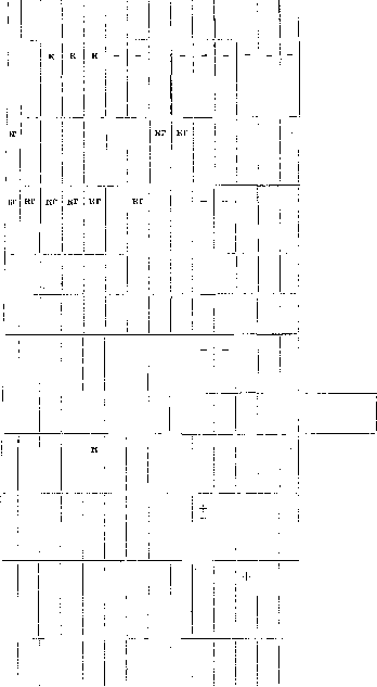
Round, whitish, with a mucous rim
with gelatin Small, round, translucent, with concentric circles Pathogenicity For Location. Where isolated. What infectious disease it causes ! in Sh I + Causative agent of infectious abortion in horses Small, translucent Round, transparent, slightly bluish, serrated edges Grayish, round + + + Causative agent of infectious abortion in cattle and typhoid-like disease in humans Soil. Detected in a number of cases of gas gangrene + i + In the intestine of humans and animals. Causative agent of food poisoning Irregular, fimbriated, curl-like Round, grayish-white, fimbriated + + Causative agent of anthrax Round, amoeboid Isolated from rotting meat and contaminated wounds Small, round, shiny, granular + | Causative agent of epizootic in wild deer and cattle f Flat, grayish, {irregular shape, granular + 'Causative agent of botulism. Found on fruits, legumes, soil Mon char Pathogenic £ ak Name Size in microns A O o e i a o 0) i l and o ev ft o o o i o And o ev ft k i 5 CO ^ I^ o in + i. is N (O ° 1 i i n a p sh yu I K f-1 s O ft Sv re o ft n o K p " ev ga ev 03 O I I a ev 03 o Yo ev ev and kg k ev O n I ev ev 03 O d k ev ev m o' a ev and ev o ev 03 o I I >E ev A and i ft i in ev E X ev S. Bac. Chauvoei(Bac. anthracis sym-ptomatici Kruse, Rauschbrandba-zillus, Charbon symptomatique du boeuf) Arloing, Cornevin & Thomas 1887 [Clostridium Chauvoei (Arloing, Cornevin & Thomas) Holland] 1.0-3.0 x 0.5 + + + an + kg kg k kg kg Vibrio cholerae. Koch 1884 [Vibrio comma (Schruter)] 0.4-0.6x X 1.5-3.0 - aer fac + k k k k Bact. cholerae gallinarum. Hueppe 1887. Bacillus avisepticus [Pasteu-rella avicida (Kitt) Trevisan] 0.25-0.4 X X0.5 - aer fac ~ K | | K - k Bact. cholerae suum, see Bact. suipestifer + ! [ | | i | ! Bact. coli. Escherich 1886 (Bact. coli commune Escherich, Bac. coli communis Sternberg) [Escherichia coli (Escherich) Castellani & Chalmers] 0.5-1.0x X2.0 aer fac kg kg kg kg kg Bact. columbense. Castellani 1914 [Salmonella Columbensis (Castellani) Castellani & Chalmers] 0.5-0.6x X2.0-5.0 + aer fac - j KG kg - KG i kg - Bac. conjunctivitidis. Koch-Weeks 1883 [Hemophilus conjunctivitidis Bergey et al.] 0.2-0.3X X 0.5-2.0 fac - 1 -| Bac. cuniculicida. Koch 1878 (Bact. lepisepticum Ferry & Hoskins) [Pasteurella cuniculicida (Koch) Trevisan] 0.6-0.7 X X 1.4-2.6 ! - ! aer |fac | - k ! 1 1 k Bac. dispar (B. Schmitzii). Andrews-Schmitz 1918 [Shigella dispar (Andrews) Bergey et al.] 0.5-1.0x X1.5 - 1 - : aer !fac , 1 k k± Bact. dysenteriae Shiga-Kruse. Shiga-Kruse 1898 [Shigella dysenteriae (Shiga) Bergey et al.] Bact. dysenteriae Flexner. Flexner 1900 [Shigella paradysenteriae (Collins) Bergey et al.] 0.4-0.6 X X1.0-3.0 " aer fac k k i k ! 1 similar to B. Shiga aer fac| k ! ~~ k Bact. dysenteriae. His-Russell 1900 [Shigella paradysenteriae (Collins) Bergey et al.] Id. aer fac k teria (continued). Pen-toses Alcohols i! co It a ev a a < Glycosides Formation Type of colonies on agar plates with gelatin j « Location. Where isolated. What infectious disease it causes + Small, grayish, slightly opalescent, sometimes lobed KG
; +
Grayish-white, round, transparent, shiny Transparent, bluish, slightly opalescent Yellowish-white, small, granular Extremely weak growth + Soil. Causative agent of gas gangrene
Causative agent of cholera Causative agent of chicken cholera and avian septicemia KG
! +
Round, whitish-yellow, sometimes wavy Round, whitish-gray, with lobed edges Round, grayish; Round, grayish, sometimes with lobed edges; Facultative parasite Common saprophyte of the human and animal intestine + On blood agar: small transparent Does not grow Isolated in typhoid-like diseases Causative agent of acute infectious conjunctivitis Whitish, shiny, round with wavy edges Small, round, white, finely granular, wavy Causative agent of infectious rhinitis in rabbits. Causes septicemia in rabbits upon artificial infection k± Flat, moist, shiny Small, grayish, homogeneous with notched lobed edges Similar to Bact. Shiga Isolated from the intestine. Causative agent of human dysentery Causative agent of human dysentery Causative agent of human dysentery Id. (Causative agent of human dysentery
Pathogenic bacteria Name (in square brackets-according to American classification, in round brackets-synonyms) Size in microns O) o I) o5 a m and o nt o o y a v a n >, ;» yu S i o o B a n o o, o S^ o n a o ov i" O a Disaccharides l "3 ev U! a n a t- p O! « tl k Monosaccharides 03 fcf Polysaccharides 03 X ----.- ev and co o a i o < l y ft p £ Bact. dysenteriae Strong. Strong 1906 [Shigella paradysenteriae (Collins) Bergey et al.] Bact. dysenteriae Sonne. Sonne 1914-15 [Shigella paradysenteriae var. Sonne] Similar to B. Shiga Id. fac aer | fac:
K
K : K j K k ! k ! k ' k k Bact. dysenteriae Schmitz, see Bac. dispar. Vibrio El Tor. Gotschlich 1905 Bact. enteritidis. Gartner 1888 [Salmonella enteritidis (Gartner) Castellani & Chalmers] Morphologically and biologically similar to "Vibrio cholerae Bact. erysipelatos suum. Loffler 1885 (Bac. rhusiopathiae suis Kitt) [Erysipelothrix
rhusiopathiae (Kitt) Holland] Bact. faecalis alcaligenes. Petruschky 1896 [Alcaligenes faecalis Castellani & Chalmers] 0.6-0.7 x X2.0-3.0 0.2- 0.3X X0.5-1.5 - aer j fac 0.5-1.0x X2.0 - | aer fac Bac. filamentosus putrificus. Distaso 1911 [Clostridium filamentosum Bergey et al.] an + k + - ! : Coccobacillus foetidus ozaenae. Perez 1899 [Escherichia foetida Bergey et al.] 1.0-4.0 aer fac kg Bact. typhi gallinarum. Klein 1899 [Salmonella gallinarum (Klein) Bergey et al.] 0.4-0.6 x X 0.8-1.6 i iz; (fac; k : k : k ; k Bact. Glasser-Voldagsen. Dammann-Glasser + - aer I fac the rest, as in B. teria (continued). Pentoses Alcohols | Glycosides Formation and and a o n o n o> co (-C Type of colonies on plates Pathogenicity for with agar with gelatin Location. Where isolated. What infectious disease it causes I
!
KG KG | KG K ! K ! K paratyphi B Similar to Bact. Shiga Causative agent of human dysentery Grayish-white, smooth, shiny Id. Round, grayish, smooth Round, grayish, granular, translucent Delicate, slightly elevated, turbid, gray-bluish, brush-like + Causative agent of food poisoning
Pathogen of swine erysipelas. Transparent, round, grayish, translucent center, with wavy edges. Moist, round, translucent, yellowish-brown. Brown granular with a greenish tint. Surface colonies, gray; deep, reddish-brown. Found in the human intestine. Intestine. Isolated from the intestine of patients with cholera nostras. Pathogen of chronic foul-smelling rhinitis (ozaena). Moist, small, grayish-white, round, finely granular, round. Grayish-white colonies, without a ridge. Pathogen of fowl typhoid. Pathogen of a typhoid-like disease in humans. Pathogenic bacteria. Name (in square brackets—according to American classification, in parentheses—synonyms). Size in microns. Monosaccharides. Disaccharides. Polysaccharides.
Bact. hemophilus influenzae, see Bac. influenzae. Bac. histolyticus. Weinberg & Seguin 1916 [Clostridium histolyticum (Weinberg & Seguin) Bergey et al.]. 0.5-0.7 x 3.0-5.0. Bac. hydrophilus fuscus. Sanarelli 1891 [Proteus hydrophilus (Sanarelli) Bergey et al.]. 0.6 x 1.3. Bact. icteroides. Sanarelli 1897 [Salmonella icteroides (Sanarelli) Bergey et al.]. 0.5-0.6 x 1.0-3.0. Bact. influenzae. Pfeiffer 1892 [Hemophilus influenzae (Pfeiffer) Committee S. A. B.]. 0.2-0.3 x 0.5-2.0. Bact. Jeffersonii. Hadley 1918 [Shigella Jeffersonii (Hadley) Bergey et al.]. 0.5-0.8. Bact. lactis aerogenes. Escherich 1886 [Aerobacter aerogenes (Kruse) Beijerinck]. Bact. lepisepticum. Ferry-Hoskins 1920, see synonym Bac. cuniculicida. 0.5-0.8 x 1.0-2.0. Bact. mallei. Löffler 1886 [Pfeifferella mallei (Löffler) Buchanan]. 0.25-0.4 x 1.5-3.0. Bact. melaninogenicum. Oliver-Wherry 1921 [Hemophilus melaninogenicus (Oliver & Wherry) Bergey et al.]. 0.8-1.0 x 3.0. Bact. melitense (Micrococcus melitensis, Brucella melitensis). Bruce 1893 [Alcaligenes melitensis (Bruce) Bergey et al.]. 0.3-0.4. Vibrio Metschnikovi. Gamaleia. Bacteria (continued). Pentoses. Alcohols. Glycosides. Formation. Type of colonies on agar plates with gelatin. Pathogenicity for.
Surface colonies, delicate, irregular; deep, coral-like. Whitish, moist, elevated. Gray, smooth, round. Delicate, bluish, with irregular jagged edges. Small, round, grayish, transparent. On blood agar: small, round, translucent, homogeneous. Small, grayish, translucent. Small, gray, smooth, translucent. Isolated from wounds. Causes tissue necrosis. Has a nauseating odor. Isolated from frogs that died of septicemia. First isolated from the corpses of those who died of yellow fever, but is not the pathogen of this disease. Dense, moist, opaque, smooth, white. Glycerin agar: mucous, gray, translucent, with smooth edges. Porcelain-white, moist, opaque. Found in the respiratory organs of humans. Pathogen of influenza. Isolated in a typhoid-like disease in domestic birds. Intestine of humans and animals. Irregular shape, round, with wavy edges, whitish, shiny, translucent. Pathogen of glanders.
Small, round, with a convex dark center. Small, transparent, with even edges. Similar to colonies of V. cholerae. Genital organs and oral cavity. Pathogen of Malta fever. Pathogen of cholera in chickens. Pathogenic bacteria. Name (in square brackets—according to American classification, in parentheses—synonyms). Size in microns. Monosaccharides. Polysaccharides. Bact. minutissimum. Kruse-Flügge 1886 [Shigella minutissima (Kruse) Bergey et al.]. 0.5-1.0. Diplobacillus Morax. Morax 1896, Axenfeld 1897 [Hemophilus lacunatus (Morax-Axenfeld) Holland]. 0.4-0.5 x 2.0. Bact. Morgani. Castellani-Chalmers 1911-1919 (Bac. metacoli) [Salmonella Morgani Castellani & Chalmers]. 0.4-0.6 x 1.0-2.0. Bac. mucosus ozaenae. Abel 1893 [Klebsiella ozaenae (Abel) Trevisan]. 1.25 thickness and various lengths. Bact. murisepticum. Flügge 1886 [Erysipelothrix muriseptica (Flügge) Bergey et al.]. 0.5-0.8 x 1.0. Bact. neapolitanum. Emmerich 1885 [Escherichia neapolitana (Emmerich) Bergey et al.]. 0.5-0.6 x 1.0-3.0. Bac. Novyi (Bac. oedematis maligni II Novy, Bac. oedematiens Weinberg, Seguin). Novy 1894 [Clostridium Novyi Bergey et al.]. 0.8-0.9 x 2.5-5.0. Bac. oedematis maligni II Novy, see Bac. Novyi.
Bac. oedematis maligni Koch (Vibrion septique Pasteur). Koch 1877 [Clostridium oedematis maligni (Koch) Bergey et al.]. 0.6-1.0 x 3.0-8.0. Bact. oxygenes Ford. Ford [Shigella oxygenes (Ford) Bergey et al.]. 0.5-2.0 x 3.0. Bac. oxytocus perniciosus. Wyssokovitsch-Flügge 1886 [Aerobacter oxytocum (Migula) Bergey et al.]. Bact. paradysenteriae no Kruse.
Bacteria (continued). Pentoses. Alcohols. Glycosides. Formation. Type of colonies on plates with agar with gelatin. Pathogenicity for. Round, small, grayish, with smooth edges. Small, round, pale yellowish-brown. On blood agar: small, round, translucent. Gray-white-blue, round, with smooth edges. Does not grow. Bluish-gray, homogeneous, with even edges. Small, whitish, convex, transparent. Small, whitish, in the form of dew drops. Small, whitish, with a dark center. Delicate, translucent, grayish. Location. Where isolated from. What infectious disease it causes. Intestine. Isolated by Kruse from a brain abscess. Causes subacute conjunctivitis. Intestine of healthy people and animals. Pathogen of enteritis and typhoid-like diseases. Isolated during ozaena from nasal mucus. Found in septicemia of white mice.
Fuzzy, bluish, translucent. Grayish-white, smooth, shiny, with wavy edges. Irregular, brownish. Grayish-white, round. In the human intestine. Erroneously described as the causative agent of cholera. First isolated by Novy from milk nuclein, then isolated in cases of malignant edema in humans. Causative agent of malignant edema. Intestine of patients. Dairy products, soil and human intestine. Causative agent of human dysentery. Pathogenic bacteria. Name (in square brackets—according to American classification, in parentheses—synonyms): Bact. paradysenteriae according to Bergey, see Bact. dys. Hiss, Flexner, Strong. Bact. paramelitense Bruce. Size in microns. Monosaccharides. Disaccharides. Polysaccharides. Bac. parasarkophysematos Kitt, Miessner [Pararauschbrandbacillus, Bradsotbacillus Jensen], poorly agglutinating, identified by Zeissler with Bac. parasporogenes. Mc Intosh 1917 [Clostridium parasporogenes (Mc Intosh) Bergey et al.]. Bact. paratyphi A. Brion-Kayser 1902 [Salmonella paratyphi (Brion & Kayser) Seiffert]. Bact. paratyphi B. Schottmüller 1900 [Salmonella Schottmülleri (Winslow et al.)]. 0.6-3.0 x 0.4-0.6. 0.6-0.7 x 2.0-3.0. Aerobic, facultative. Bact. paratyphi C Weil = B. Erzindjan, Neukirch = Bact. paratyphi C Bitter. Bact. paratyphi N. Iwaschenzow, 1921 (Bact. paratyphi C Andrewes-Bitter), a bacterium similar to paratyphoid, isolated by Weil and a bacterium similar biochemically and culturally to paratyphoid, but serologically possessing special receptors. Two varieties are distinguished: X2 corresponding to C2 Andrewes-Bitter, N2 corresponding to C1 Andrewes-Bitter. Bact. paratyphi C Uhlenhuth, a non-agglutinating bacillus, isolated from sick and healthy pigs and calves. Bact. paratyphi abortus ovis. Schermer-Ehrlich-Stephan-Geiger, a paratyphoid-like bacterium with special receptors. Bact. paratyphi alvei Bahr. Bac. perfringens. Welch-Nutall-Fraenkel 1892 (Bac. Welchii Migula) (Bac. phlegmonis emphysematosae Fraenkel) [Clostridium Welchii (Migula) Holland]. Type I. Aerobic, facultative. 1.0-1.5 x 4.0-8.0. [Hemophilus pertussis (Bordet & Gengou) Holland] (Microbe de Coqueluche). Bordet-Gengou 1906. 0.2-0.3 x 0.5-2.0. Bact. pestis. Yersin-Kitasato 1894 [Pasteurella pestis (Yersin & Kitasato) Bergey et al.]. 1.0-2.0. Aerobic, facultative. Bacteria (continued).
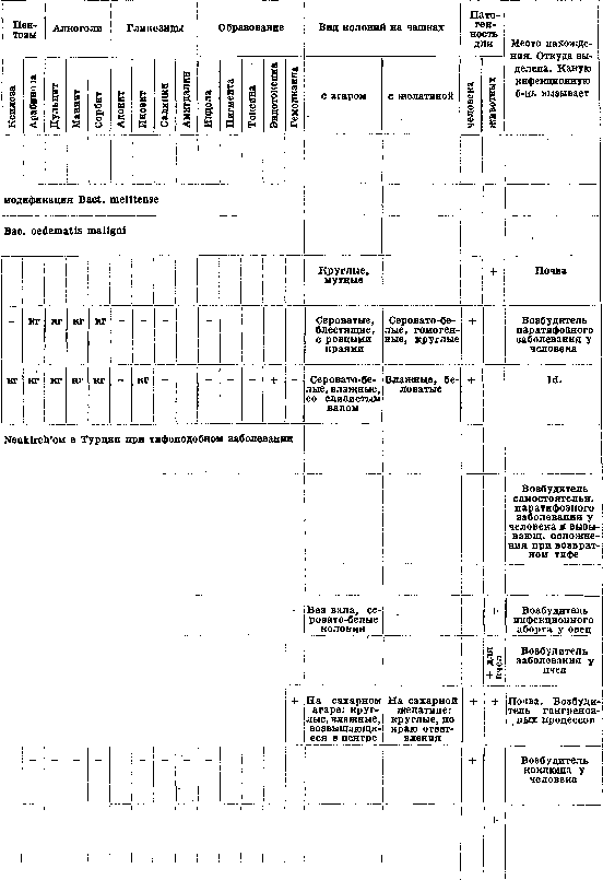
similar to paratyphoid, but serologically possessing special receptors. Two varieties are distinguished: X2 corresponding to C2 Andrewes-Bitter, N2 corresponding to C1 Andrewes-Bitter.
from sick and healthy pigs and calves, with special receptors. On blood agar: small, transparent, round. Grayish-white, transparent, with an elevation in the center, with wavy edges. Small, flat, gray, with granular edges. Causative agent of plague in humans and rodents. Found in the organism of healthy rodents. Pathogenic bacteria. Name (in square brackets—according to American classification, in parentheses—synonyms). Size in microns.
Bact. pfaffi. Hadley 1918 [Shigella pfaffi (Hadley) Bergey et al.]. [Vibrio piscium]. David 1927. Bact. pneumoniae. Friedländer 1882 (Pneumoniococcus Friedländer) [Klebsiella pneumoniae Trevisan]. Bact. pneumosintes. Olitsky-Gates 1921-22 [Dialister pneumosintes (Olitsky & Gates) Bergey et al.]. Bac. proteus vulgaris (Bact. vulgare). Hauser 1885 [Proteus vulgaris Hauser]. Bact. proteus vulgare. Var. Weili (Bac. proteus X 19) Weil-Felix. Vibrio proteus. Finkler-Prior 1884 (V. finkleri) [Vibrio proteus Buchner]. Bac. proteus fluorescens. Jaeger 1892 [Pseudomonas jaegeri Migula]. 0.4-1.4. 0.3-0.5 x 2.0. 0.3-0.6 x 5.0. 0.15-0.3 length. 0.5-1.0 x 1.0-3.0. Aerobic, facultative. Monosaccharides. Disaccharides. Polysaccharides. Similar to the previous one, isolated by Weil and Felix from typhus patients, with whose serum it is agglutinated. 0.4-0.6 x 2.4. Aerobic, facultative. Bact. pseudodysenteriae, a collective name given by Kruse. Bac. pseudomallei. Whitmore 1913-14 [Flavobacterium pseudomallei (Whitmore) Bergey et al.]. Bact. psittacosis. Nocard 1893 [Salmonella psittacosis (Nocard) Seiffert]. Aerobic, facultative. 0.4-0.6 x 1.0-1.5. Bacteria (continued). Pentoses. Alcohols. Glycosides. Formation. Type of colonies on plates with agar with gelatin. Pathogenicity for. Small, grayish, homogeneous, translucent. Yellowish, round, smooth. White, convex, smooth, shiny. Small, grayish, translucent. Location. Where it was isolated from. Which infectious disease it causes. Causative agent of typhoid-like diseases in birds. Round, granular, opaque. Dirty-white, opaque, smooth. Blood agar: small, round, shiny. For B. dysenteriae, Hiss-Russel, Flexner, Strong, etc. Gray, opaque, giving irregular outgrowths. Yellowish in the middle. Irregular shape, giving surface spreading.
Round, dense, opaque, yellowish in color, with irregular edges + a Small, grayish, round, granular Small, transparent, similar to B. proteus Causative agent of epidemic diseases in fish. Pathogenic for frogs Respiratory tract. Causative agent of pneumonia Passes through a filter. Nasal mucus in patients with influenza Intestine. Causes putrefactive decomposition of proteins Intestine and faeces of healthy people and patients with diarrhea. Isolated from patients suspected of having cholera Isolated by Jaeger in Weil's disease Small, gray, with smooth or wavy edges Small, bluish, shiny, as if porcelain Causative agent of rat-bite disease in rats, in humans (India) Until the last epidemic of psittacosis (1929-30) was considered the causative agent of psittacosis Pathogenic bacteria Name (in square brackets - according to American classification, in round brackets - synonyms) Size in microns Bact. pullorum. Rettger 1900 [Salmonella pullorum (Rettger) Bergey et al.] Bac. putrificus. Bienstock 1906 (Bac. verrucosus Zeissler) [Clostridium putrificum (Bienstock) Bergey et al.] Bact. pyocyaneum. Schroter 1872, Gessard 1882 [Pseudomonas aeruginosa (Schroter) Migula] 0.3-0.5 X 1.0-2.5 0.6-0.8 X 5.0-6.0 0.5-0.6 X 1.4-1.5 Bact. pyosepticum (Bact. pyosepticum viscosum equi). Andersen-Magnusson 1923 [Shigella viscosa (Magnusson) Snyder] 0.3-0.4 x 0.4-0.8 Bact. rettgeri. Hadley 1918 [Shigella rettgeri (Hadley) Bergey et al.] 0.5-0.8 length ae fac an | + Monosaccharides Disaccharides fac ae fac Bact. rhinoscleromatis. Fritsch 1882 [Klebsiella rhinoscleromatis (v. Fritsch) Trevisan] Bac. rhusiopathiae suis, see synonym Bact. erysipelatos suum [Erysipelothrix rhusiopathiae] Bac. saccharobutyricus (Vibrion butyrique. Pasteur 1861). Bac. amylobacter v. Tieghem 1887. Bac. butyricus Botkin proparte 1892. Bac. saccharobutyricus v. Klecki 1896 [Clostridium butyricum Prazmowski] 0.8 thickness 0.75-1.0 X + + X 3.0-10.0 ae fac ae fac Polysaccharides Bact. Salmonella, see synonyms: Bact. paratyphus-enteritidis-Hog-cholera [Genus Salmonella Lignieres 1900] Bac. septicaemiae anserum exsudativae. Riemer 1904 [Shigella septicaemiae Bergey et al.] 0.1-0.3 x 1.0 Bac. sporogenes. Metchnikoff 1908 [Clostridium sporogenes (Metchnikoff) Bergey et al.] an - kg 0.6-0.8 x 3.0-7.0 + + ae + fac an + bacteria (continued). Pentoses Alcohols Glycosides Type of colonies on plates with agar with gelatin Pathogenicity for Grayish-white, smooth, shiny, with even or wavy edges Grayish-white, moist, lobed in the form of a grape leaf Small, round, flat Lush, mucous, yellowish or yellow-green color. Around the colonies a green-blue zone Mucous, grayish-white, semicircular, smooth, shiny Location. Where isolated from. What infectious disease it causes Causative agent of diarrhea and septicemia in chicks Permanent inhabitant of the intestine. Causative agent of putrefactive processes. Symbiont in malignant edema Yellowish or green-yellow, irregular shape, granular, fringed Grayish-white, round, transparent Small, grayish, homogeneous, transparent Small, grayish, transparent Intestine, open wounds Causative agent of septicopyemic processes in foals White, shiny, smooth Round, white, convex On serum agar: small, flat, irregular shape, grayish, translucent Round, transparent, smooth, homogeneous Small, white, round Causative agent of typhoid- and cholera-like diseases in birds Causative agent of rhinoscleroma Soil, faeces; water, milk, cheese, contaminated wounds, also in malignant edema Small, transparent, fringed, whitish-yellow color Causative agent of septicemia in goslings Soil, intestine! Pathogenic bacteria Name (in square brackets - according to American classification, in round brackets - synonyms) Bact. suicida. Loffler-Schutz 1886 (Bac. suisepticus Kruse) [Pasteurella suiseptica (Loffler-Schutz) Trevisan] Bact. suipestifer. Salmon-Smith 1885, Kruse-Flugge 1896 (Bac. Hog-cholerae) [Salmonella suipestifer (Kruse) Lignieres] Size in microns 0.4-0.5 x 1.0-1.5 0.6-0.7 X 2.0-3.0 Monosaccharides Disaccharides Polysaccharides fac [Eberthella talavensis (Castellani & Chalmers 1919) Bergey et al.] 0.5-1.0 x
Eberthella tarda (Assis 1928) 0.2-0.4 x 1.8-2.0, facultative anaerobe. Clostridium tertium (Henry 1916) (Henry 1916) 0.4-0.6 x 3.0-6.0, pathogenic. Clostridium tetani (Nicolaier 1884) (Nicolaier 1884). Pasteurella tularensis (Mc Coy & Chapin 1910) 0.4-0.6 x 0.7. Eberthella typhi (Eberth 1880, Gaffky 1884) 0.6-0.7 x 2.0-3.0, facultative anaerobe. Salmonella typhimurium (Loffler 1892) 0.5-1.0 x 1.5. Hemophilus ducreyii (Ducrey 1889) 0.5-1.5 x 2.0, facultative anaerobe. Plocamobacterium vaginae (Doderlein 1892) (Acidobacterium Doderleinii) (Bacillus vaginae Kruse, Bacillus vaginalis longus Maunu, Bacillus crassus) 1.2 thickness, aerobe.
Regarding the continuation of the table for pathogenic bacteria: The columns include pentoses, alcohols, glycosides, formation of gas, colony appearance on agar and gelatin plates, pathogenicity, and location/source of isolation and the infectious disease caused. For example, the agent of so-called 'swine plague' (grayish, smooth, flat, shiny, with irregular edges) was isolated from swine plague and mistakenly taken for the causative agent; it is a cause of food poisoning. The intestine is the location for the suspected causative agent of enteritis. The intestine of patients with dysentery is the source for the dysentery bacillus. Bacillus tertius was isolated from wounds during the war; Zeissler identifies Bacillus tertius with Bacillus saccharobutyricus. Soil and dust are the sources for the causative agent of tetanus (on serum agar: small, transparent, with serrated edges).
Bacillus verrucosus (Zeissler), see synonym Bacillus putrificus (Bienstock) [Clostridium putrificum Bergey et al.]. Bacterium viscosum equi, see Bacterium pyosepticum. Bacterium Voldagsen, see Bacterium Glasser-Voldagsen. Bacterium vulgare, see synonym Bacterium proteus vulgaris. Bacillus Welchii, see synonym Bacillus perfringens. Microspira Wolfii (Migula, Wolf 1893) [Vibrio wolfii (Migula) Bergey et al.]. Diplococcus pneumoniae (Frankel-Weichselbaum 1886) (Pneumococcus Frankel) (Streptococcus lanceolatus Pasteuri Gamaleia) (Microbe septique du salive Pasteur 1881) (Diplococcus capsulatus) [Diplococcus pneumoniae Weichselbaum]. Diplococcus mucosus (Lingelsheim 1908), see Streptococcus mucosus [Diplococcus mucosus v. Lingelsheim]. Diplococcus gonorrhoeae, see synonym Micrococcus gonorrhoeae. Diplococcus intracellularis meningitidis, see synonym Micrococcus intracellularis [Micrococcus aerogenes (Schottmuller) Bergey et al.] (Staphylococcus aerogenes) Schottmuller 1912. Micrococcus catarrhalis (Pfeiffer 1896) [Neisseria catarrhalis (Pfeiffer) Holland].
Regarding the continuation of the table for pathogenic bacteria: The columns include monosaccharides, disaccharides, polysaccharides, colony appearance on agar and gelatin plates, pathogenicity, and location/source of isolation and the infectious disease caused. For Diplococcus pneumoniae: small, transparent, grayish, with even edges. On blood agar, colonies have a bluish-greenish color. It is the causative agent of pneumonia in humans and forms 4 serological types. For Micrococcus aerogenes: on serum-dextrose agar, small, yellowish-white, with gas bubbles. It was isolated during puerperal sepsis from uterine secretions. For Micrococcus catarrhalis: small, round, grayish-white with eroded edges. It is found in the mucous membrane of the respiratory tract; it often joins inflammatory processes (bronchitis, pneumonia) and is sometimes an independent causative agent.
Micrococcus gonorrhoeae (Neisser 1879) (Gonococcus) (Diplococcus gonorrhoeae) [Neisseria gonorrhoeae (Neisser) Trevisan] 0.6-1.0, aerobe/facultative anaerobe. Micrococcus intracellularis (Weichselbaum 1887) (Diplococcus intracellularis meningitidis Weichselbaum) (Streptococcus intracellularis meningitidis Lehmann & Neumann) (Micrococcus meningitidis cerebrospinalis Albrecht & Ghon) (Meningococcus intracellularis Jager) [Neisseria intracellularis (Weichselbaum) Holland] 0.6-0.8, aerobe/facultative anaerobe. Micrococcus melitensis (Bruce), see synonym Bacterium melitense. Micrococcus pyogenes aureus (Rosenbach 1884) [Staphylococcus aureus Rosenbach] 0.8-1.0. Micrococcus pyogenes citreus (Passet 1885) [Staphylococcus citreus Migula] 0.9 in diameter. Micrococcus pyogenes albus (Rosenbach 1884) [Staphylococcus albus Rosenbach] 0.6-0.8 in diameter. Micrococcus tetragenus (Gaffky-Koch 1883-84) (Sarcina tetragena) (Micrococcus tetragenus septicus Boutron) [Gaffkya tetragena (Gaffky) Trevisan] [Staphylococcus pharyngis Bergey et al.] 0.6-0.8. Staphylococcus minimus (Gioelli 1907) [Micrococcus minimus (Gioelli) Bergey et al.] 0.2-0.3. Streptococcus agalactiae, see synonym Streptococcus mastitidis.
Id. Mucosa of the respiratory tract. Isolated from the sputum of tuberculosis patients, as well as from the air and skin. Isolated from the nasopharynx during acute inflammatory processes. Does not grow | + I Isolated during purulent inflammation in the pelvis. Pathogenic bacteria. Name (in square brackets—according to American classification, in parentheses—synonyms) g Streptococcus cardioarthritidis Small 1927. Size of microbes 0.7-1.2. Streptococcus cuniculi Ross 1923 [Streptococcus cuniculi Ross]. Streptococcus equi Kitt-Schütz 1898 [Streptococcus equi Schütz] 0.7-0. Streptococcus erysipelatis (Strept. pyogenes) Fehleisen 1882 [Streptococcus erysipelatis Fehleisen]. Streptococcus haemolyticus [Streptococcus mixtos Bergey et al.]. Streptococcus mucosus Howard-Perkins (Strept. mucosus Schottmüller, Strept. mucosus capsulatus Buerger, Str. lanceolatus var. mucosus Park-Williams, Diplococcus mucosus v. Lingelsheim) 0.6-0.8 0.6-0.8. Streptococcus mastitidis Nocard-Guillebeau 1887 (Str. mastitis contagiosae, Str. agalactiae) [Streptococcus mastitidis (Guillebeau) Migula]. Streptococcus mitior Schottmüller 1905 (Str. viridans) [Streptococcus mitior Schottmüller]. Monosaccharides.

Polysaccharides. Morphologically similar to Str. pyogenes, serologically not identical. Morphologically similar to Str. pyogenes. Morphologically similar to Str. pyogenes. Streptococcus morbilli Ferry-Fischer 1929 [Streptococcus morbilli Ferry & Fischer]. Pathogenic bacteria (continued). Pathogenicity for. Pentoses. Alcohols. Glycosides. Formation of. Type of colonies on plates with agar. With gelatin. On blood agar: small, gray-white, cloudy, sometimes red-brown. Location. Where isolated from. What infectious disease it causes. On blood agar around small colonies a wide zone of hemolysis, biochemically somewhat different. In the form of dewdrops, mucous, transparent and cloudy, merging into a mucous mass, even edge, coarsely granular, serologically different. On blood agar: small, smooth colonies surrounded by a green zone. Isolated from the throat of patients with articular rheumatism. + Causative agent of an infectious disease in rabbits.
Pathogen of strangles in horses. Found as an incidental finding in inflammatory processes in humans. Pathogen of erysipelas. Found in various inflammatory processes. Small, white, opaque colonies. Isolated in fibrinous pneumonia, in otitis, in meningitis of aural origin, in whooping cough, and generally in mucopurulent processes. Pathogen of infectious mastitis in cows. Orla-Jensen found this same microbe in mastitis in women. Found on the mucous membranes of the nose and throat. Isolated from abscesses, from blood in endocarditis. Isolated from the blood, nose, throat, and conjunctiva of patients with measles. Pathogenic bacteria. Name (in square brackets—according to the American classification, in round brackets—synonyms). Size in microns. Monosaccharides. Disaccharides. Polysaccharides. Alcohols. Glycosides. Formation. Type of colonies on agar plates. Pathogenicity for. Location. Where isolated from. What infectious disease it causes. Streptococcus ovalis. Escherich-Thiercelin 1902 (Enterococcus) [Streptococcus faecalis Andrewes & Horder]. 0.6-1.0 in diameter. + aer. fac. On blood agar: whitish colonies with strong blackening, and sometimes greening of the agar. Human intestine. Isolated in septic abortions. Streptococcus putridus. Schottmüller 1911. + anaer. Streptococcus pyogenes. Rosenbach 1884 [Streptococcus pyogenes Rosenbach]. 0.6-1.0 in diameter. + aer. fac. On blood agar: small, round colonies surrounded by a zone of hemolysis. Soil, air. In a healthy organism on the tonsils, in the oral cavity. Pathogen of suppuration, phlegmon, abscesses, angina, pyemia, septicemia, etc. Streptococcus scarlatinae [Streptococcus scarlatinae Klein]. + aer. fac. Small, round, whitish, flat colonies. Isolated from the throat of patients with scarlet fever. [Streptococcus puerperalis Arloing] Arloing 1882. 0.6-0.8. + aer. fac. Somewhat larger than Str. pyogenes, opaque. Pathogen of puerperal sepsis. Corynebacterium acnes. Gilchrist 1901 (Acne-bacillus) [Corynebacterium acnes (Gilchrist) Bergey et al.]. 0.5x2.0. + - microaer. Very small, transparent, round, pink. Isolated from sweat glands, hair follicles, and from acne. Actinomyces asteroides. Eppinger (Cladothrix asteroides) [Actinomyces asteroides Eppinger]. 0.2 thickness. + + aer. Round, yellow-white, finely granular in the center, narrow, pale, concentric zone at the edge. Isolated in one case of abscess in humans. [Mycobacterium avium (Strauss u. Gamaleia) Lehmann & Neumann] Strauss-Gamaleia 1891 (Mycobact. tuberculos. typus gallinaceus, Bac. tubercul. avium Maffuci). aer. Very similar to Bac. tuberc. bovin. On glycerin serum: small, white, wrinkled, shiny, yellowish. Pathogen of tuberculosis in birds. Actinomyces bovis. Harz 1877-78 (Discomyces bovis (Harz), Rivolta, Act. bovis sulfureus, Nocardia actinomycis Trevisan, Streptothrix Actinomyces Rossi) [Actinomyces bovis Harz]. 0.4-0.6. + aer. fac. Grass, cereals. Pathogen of actinomycosis in humans and animals. [Actinomyces candidus Petruschky]. aer. + [Mycobacterium chelonei Bergey et al] Friedmann 1903 (Turtle-bacillus, Bac. Tuberc. d. Schildkröten). 0.3-0.5 x 4.0. Small, grayish, granular, with irregular edges. Isolated from the lungs of a turtle. Corynebacterium diphtheriae. Klebs-Löffler 1883-1884 (Bac. diphtheriae) [Corynebacterium diphtheriae (Klebs-Löffler) Lehmann & Neumann]. 0.3-0.8 x 1.0-8.0. + aer. fac. Small, transparent, gray. Pathogen of diphtheria. Bac. diphtheroides gallinarum. Graham-Smith 1901 [Corynebacterium gallinarum (Graham & Smith) Bergey et al.]. On agar with dextrose, bacillary forms form very small colorless colonies; coccal forms—white-yellow. Isolated from the throat of chickens. Bac. enzymicus. Mellon 1916 [Corynebacterium enzymicum (Mellon) Bergey et al.]. 0.2-0.3 x 2.0-3.0. + aer. fac. Isolated from the lungs, blood, and joints of humans. Corynebacterium equi. Miessner-Wetzel 1923 (Corynebact. pyogenes equi). 0.25 thickness. + aer. On glycerin gelatin: nodular, granular with radial edges. Pathogen of epid. diseases in horses. Actinomyces farcinicus. Nocard 1888, Trevisan 1889-92 (Bacille du farcin Nocard, Nocardia farcinica Trevisan) [Actinomyces farcinicus (Trevisan) Gasperini]. 0.75-1.0 x 3.0-5.0. + aer. Very small, grayish, with lobed edges. Bact. flavidum. Morse 1912 [Corynebacterium flavidum (Morse) Bergey et al.]. 0.8-1.0 x 5.0-10.0. + aer. fac. Small, round, transparent. Fusobacterium Plaut-Vincenti. Hölling-Babes 1889 [Fusiformis dentium Hölling]. + anaer. Yellowish-white, irregular, shiny, light-refracting. [Corynebacterium Hodgkinii (Bunting & Yates) Bergey et al.] Bunting-Yates 1914. + aer. fac. Yellowish, wrinkled, densely growing. [Actinomyces hominis Bostrom]. + aer. On serum agar with dextrose: yellow-white, with a dark center, delicately granular. Isolated from the lungs in tuberculosis of them. Small, round, transparent, shiny.
Pathogen of cattle scab in Guadeloupe and Northern France. The disease resembles tuberculosis of the cutaneous lymph nodes. Nasal and pharyngeal mucosa. Analogous microbes encountered in mastitis in cows. Oral cavity, pathogen of Vincent's angina together with the symbiont Spirochaeta Plaut-Vincenti. Isolated from lymph nodes in Hodgkin's disease. Pathogen of actinomycosis in humans.
Pathogenic bacteria. Name (in square brackets—according to American classification, in round brackets—synonyms). Size in microns. [Corynebacterium kutscheri (ligula) Bergey et al.] Kutscher 1894 (Bacillus pseudotuberculosis murium). Bacillus leprae. Hansen 1879 [Mycobacterium leprae (A. Hansen) Lehmann & Neumann]. Bacillus lymphophilus. Torrey 1916 [Corynebacterium lymphophilum (Torrey) Bergey et al.]. [Actinomyces madurae (Vincent) Lehmann & Neumann] Vincent. [Mycobacterium marinum Aronson] 1926. [Actinomyces necrophorus (Löffler) Lehmann & Neumann] Löffler 1884 (Bacillus d. Kälberdiphtherie, Bacillus diphtheriae vitulorum Flügge, Necrosebacillus Bang, Streptothrix necrophora Kitt). [Mycobacterium paratuberculosis (Johne) Bergey et al.] Johne-Frothingham 1895 (Johne's bacillus, Mycobacterium enteritidis Lehmann). Bacillus pseudodiphtheriae. Hoffman-Wellenhof 1887 [Corynebacterium pseudodiphthericum Lehmann and Neumann]. Bacteria (continued). Monosaccharides. Disaccharides. Polysaccharides. Pentoses. Alcohols. Glycosides. Formation. Appearance of colonies on plates with agar, with gelatin. Pathogenicity for. Location. Where isolated from. What infectious disease it causes. Small, delicate, yellowish-white, transparent, with serrated edges. Small, white, transparent. Isolated from caseous degenerated lungs of a mouse. Grayish, round, brownish, edges serrated or fringed. Grayish-white, dense, granular. Pathogen of leprosy. Isolated from lymph nodes in Hodgkin's disease, but is not its pathogen. On synthetic agar: yellowish, with outgrowths. Pathogen of the disease 'Madura foot'. On Dorset and Petroff media: grayish-white, moist, raised, with irregular edges, similar to colonies of avian tuberculosis. Isolated from a necrotic focus in the liver of marine fish. Pathogen of tuberculosis in marine fish. Small, dirty-white, round, turbid, with a yellowish center, with fringed edges. Pathogen of diphtheria in cats with multiple abscesses, gangrenous dermatitis in horses. One case of human infection described. Pathogen of chronic diarrhea in cattle. Turbid, grayish, smooth, homogeneous. Small, grayish, sometimes pale yellow, smooth, homogeneous. Pharynx of healthy people.
Pathogenic bacteria. Name (in square brackets—according to American classification, in round brackets—synonyms). Size in microns. Bact. pseudotuberculosis murium, see Corynebacterium kutscheri. Bact. pseudotuberculosis rodentium. Pfeiffer 1889, Eisenberg 1891, Preisz 1894 [Corynebacterium pseudotuberculosis (Eisenberg) Bergey et al.]. Bac. pseudotuberculosis ovis. Nocard 1885 [Corynebacterium ovis Bergey et al.]. [Mycobacterium piscium (Kral & Dubard) Bergey et al.] Kral-Dubard 1897. Mycobacterium tuberculosis var. poicilothermorum Lehmann & Neumann (Mycobact. tuberc. y-piscicola, a-testudinis, e-ranicola, f-anguicola) [Mycobacterium ranae (Kuster) Bergey et al.] Kuster 1905 [Mycobacterium tropidonatum (Sibley) Bergey et al.] Sibley 1892. [Mycobacterium tuberculosis bovis (Smith) Lehmann & Neumann] Smith 1896 (Bacillus tubercul. bovis). Mycobacterium tuberculosis typus gallinaceus, see Mycobact. avium. [Mycobacterium tuberculosis (hominis) (Koch) Lehmann & Neumann] Koch 1884 (Bacillus tuberculosis, Bacillus Kochii, Sclerothrix Kochii Metschnikoff). Bacteria (conclusion). Monosaccharides. Disaccharides. Polysaccharides. Pentoses. Alcohols. Glycosides. Formation. Appearance of colonies on plates with agar, with gelatin. Pathogenicity for. Location. Where isolated from. What infectious disease it causes. Round, convex, homogeneous, grayish-white, moist. Delicate, grayish-white, serrated. Round, transparent, with concentric circles. Weak growth. Small, round, white, moist. Weak growth, opaque, dry, wrinkled. Dry, whitish-yellow, folded. Pathogen of spontaneous disease in rabbits, guinea pigs. Isolated from kidneys of sheep. Pathogen of disease with formation of nodules and tumors in carp. Pathogen of tuberculosis in cold-blooded animals. Isolated from liver of frogs. On egg medium: small, flat, grayish, moist, shiny. On glycerin agar: small, crumbly, irregular, whitish-yellow, in old cultures brownish, moist, later dry. Pathogen of disease with formation of nodules and tumors in snakes. Pathogen of nodules and tumor-like formations in snakes. Pathogen of tuberculosis in cattle, cats, humans, pigs. Pathogen of tuberculosis in humans.
MICROORGANISMS
Name (in square brackets—according to American classification, in parentheses—synonyms) Size in microns Location. Where isolated from. What infectious disease it causes Pathogenicity for humans. Spirochaeta anserina. Sakharoff 1891 [Borrelia anserina (Sakharoff) Bergey et al.] 0.3-10.0x20.0 Isolated from geese with a febrile disease. Spirochaeta argentinensis. Kuhn-Steiner (Spirochaete of multiple sclerosis) Isolated from rabbits and guinea pigs infected with blood and lumbar fluid of patients with multiple sclerosis. [Borrelia berbera (Sergent & Foley) Bergey et al.] Sergent-Foley 1908 0.2-0.3x12.0-18.0 Causative agent of relapsing fever in Algeria, Tunisia, and Tripoli. Spirochaeta bronchialis. Castellani 1907 4.0x30.0 Isolated from humans with asthma, bronchitis, and chronic bronchopneumonia. [Borrelia carteri (Mackie) Bergey et al.] Mackie 1907-08 0.2-0.5x10.0-30.0 Causative agent of Indian relapsing fever. Transmitted by the bedbug (Cimex rotundatus). Spirochaeta cuniculi. Kolle Causative agent of a spontaneous disease in rabbits similar to syphilis. Spirochaeta Dengue. Couvy-Broquet Isolated from patients with dengue fever. Spirochaeta Duttoni. Dutton-Todd 1905 [Borrelia duttoni (Dutton-Todd) Bergey et al.] 0.2-0.5x14.0-16.0 Causative agent of African relapsing fever. Transmitted by the tick Ornithodorus moubata. Spirochaeta eurygyrata. Hogue Isolated in one case of chronic diarrhea in humans. Spirochaeta forans. Reiter 1916 Isolated from blood in a case of severe articular rheumatism. Spirochaeta gallinarum. Marchoux-Salimbeni 1903; Swellengrebel 1907 [Borrelia gallinarum Swellengrebel] 0.25-0.3x8.0-16.0 Causative agent of chicken septicemia. Transmitted by ticks Argas persicus, A. miniatus, etc. Spirochaeta hebdomadis. Ido-Ito-Wani 1918 [Leptospira hebdomadis Ido-Ito-Wani Noguchi] Causative agent of seven-day fever in Japan. Transmitted by the field mouse Microtus montebelli. [Borrelia hyos Bergey et al.] King-Baeslach 1912 Found in the blood and intestinal ulcers of patients with swine plague. Spirochaeta icterohaemorrhagiae Inado-Ido 1916 (Spir. icterohaemorrhagica, Spir. icterogenes) [Leptospira icterohaemorrhagiae (Inado & Ido) Noguchi] 0.25-0.3x6.0-9.0 Causative agent of infectious jaundice (Weil's disease). Found in apparently healthy rats. Spirochaeta icteroides. Noguchi 1918 [Leptospira icteroides Noguchi] 0.2-0.25x4.0-9.0 Noguchi isolated it during yellow fever and considered it the causative agent of this disease. Transmitted by the mosquito Aedes aegypti (Stegomyia calopus). [Borrelia Kochii (Novy) Bergey et al.] Novy 1906 Morphologically similar to Sp. Duttoni, but serologically distinct. Causative agent of African relapsing fever. Spirillum minus Carter. Futaki 1917 (Spirochaete morsus muris Futaki) Causative agent of sodoku disease. [Borrelia novyi (Shellack) Bergey et al.] Norris 1906, Shellack 1908 0.3-17.0x20.0 Causative agent of African relapsing fever. Spirochaeta pallida. Schaudinn-Hoffmann 1905 [Treponema pallidum Schaudinn & Hoffmann] 0.25-0.3x6.0-14.0 Causative agent of syphilis. Spirochaeta pappataci. Couvy-Wittingham Isolated during pappataci fever. Spirochaeta parotidis. Kermogrant 10.0-14.0 Isolated from oral mucus during infectious parotitis, considered the causative agent of this disease. Spirochaeta pertenuis. Castellani 1905 [Treponema pertenuis Castellani] 0.25-0.3x18.0-20.0 Causative agent of tropical yaws. [Borrelia phagedenis (Noguchi) Bergey et al.] Noguchi 1913 0.7-0.8x10.0-15.0 Isolated from a phagedenic ulcer. Causative agent of inflammation in rabbits and monkeys (Macacus rhesus). Spirochaeta Plaut-Vincenti. Blanchard [Borrelia vincenti (Blanchard) Bergey et al.] 0.3x12.0-25.0 Found in the oral cavity. In symbiosis with Bac. fusiformis, they cause Vincent's angina. Spirochaeta recurrentis. Obermeier 1868 [Borrelia recurrentis (Obermeier) Bergey et al.] 0.35-0.5x8.0-16.0 Causative agent of European relapsing fever. Transmitted by the body louse. Also found in the bedbug Cimex lectularius. Spirochaeta sporogona rheumatismi Rask Isolated during erythema multiforme and nodosum. III. Lactic acid bacteria. Name (in square brackets—according to American classification, in parentheses—synonyms) Size in microns Monosaccharides Disaccharides Polysaccharides Pentoses Alcohol Glycosides Formation Type of colonies on plates Location. Where isolated from. Agar Gelatin. Bact. acidi lactici. Hueppe 1884 [Escherichia acidi lactici (Zopf) Bergey et al.] 0.3-0.4x1.0-1.4 Small, white, shiny Milk, cheese, intestine. Streptococcus acidi lactici. Lister 1873, Lohnis 1909 [Streptococcus lactis (Lister) Lohnis] 0.5-1.0 Small, round or oval, gray Milk; causes it to sour. Micrococcus ac. lactici. Kruger 1891 [Micr. freudenreichii Guillebeau] 0.6-1.2 Milk. Bac. acidophilus. Moro 1900 [Lactobacillus acidophilus (Moro) Holland] length 4.0-5.0 Small, mucous. Plocamobacterium bulgaricum. Grigoroff 1905, Luerssen-Kuhn (Bac. bulgaricus) [Lactobacillus bulgaricus (Grigoroff) Holland] 1.0-2.0 Small, white, opaque. [Lactobacillus caucasicus (Kern) Beijerinck] Kern 1881 (Dispora caucasica, Bac. lebenis Rist-Khouri, Bact. mazuni) Delicate, irregular shape, transparent. Bact. casei. Freudenreich 1891 [Lactobacillus helveticus (Orla-Jensen) Bergey et al.] 0.3-1.0x5.0-6.0 Gray, fimbriated Intestine. [Flavobacterium lactis Bergey et al.] Grimm 1902 (Bac. aromaticus lactis Grimm) 0.7-0.9x2.0-6.0 Isolated from Bulgarian sour milk-yogurt. [Flavobacterium harrisonii Bergey et al. 1905] (Bac. lactis Harrisonii Conn) 0.7-1.0x3.5-4.0 On serum agar: round, irregular, filamentous. Bac. lactis erythrogenes. Hueppe 1889 [Serratia lactica Bergey et al.] 0.25-0.75x0.3-3.5 Small, flat, grayish, round, wavy Kefir. Bact. lacticum. Kramer 1892 [Achromobacter lacticum (Kramer) Bergey et al.] 0.4-0.5x1.0-1.4 On lactose agar: small, grayish, viscous Sour milk, cheese. [Bac. lactis. Flugge 1894] (Bac. cylindrosporus Burchard) 0.2-1.4x3.5-4.0 Round, slightly yellowish, mucous Milk. Bac. lactis acidi. Leichmann 1920 [Lactobacillus lactis acidi (Leichmann) Holland] 0.5-0.8x0.8-1.2 Small, gray, lobed, sometimes brownish Milk. Bac. lactis viscosus. Adametz 1891 (Lactobacillus viscosus Bergey et al. 1923) [Achromobacter viscosum (Adametz) Bergey et al.] 0.5-1.2x0.5-2.5 Small, gray-white, later yellowish, center pink Milk. Bac. Leichmanni. Henneberg 1903 [Lactobacillus Leichmanni (Henneberg) Bergey et al.] 0.6-2.0x4.0 Round, grayish Milk. Bac. pabuliacidi. Weiss 1899 [Lactobacillus pabuliacidi Bergey et al.] 0.8-1.0x3.0-8.0 Grayish-white, homogeneous Milk. [Lactobacillus plantarum (Orla-Jensen) Bergey et al.] Orla-Jensen 1919 (Streptobacterium plantarum) Small, with wavy edges Milk. [Lactobacillus thermophilus. Ayers & Johnson 1924] 0.5-3.0 Small, flat brownish. Small, grayish, later yellowish Milk. Small, whitish-transparent Sour milk. Small, transparent Cheese. Small, translucent Cheese, sour dough. Small, like pinpricks Pasteurized milk.
The most important species of bacteria causing acetic acid fermentation. Names (in square brackets - according to the American classification, in parentheses - synonyms). [Table content: Bact. acetosum Henneberg 1898 (Acetobacter acetosus (Henneberg) Bergey et al.), size 0.4-0.8 x 1.0 microns; Mycoderma aceti Thomsen 1852 (Acetobacter aceti (Thomsen) Committee S. A. B. 1920), size 0.4-0.8 x 1.0-2.0 microns; Bact. acetigenum Henneberg 1898 (Acetobacter acetigenus (Henneberg) Bergey et al.); Bact. ascendens Henneberg 1898 (Acetobacter ascendens (Henneberg)); Bact. industrium Henneberg 1898 (Acetobacter industrius (Henneberg) Bergey et al.), size 0.3-0.8 x 2.4-20.0 microns; Bact. kutzingianum Hansen 1894 (Acetobacter kutzingianus (Hansen) Bergey et al.); Bact. Lindneri Zeidler 1896 (Acetobacter Lindneri (Zeidler) Bergey et al.); Bact. oxydans Henneberg 1897 (Acetobacter oxydans (Henneberg) Bergey et al.), size 0.8-1.2 x 2.4-2.7 microns; Bact. pasteurianum Hansen 1879 (Acetobacter pasteurianus (Hansen) Beijerinck), size 0.4-0.8 x 1.0 microns; Acetobacter plicatus Fuhrmann 1906, size 0.4-0.6 x 1.4-4.6 microns; Bact. xylinum Brown 1886 (Acetobacter xylinus (Brown) Bergey et al.). Colony appearance on gelatin plates and location/source: Bact. acetosum: on gelatin with beer, shiny, large; source: beer. Mycoderma aceti: on gelatin with dextrose, grayish, shiny; source: vinegar. Bact. acetigenum: on gelatin with glucose, dry, white, with a white zone around the colonies; source: vinegar and red wine. Bact. ascendens: source: beer. Bact. industrium: on gelatin with beer, small, round, with a worm-like surface; source: beer. Bact. kutzingianum: on gelatin with wort, small, round, slightly granular, yellowish-brown; source: beer wort. Bact. Lindneri: round, irregular, with branches; source: beer. Bact. oxydans: on gelatin with wort, small, round, gray, shiny; source: vinegar, beer, and beer wort. Bact. pasteurianum: on gelatin with wine, round, convex, moist, shiny, slightly yellowish; source: wine. Bact. xylinum: source: vinegar.]
Protozoa. Class. Name of genus and species. Year of description. Author. Size of vegetative forms in microns. Motility of vegetative forms in the host organism. Size of cyst in microns. [Table content: Entamoeba histolytica, 1875, Losch, 10-20, +, 1-, 2-, 4-nucleated; 10-20. Entamoeba coli, 1903, Schaudinn, 20-30, +, 1-, 2-, 4- and 8-nucleated; 16-18. Endolimax nana, 1903, Wenyon and O'Connor, 6-10-12, +, 5-9. Iodamoeba butschlii, 1912, v. Prowazek, 6-20, +, 8-13. Dientamoeba fragilis, 1918, Jepps and Dobell, 3.5-12, +, not detected. Coprozoic: Hartmanella hyalina, 1912, 8-17-20-33, not detected.]
Class Mastigophora. [Table content: 10-15 microns.]
Related articles
Mentioned in
Cite this page
“Microorganisms.” Soviet Medical Encyclopedia. English translation of Bolshaya Meditsinskaya Entsiklopediya, 1st ed. (Moscow, 1928–1936), ed. N. A. Semashko. https://sovietmedicalencyclopedia.pages.dev/article/microorganisms/