Encephalitozoon Cuniculi
Historical document, translated for reference. It reflects medical knowledge of the 1920s–30s and is not medical advice.
Summary
This article from the 1928–1936 Great Medical Encyclopedia discusses Encephalitozoon cuniculi, a microsporidian parasite causing spontaneous epizootic encephalitis in rabbits and mice. It details the morphology, developmental cycle, pathology, and potential links to similar infections in dogs and humans.
Encyclopedia article (1928–1936)
ENCEPHALITOZOON CUNICULI et muris, a microorganism causing a spontaneous disease of rabbits and mice, chronic epizootic encephalitis. This disease, described in rabbits by Bull (1917), Oliver and Twort (1922), and in mice by Levaditi (1924), runs a chronic course, often with a fatal outcome preceded by a period of paralysis and convulsions. Wright and Craighead were the first to observe this parasite, describing in 1922 infectious paralysis in young rabbits, apparently identical with spontaneous epizootic encephalitis, and discovering in it formations both in the brain and in the kidneys which they considered to be the causative agent and classified among protozoa. In 1923, the causative agent was described in detail by Levaditi with co-workers Nicolau and Schoen, and simultaneously by Doerr and Zdansky, and was named Encephalitozoon cuniculi (Levaditi). The parasite is most often found in the brain, as well as in the kidneys and urine in both rabbits and mice. The identity of the parasite in both is proven by the fact that inoculation of the brain of an infected rabbit into mice causes in the latter a disease corresponding to murine spontaneous encephalitis, and vice versa, as well as by identical morphology and pathohistological changes. There are data on the existence of similar parasites in the dog and man. Kantorowicz and Lewy (1926) in one of 22 examined dogs with the nervous form of distemper found in the brain cyst-like accumulations of parasites similar to E. c.; Torres in 1927 described in a congenital disease in an infant, presenting phenomena of muscular contracture and dying 2 days after birth, intracellular parasites in the muscles (in the myocardium), subcutaneous tissue, and also in the nervous system, morphologically close on the one hand to E. c. and on the other to Toxoplasma. Provisionally this parasite was assigned by the author to the group Encephalitozoon under the name Encephal. Chagasi. Cyst-like accumulations of parasites close to E. c. have been described (Jankii) in the retina of an eleven-month-old infant with hydrocephalus. Morphology of E. c., its relation to brain tissue and pathohistological changes. Most often the parasite is found in the spore-formation stage in the form of cyst-like accumulations. The number of spores ranges from 1-2 dozens to many hundreds. The size of the cysts reaches 200 µ in diameter. Around these accumulations a thin membrane is sometimes visible (see figure). Spores represent very small formations of oval or egg-shaped form, measuring 1.5-2.5 x 0.4-1 µ. In them a membrane, a centrally located nucleus, and 1-2 vacuoles are visible.
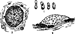
Encephalitozoon cuniculi: I - spores; II - pansporoblast in the mouse brain. The thin membrane clothing it and individual spores are visible; III - multinucleated trophozoite.
and metachromatic granules, often located at the poles. Regarding coloring substances, it should be noted that with some stains the spores of E. c., or rather their membrane, stain metachromatically, just like Negri bodies in rabies stain by the Mann, Kulchitsky-Epstein, Turevich method in red color, alkaline methylene blue in violet color, etc. Foci are encountered where spores partly lie freely in the tissue; for the most part they are enclosed in mononuclear cellular elements, with all transitions visible from a cell with 1-2 spores to large cells filled with many dozens of spores. Spores are also encountered in characteristic nodules that are typical for the brain affected by E. c. The central part of the nodules is often necrotized. The development cycle of E. c. is not completely elucidated. Levaditi in the peritoneal cells of a rat infected intraperitoneally with E. c. discovered chromatin granules arranged in pairs, often connected to each other by a thin thread, and defined them as the stage of a pansporoblast. Verrate and Sala in rabbits killed 6-10 months after experimental infection with E. c. found formations 25-60 µ in diameter consisting of a protoplasmic mass and a large number of small nuclei. Muratova discovered in the walls of small vessels and in the choroid plexus of white mice spontaneously infected with E. c. elongated oval forms, 10-40 µ in diameter, with a large number of nuclei of a structure usual for microsporidia. Apparently these formations represent a multinucleated trophozoite leading to the formation of "cysts," which is confirmed by the finding in some cysts of nuclei not entering into the process of sporogenesis. Presumably the mechanism of infection and the developmental cycle of the parasite are represented in the following way: fodder is contaminated with spores found in the urine upon kidney damage; from a spore that has entered with food into the gastrointestinal tract of a new host, an ameboid emerges which penetrates through the mucous membrane, enters the circulatory system, is carried by the blood flow to the brain and kidneys, is caught between endothelial cells, divides, while protoplasm does not participate in division; a multinucleated trophozoite is formed, further a pansporoblast, which then ruptures, followed by the accumulation of macrophages engulfing the spores, as well as the formation of nodules. The majority of authors assign E. c. to microsporidia or haplosporidia. This is not recognized by all authors: Wenyon suggests the possibility of a fungal nature of the parasite, and Goodpasture assigns it to bacteria. Recently, the proximity of E. c. to Toxoplasma cuniculi (Splendore) has been pointed out. The percentage of rabbits and mice affected by E. c. in various breeding facilities is not uniform, in some cases reaching 33% (Goodpasture, Muratova). Practical significance. E. c. 1. In all experiments related to the study of the brain and kidneys of rabbits and mice, it is necessary to keep in mind the possibility of the presence of spontaneous encephalitis caused by E. c. The parasite itself often escapes study due to its small quantity or unsuitable staining, but the histological changes caused by it are distributed throughout the brain, attract attention, and can give rise to an incorrect conclusion. 2. It appears important to clarify the question of the possible consequences of E. c. getting into the vaccine emulsion during antirabic vaccinations. E. rabiei was described by Manouelian and Viala as the causative agent of rabies. Negri bodies, according to the author, are a product of the degeneration of agglutinated E. rabiei in nerve cells. According to Paul and Schweinburg, E. rabiei does not differ in any way from small Negri bodies.
Related articles
Mentioned in
Cite this page
“Encephalitozoon Cuniculi.” Soviet Medical Encyclopedia. English translation of Bolshaya Meditsinskaya Entsiklopediya, 1st ed. (Moscow, 1928–1936), ed. N. A. Semashko. https://sovietmedicalencyclopedia.pages.dev/article/encephalitozoon-cuniculi/