Hymenolepis
Historical document, translated for reference. It reflects medical knowledge of the 1920s–30s and is not medical advice.
Summary
Hymenolepis is a genus of tapeworms characterized by three testes in mature segments. Two species infect humans: H. nana (dwarf tapeworm) and H. diminuta (rat tapeworm). The article describes their morphology, biology, diagnosis, treatment, and prevention.
Encyclopedia article (1928–1936)
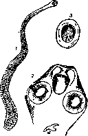
HYMENOLEPIS, a genus of tapeworms from the order of cestodes, characterized by the presence of three testes in mature segments and a sac-like uterus. In humans, two species of this genus parasitize: H. nana and H. diminuta. Hymenolepis nana (Siebold, 1852, synonym Taenia nana), the dwarf tapeworm (see figures 1, 2). Morphology: the strobila is about 15 mm long, rarely up to 30-40 mm. The width of the posterior segments is 0.5-1.0 mm. On the rostellum there is a single row of 24-30 hooks, 0.014-0.018 mm in length (see figures 1, 2). The eggs of Hymenolepis nana are oval in shape (see figure 1, 3), about 0.068 mm in length, and the oncosphere reaches 0.029-0.030 mm in diameter. The oncosphere capsule has small swellings at both poles, from which several winding filaments extend. Biology: Host - human; localization - small intestine. For this parasite, humans serve sequentially as both intermediate and definitive hosts. When a human swallows the egg, or rather the oncosphere of the parasite, it initially enters the intestinal villus, where it transforms into a cysticercoid. The formed cysticercoid eventually necrotizes the villus, falls into the intestinal lumen, attaches with its scolex to a new area of the mucosa, and transforms into a sexually mature individual. Saecki (Saeki, 1920), who elucidated the developmental cycle of H. nana, experimented on a 4-year-old child who was infected with eggs of this parasite. On the 19th day, the eggs of this parasite were already found in the child's feces, and after 62 days, 97 mature cestodes were expelled by deworming. The source of infection with H. nana is humans, who through untidiness have the opportunity to repeatedly infect themselves as well as others. Saecki's experiment, which showed that H. nana develops without changing hosts, brought this parasite biologically closer to the mouse cestode - Hymenolepis murina. However, the question of the identity of these parasites has not been finally proven. H. nana, first discovered only in 1851 in Cairo, was long considered extremely rare, and indeed was not found again until 1885. Afanasiev (1890) was the first to report finding this parasite in Russia. At present, H. nana in the USSR, as well as abroad, is a very common parasite, affecting however the child population more often than adults. For children in Moscow and its vicinity, some authors indicate a 3% infection rate. In the Donbas region, out of over 7,000 examinations (mainly of adult men), H. nana was found in 1.8%, and in one of the orphanages in Lugansk, the infection rate reached 12.5% (Skryabin and Shultz, 1927). - As for the clinical picture, as with other cestodiasis, cases of infection that occur completely without symptoms are observed, while in other cases there are severe, mostly nervous disorders. The number of parasites here is not a decisive factor, which is proven by cases of very intense infection with the absence of any disorders or their mild manifestation, and conversely, cases of severe disorders with a small number of cestodes. Grassi observed serious illnesses in 5 out of 14 cases: epileptoid seizures, decline in mental abilities, melancholy. Other authors note diarrhea, constipation, abdominal pain, variable appetite, emaciation, headaches, insomnia, and elevated temperature. Sometimes there is a pronounced eosinophilia. - Diagnosis is made by finding eggs in fecal masses. - Therapy is conducted with male fern extract or thymol. - Prevention. The main preventive measure is observing the rules of cleanliness. While the question of the independence of the species H. nana and H. murina has not yet been finally resolved, a reasonable auxiliary measure in the fight against the dwarf tapeworm should be considered deratization and the preservation of foodstuffs in such a way that they cannot be contaminated by rat and mouse excrement. Hymenolepis diminuta (Rud., 1819), the rat tapeworm, a tapeworm of rats and mice, which can also parasitize in the small intestines of humans. The length of the strobila is 200-600 mm with a width of mature segments of about 3.5 mm. The scolex (see figure 2, 1) is devoid of hooks. The egg is 0.059-0.077 mm in length and 0.049-0.069 mm in width. The length of mature segments reaches 0.66 mm. The arrangement of the sex glands in them is as follows: the female sex glands are located along the midline, on one side of them is one testis, and on the other - two, one somewhat in front of the other (see figure 2,2). The eggs are oval in shape (see figure 2,3). The egg shell is yellowish in color with indistinct radial striations. The oncosphere is 0.028-0.036 mm in diameter, covered with a double shell, with the outer one somewhat pointed at the poles. Definitive hosts: rodents of the family Muridae (rats, mice); a facultative host is humans, in whose small intestine the parasite can reach the mature stage. Intermediate hosts are various insects, including Asopia farinalis (larva of the meal moth), the mealworm - Tenebrio molitor, cockroaches - Blatta orientalis, Phillodromia germanica. Wu f2. [ЧэШкгТ -/
f

Fig. 2 Hymenolepis diminuta 1-scolex; 2-hermaphroditic segment: a-testis, f-ovary, b-bursa cirri, n-genital pore; 3-egg. di- two
In recent times, cysticercoids of this parasite have been found in the bodies of rat fleas: Xenopsylla cheopis and Ceratophyllus fasciatus. - Statistics. On the territory of the USSR, H. diminuta is found in rats in 22% of cases (Polyapolskaya, 1924). In humans, this cestode is relatively rarely found: only about 80 cases have been described in total. In the USSR, 6 cases have been described; it can be assumed that Hymenolepis diminuta is sometimes by physicians confused with Hymenolepis nana. - The pathogenesis and clinical picture of this form have been studied very poorly. In the available indications, emaciation, intestinal disorders (irregular stool, diarrhea, abdominal pain) and nervous phenomena are noted. According to the research of Kamada (Kamada, 1924), a hemolytic substance is contained in the body of H. diminuta. - Therapy. The parasite is removed with male fern extract. - Prevention consists of measures against the definitive host (rodents) and intermediate hosts (insects) and includes: 1) protecting dwellings and, especially, food substances from insects, 2) sufficiently baking bread and other flour products, 3) systematic deratization.
Related articles
Mentioned in
Cite this page
“Hymenolepis.” Soviet Medical Encyclopedia. English translation of Bolshaya Meditsinskaya Entsiklopediya, 1st ed. (Moscow, 1928–1936), ed. N. A. Semashko. https://sovietmedicalencyclopedia.pages.dev/article/hymenolepis/