Hyoid Bone
Historical document, translated for reference. It reflects medical knowledge of the 1920s–30s and is not medical advice.
Summary
The hyoid bone is a U-shaped bone located in the neck that serves as an attachment point for muscles of the tongue, neck, and pharynx. It is unique in that it does not articulate directly with any other bone and is suspended by muscles and ligaments.
Encyclopedia article (1928–1936)
HYOID BONE (os hyoideum), an unpaired bone symmetrically located along the midline of the neck, below and behind the chin, precisely at the apex of the angle formed by the submandibular region and the anterior surface of the neck (with the head in normal position): The hyoid bone is horseshoe-shaped, curved forward with convexity and backward with concavity, and consists of five parts: the middle part - the body (corpus) and two pairs of horns on the sides - the greater and lesser (fig. 1-5). The greater horns are also called thyroid horns (cornu thyreoideum), while the lesser horns are called styloid horns (cornu styloideum). In children and even adults, the greater and lesser horns are separate bones, articulated with the body via a joint, but with age their mobility is lost, and they fuse with the body of the hyoid bone to form a single bone. The body of the hyoid bone is oval-shaped, with its major axis positioned across the neck, and has two surfaces, two borders, and two ends. The anterior surface of the body is convex, divided by a transverse ridge into two parts: the upper part, directed upward, and the lower part, directed forward. Each of these surfaces is in turn divided by a low vertical ridge into two small lateral facets. These four facets serve as the attachment site for numerous muscles (see Neck). The body of the hyoid bone consists of compact bone, while the greater horns contain a small amount of spongy substance. The hyoid bone develops from the fusion of the II and III branchial arches, the lesser horns from the II, and the greater horns from the III branchial arch. - There are 6 ossification centers in the hyoid bone. The hyoid bone receives blood from the lingual and superior thyroid arteries through their hyoid branches (rami hyoidei).
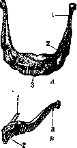
Figure 1. Figure 1. Hyoid bone. A-from above: 1- cornu majus; 2-cornu minus; 3-corpus. B-from below left: 1- cornu minus; 2-corpus; 3--cornu majus.
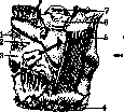

Figure 2.
Figure 3.
Figure 1. Serous bursa of the sternohyoid muscle (Bouger) (front view): 1-a. et n. laryngeus sup.; 2-membrana thyreo-hyoidea; 3-thyroid cartilage; 4-aponeurosis; 6-m. thyreo-hyoideus; 6-os hyoideum; 7-epiglottis. (After Testut-Jacob.) Figure 3. Serous bursa of the sternohyoid muscle in sagittal section: 1-bursa; 2-aponeurosis of the neck; 3-cricoid cartilage; 4-posterior wall of the bursa; 5-epiglottis; 6-os hyoideum. (After Testut-Jacob.) The hyoid bone is suspended directly to the skull by the stylohyoid ligament and muscle, and the digastric muscle. A group of muscles suspending it to the skull via the mandible are the geniohyoid (m. genio-hyoideus) and mylohyoid (m. mylo-hyoideus) muscles. The hyoid bone serves as the attachment site for the muscles of the tongue (m. hyo-glossus, part of the m. genio-glossi, m. longitudinalis inf. linguae, m. longitudinalis sup. linguae), and thus serves as its skeleton. Part of the pharyngeal musculature, the middle constrictor of the pharynx (m. constrictor pharyngis medius), also finds a point of leverage on the hyoid bone. From below, a series of muscles approach the hyoid bone, which when contracted lower the hyoid bone and the larynx suspended from it - the omohyoid, thyrohyoid, and sternohyoid muscles (mm. omo-hyoidei, thyreo-hyoidei, sterno-hyoidei). Through the combined action of all these muscles on both sides, the hyoid bone is maintained in a position that is more stable and firm the stronger these muscles contract and the more coordinated their action (Lesgaft). The entire muscular apparatus of the hyoid bone receives its innervation from four sources - from the V, VII, and XII pairs of nerves and from the superior cervical plexus (for the innervation of each muscle, see Human Muscles). When any of these nerves are affected, the harmony of action of the entire muscular apparatus of the hyoid bone is disrupted, which leads
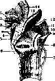
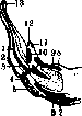
Figure.
Figure 5. Figure 4. Sagittal section of the larynx (right): 2-muscles of the tongue; 2-hyoid bone; 3-adipose tissue; 4-serous bursa (Bouger); 5 and 10-thyrohyoid plate; 6-thyroid cartilage; 7-cricoid cartilage; 8-t. aryth. post.; 9-superior horn of the thyroid cartilage; 11-thyrohyoid lateral ligament; 12-greater horn of the hyoid bone; 13-epiglottis; 14-root of the tongue. (After Testut-Jacob.) Figure 5. Hyoid bone with sites of muscle attachment: 1-m. hyo-glossus; 2-m. thyreo-hyoideus; 3-m. digastricus; 4-m. stylo-hyoideus; 5-m. omo-hyoideus; 6-m. sterno-cleido-mastoideus; 7-m. mylo-hyoideus; 8-m. genio-hyoideus; 9-m. genio-glossus; 10-m. stylo-hyoideus; 11, 12 and 13-m. constrictor pharyngis. (After Testut.) to changes in the position of the tongue and soft palate, changes in voice and speech, and changes in swallowing and chewing. Clinical picture. Fractures of the hyoid bone are very rare and most often occur under the influence of direct violence, a push, a blow, a fall on the anterior surface of the neck, being run over by a carriage wheel, or during hanging, strangulation, and rarely from muscular traction. Clinically, a fresh fracture of the hyoid bone manifests with severe pain during all acts accompanied by movement of the hyoid bone. At the site of the fracture, there is a noticeable external blood tumor (hematoma), crepitus, and mobility of the fragments; in fractures of the hyoid bone with rupture of the pharyngeal mucosa, severe bleeding from the mouth is observed. The prognosis is doubtful due to frequent complications from the respiratory organs and the danger of suffocation. Healing usually occurs by callus formation, rarely one of the fragments becomes necrotic. Mortality reaches 50% (Platt, Fischer). Treatment first consists of eliminating displacement of the fragments: this can be achieved by pressure from the pharynx and from the outside. For rest, the patient's head and neck should be supported on the sides with sandbags or the head, neck, and shoulders should be placed in a plaster cradle. Maintaining the fragments in the correct position is achieved only by suturing in most cases. - Dislocations of the hyoid bone are extremely rare. - Tumors of the hyoid bone are an exceptional rarity. According to Tikhov, no more than 6 cases are known: 2 sarcomas, 2 chondromas, 1 osteoma, and in 1 case the nature was not determined.
Related articles
Mentioned in
Cite this page
“Hyoid Bone.” Soviet Medical Encyclopedia. English translation of Bolshaya Meditsinskaya Entsiklopediya, 1st ed. (Moscow, 1928–1936), ed. N. A. Semashko. https://sovietmedicalencyclopedia.pages.dev/article/hyoid-bone/