Facial Nerve (FACIAL NERVE, see Facialis)
Historical document, translated for reference. It reflects medical knowledge of the 1920s–30s and is not medical advice.
Summary
This historical article from the 1928–1936 Soviet Great Medical Encyclopedia details the anatomy, ontogenetic and phylogenetic development, and structural characteristics of the human face.
Encyclopedia article (1928–1936)
FACIAL NERVE, see Facialis nervus. FACE (lat. facies, also vultus, ops) is represented by the forward-facing, hairless part of the head (with the exception of the mustache, beard, and eyebrows), corresponding to the facial part of the skull with the inclusion of the frontal region of the cerebral cranium. Here are concentrated the peripheral apparatuses of the sense organs of vision, hearing, smell, taste, and the respiratory and digestive tracts open with external or entrance orifices. The bony parts of the facial skeleton are covered by the facial and masticatory musculature and a subcutaneous fat layer enclosing vessels and nerves, over which tightly stretched skin is located. The boundaries of the soft parts of the face, almost coinciding with the bony boundaries, should be considered: above—the base of the nose and eyebrows (sometimes the beginning of the hair-bearing part of the head), externally—the external ear, below—the boundary line of the lower jaw, separating the region of the face from the neck region and having sometimes, especially in full subjects, the character of a furrow (sulcus mento-cervicalis) (Fig. 1). In addition to the enumerated soft tissues (skin, subcutaneous fat layer, muscles, vessels, and nerves), the face also includes the organ of vision with accessory apparatuses, the organ of smell (nasal cavity and paranasal sinuses), the organ of taste (tongue), and a number of salivary glands.
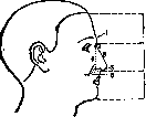
Figure 1. Division of the facial profile: 1—superciliary arches; 2—root of the nose; 3—bridge of the nose; 4—height of the nose; 5—base of the nose; 6—rise of the nose; I—frontal part; II—nasal part; III—oral part of the face.
The forms of the face of a living person are determined by the shape of the bony skeleton of the face. The right and left sides of the face very often present the phenomenon of asymmetry. In view of the complexity and extent of the boundaries of the face, it is customary to divide it into a number of regions. Due to the fact that the ends of some muscles are firmly woven into the skin on the chin, at the wings of the nose, and at the corners of the mouth, constantly existing skin folds are formed. Thus, from the wing of the nose to the corner of the mouth runs the nasolabial fold (plica naso-labialis); between the lip and the chin is located the transverse mentolabial fold (plica mento-labialis). The muscles of the face are attached by both ends to the skin or by one end to the bones and the other to the skin, which is why upon their contraction the skin gathers into characteristic folds and somewhat draws closer. If the activity of a known group of muscles occurs frequently and moreover for a long time, an prevailing main feature is formed, remaining forever. With a moderate development of soft tissues and especially the subcutaneous fat layer, it is quite easy to palpate individual bony parts serving for orientation. Thus, one can easily feel the supraorbital margins, delimiting the face from the forehead, the bony margins of the entrance into the orbit, the zygomatic arches, the margins of the nasal aperture, the bridge of the nose, the lower margins of the mandible up to the angle, the masseter muscle and the external maxillary artery looping over its anterior edge for examining the pulse, and finally the mass of the parotid salivary gland. Ontogenetic development of the face (of man). In the embryo at an early stage of development in the head region, a depression is formed, the so-called oral bay, along the lateral walls of which under the ectoderm lining them appear, thanks to the thickening of the mesoderm, branchial arches separated from each other by branchial clefts. The cavity of the oral bay is bordered by parts of the first branchial arch, represented by the so-called upper and lower jaw processes. Below and somewhat posteriorly are located the second and third branchial arches (see Visceral skeleton). Laterally above the oral bay with the first branchial arch bordering it appears the so-called frontal process, representing a continuation of the frontal part of the base and passing on each side into the outer and inner nasal processes; between the latter remain the so-called nasal pits, communicating with the oral bay and the oral cavity developing from it later by two openings, which Kölliker calls internal nasal openings. Between the outer nasal and maxillary processes one can see a small longitudinal groove, which ascends upwards and backwards to the eye vesicle and is called the lacrimal groove (sulcus lacrimalis). Into the still undifferentiated nasolacrimal cavity protrudes the nasal septum with a thickening at its anterior end. Subsequently, the internal nasal processes fuse with each other, as well as the frontal parts of the facial skeleton. Somewhat later, the outer nasal processes are involved in the fusion process, fusing with the maxillary processes of the first branchial arch. Due to this, the lacrimal groove lying between them closes and turns into the nasolacrimal canal. The outer nasal processes, now fused with the maxillary processes of the first branchial arch, approach each other along the midline and fuse with the margins of the median jaw, due to which the alveolar margins of the jaw bones are formed and above them the external openings of the nasal cavity, located between the nasal processes. All the above-indicated changes within the first branchial arch occur during the 5th, 6th, and 7th weeks of intrauterine life (see vol. X, p. 560, figure 1). The soft tissues of the face develop from all sides in the form of folds, forming cheeks on the sides, and lips in front, between the unfused edges of which the oral opening remains. Phylogenetic development of the face is closely connected with the general development of the cerebral cranium, wherein changes in one part correlatively entail a series of changes in another. Comparing the skulls of modern man with the skulls of primitive man and apes, as well as the face of man with the muzzle of animals, one can see that along with the progressive development of the cranial vault went a regressive development of the middle and lower divisions of the facial skeleton. The progressive development of the cerebral cranium over the centuries, accompanied by corresponding
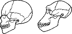
Fig.
Outline of the skull of a Sinhalese (a); the same of a chimpanzee (b). Progressive development of the cerebral cranium, accompanied by corresponding progressive growth of the brain, conditioned the paths of regressive development of the nasal cavity as a receptacle of the organ of smell and the masticatory apparatus, represented by strong and strongly developed jaws and teeth, which served our animal ancestors as a powerful weapon of attack and defense (fig. 2). And indeed, even upon superficial inspection, it is visible that the facial part of the head in man is an insignificant appendage of the head region, while in apes, even anthropoid ones, the facial skeleton is developed massively, and the head region is as if an appendage to it. Hence the radical difference between the face of man and the forward-protruding muzzle of apes and other animals. According to Stratz's calculations, the facial part of the skull (splanchnocranium) of a European on average equals 42.8% of the cerebral part of the cranial vault (neurocranium) and gives fluctuations for different races from 35.6% to 48.5% in men. In women, the facial part
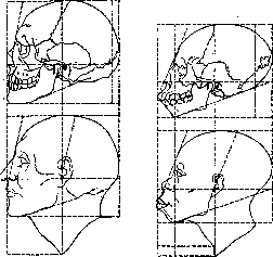
aъ
Figure 3. Facial angle: a—skull and face of a European; b—the same of a Negro. The facial part is significantly smaller and on average constitutes 30% of the cerebral division of the cranial vault. According to Stratz's calculations, in the horse the facial part of the skull (muzzle) constitutes 450%, in the orangutan 102%, and in the chimpanzee 90.4–94.2% of the cerebral division of the cranial vault. Klaatsch, comparing the profile outlines of the skulls of the gorilla, the Java pithecanthropus, and primitive man with the skulls of representatives of modern humanity from the so-called "lower" races to the civilized European, established with certainty the fact of the progressive development of the cranial vault, accompanied by the reduction of the jaws and teeth and the gradual retraction of the middle division of the facial skeleton (fig. 3). In the phylogenetic process of the development of the face, it is necessary to note the smoothing of the superciliary arches and the changing architecture of the forehead, which from low, sloping, as if flattened makes a transition to high, steep, rising upwards and open, which along with the progressive protrusion of the lower division of the body of the mandible corresponding to the chin sharply changed the profile outlines and the general aspect of the facial skeleton and the face of modern civilized man. Walkhoff was the first to note the evolutionary development of the chin in connection with the development of the musculature of the tongue and lower jaw, participating in the act of uttering sounds of articulate, distinct human speech. That is why the facial skeleton of animals, as well as of primitive man, lacks a typically developed chin and closely resembles the outlines of an animal muzzle. Parallel to the indicated changes in the facial skeleton, it is necessary to note corresponding changes in the facial musculature, consisting in the regressive development of the masticatory and progressive development and physiological differentiation of the mimetic musculature of the face, in the thinning of the skin cover, characterized by depletion and sexual differentiation of the hair cover of the face. In connection with the reduction of the masticatory musculature, it is necessary to note the decrease in the extent surface of the temporal fossae and the shortening of the transverse dimension of the face, conditioned by lesser protrusion and smaller dimensions of the zygomatic arches, which along with the temporal fossae serve as the site of attachment of the strongest masticatory muscles (m. temporalis, m. masseter).
, Age-related changes of the face. In humans, the final development of the face, like that of the entire skull, is completed with the end of the general growth of the organism, approximately at the 20th–22nd year of life. During ontogeny, the proportions of the individual tiers of the facial skeleton and of the entire face in relation to the cranial vault change drastically (Fig. 4). A schematic representation of the newborn skull at the scale of the adult skull shows the paths of the ontogenetic development of the skull. The cerebral part of the skull in a child is comparatively large, because the newborn's brain reaches approximately one quarter of its final weight development, while the total body weight of the newborn is equal to 1/16–1/20 of the average adult weight. In accordance with this, the facial skull is comparatively small at birth. The child's face has a rounded, shortened shape (Fig. 5) and grows vigorously during ontogeny, approaching an elongated oval shape in which length sharply predominates over width. At the same time, the strongest growth is observed from birth to the end of the second year and from 12 years to adulthood; the face widens and lengthens by only approximately 2 mm in the course of each year, the volume of the facial part of the skull in
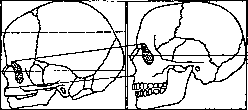
Figure 4. Growth of the facial part of the skull from birth to adulthood.
the course of the first year of life barely constitutes 13% of the volume of the cranial vault, by 8 years it grows to 18.3%, by 10 years to 20.5%, by 12 years to 21.4%, and in adulthood to 30.96%. Thus, the facial skeleton acquires more than double the volume compared to the cranial vault. The nasal bones, growing in width, are set at an angle to each other, thanks to which the gap between the eye sockets does not increase significantly. Due to the growth of the upper jaw, it protrudes further and further forward, so that the straight standing of the milk teeth (orthognathism) gradually turns into oblique (prognathism). Both halves of the lower jaw fuse along the midline, and the relief of the chin and angles appears individually distinct. The enlargement of the facial skeleton occurs due to the development of accessory sinuses, alveolar processes of the jaws and teeth, and the absolute magnitude of the width, length, and height of the jaw bones undergo continuous changes throughout ontogeny from birth to the period of growth cessation and from the latter to secondary senile changes of the face, when with the loss of teeth and the resorption of the alveolar
processes, the face once again acquires a rounded, shortened shape, bringing the faces of the elderly close to the face of a child. With the onset of puberty, the shapes of the skull and face remain almost unchanged until old age, and changes concern the hairline and skin folds of the face. In old age, the bones of the face become more brittle and thin, and the old man's skull loses 1/5 of its weight. The face decreases again in the longitudinal direction as a result of the resorption of the tooth sockets and the loss of teeth. The lower jaw, deprived of teeth, due to the resorption of the alveolar processes, forms a larger arch than the upper one and no longer meets it, but embraces it when closing the mouth, and the chin protrudes sharply forward. The rami of the lower jaw are directed obliquely backward, the nose approaches the chin, the soft parts of the cheeks, having lost their elasticity, become flabby and form wrinkles appearing on the cheeks around the mouth opening, as well as on the forehead and around the eyes. Sexual characteristics of the face. The male departs further from the infantile state both in terms of the development of the face and the facial skeleton. The ratio of the volume of the facial part to the cerebral part in women is smaller than in men. The female skull in both shape and dimensions occupies an intermediate position between the child's and the male's (see Skull). The eye sockets of the female skull have more rounded outlines, while in men they are rather quadrangular with rounded edges; at the same time, the dimensions of the female eye sockets with equal dimensions of the eyeballs appear larger. The distance between the eye sockets in women is greater than in men, thanks to which their eyes are more widely spaced. This partly explains the fact that wider noses are more frequently encountered in women, while longer ones constitute a feature of the male face. The upper jaw with its processes is developed weaker in women, and the face therefore appears narrower. The lower jaw in them is smaller than in men. The masticatory and skeletal musculature in men is developed more strongly, which is reflected on the skull by a greater prominence of tubercles and significantly greater roughness, while the female skull has a smoother surface. The superciliary arches in women are weakly expressed, the eyes are closer to the surface of the face. The mimic musculature in women is more finely developed and more often forms a tendency toward a relief expression of the lip line. The mouth opening in men is larger than in women. The skin of the face in women is thinner and more delicate, especially on the cheeks, thanks to which the vessels easily show through; the hairline of the face characteristic of the male sex is replaced in women by a light, often colorless down, and usually only in the postmenopausal period, as well as in pathological cases, can a more intensive growth of mustache and beard be observed. Ossification of the face. The process of ossification in the bones of the face begins in the 6th or 7th week of embryonic life; primary ossification centers appear in the upper and lower jaw bones. The maxillary bone ossifies from five centers. The palatine, nasal, zygomatic bones, and vomer ossify in the 7th or 8th week; the lacrimal bones by the end of the 12th week, and the inferior nasal conchae in the 5th month of embryonic life. Ossification centers of the greater horns and the body of the hyoid bone appear by the end of embryonic life, and of the lesser horns even during the first year of extraembryonic life. Of all the facial bones, only the inferior nasal conchae pass through three stages (membranous, cartilaginous, and bony); all the rest transition from the membranous stage directly into the bony one. The bony framework of the face is formed by 14 facial bones (paired—nasal, maxillary, zygomatic, lacrimal, and palatine bones, nasal conchae, and unpaired—mandible and vomer) and is partially complemented by the bones of the cerebral cranium: ethmoid, sphenoid, temporal, and frontal. The facial part of the skull, adjoining the anterior cranial fossae from below, forms a series of tier-like cavities. The upper third of the face is occupied by the openings of the eye sockets, which are separated from each other by the forward-projecting roof of the nasal cavity formed by the nasal bones and the frontal processes of the maxillae (see Skull). Externally, these openings are demarcated by the sphenofrontal processes of the zygomatic bones, forming arches characteristic of primates and especially of humans, separating the eye socket from the base of the skull. Below lies the nasal cavity, opening with the piriform aperture (apertura piriformis). The nasal cavity with its accessory cavities (sinus paranasales) significantly reduces the massiveness of the facial skeleton and occupies the central region of the face. The lower, most massive section is represented by the entire surface of the mandibular bone and, being suspended movably from the base of the skull, forms a wide slit corresponding to the opening of the oral cavity. Despite such a structure of the face, rich in cavities and paranasal sinuses, the facial skeleton, experiencing significant pressure from the lower jaw during mastication, appears extremely solidly constructed thanks to the peculiarity of its architecture. The force of mechanical shocks and concussions experienced during the movement of the lower jaw and transmitted through the facial part of the skeleton to the cerebral one is moderated and weakened thanks to the elastic, somewhat movable junc
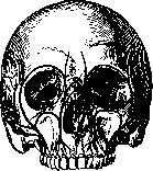
Figure 6. Anterior view of the skull with facial buttresses.
connection of the teeth in the jaw bones (see Teeth), as well as the existence of a number of facial buttresses that strengthen the facial skeleton and play the role of true struts, in the spaces between which various cavities of the facial skeleton are located, thereby achieving the structural strength of the face, which sometimes experiences strong external traumatic influences. These buttresses, three in number, have the outline of arch-curved columns resting below on the alveolar margins of the corresponding teeth, and above on various points of the facial and cerebral skull.-1. The frontonasal buttress, corresponding to the canines, rests below on the thickened walls of the canines as juga alveolaria and continues upward in the form of compact plates of the nasal processes of the maxilla, resting above on the outer edges of the nasal processes of the frontal bones.-2. The zygomatic buttress rests on the thickened edges of the sockets of the first two molars, goes upward in the lateral walls of the maxillary sinus and connects with the zygomatic bone, which itself rests outwardly on the zygomatic process of the temporal bone, and above on a similar process of the frontal, thanks to which the zygomatic buttress is the most important and strongest in the series of others.-3. The pterygopalatine buttress corresponds to the ends of the alveolar processes and is formed by the pterygoid processes of the sphenoid and the ascending branches of the palatine bones, resting against the posterior surface of the maxillary tuberosity (tuberositas maxillae) (Figures 6 and 7). The muscles of the face according to their topographical position and origin can be divided into three main groups. The first group includes the subcutaneous, or mimic musculature, originating through the progressive differentiation of the subcutaneous muscle of the neck (m. platysma, s. subcutaneus colli), which itself represents a rudiment of the broad subcutaneous muscle of animals, forming the so-called fleshy layer (panniculus carnosus). The second group includes visceral muscles associated with the jaw and hyoid bones, tongue, pharynx, soft palate, and auditory ossicles. These are the masticatory muscles, muscles of the floor of the oral cavity (usually described as cervical), intrinsic muscles of the tongue, soft palate, and finally a small group of muscles of the auditory ossicles. The third group includes the muscles of the eyeball. Around all the above-mentioned openings, the head section of the m. platysma differentiates into two layers of muscles with a definite arrangement of fibers: internal, or deep, circular and external, or superficial, radial, playing the role of sphincters and dilators of the openings (Figures 8, 9, and 10).-Standing apart is the muscle of the cranial vault pushed forward like a helmet (m. epicranius), the anterior muscular section of which in the form of the frontal muscle (m. frontalis) descends along the entire surface of the forehead, merging with the subcutaneous musculature of the orbits and eyebrows. Its muscle fibers begin below on the nasal process of the maxilla, from the skin of the eyebrows, from the supraorbital margin and superciliary arches, then ascending upwards, they pierce the thickness of the orbicularis oculi muscle and at the level of the frontal tubers pass imperceptibly into the tendinous part of the helmet (galea aponeurotica cranii). At the same time, a significant longitudinal section remains along the midline, having no muscle bundles. A small bundle, standing out from the mass of the muscle in the lower-anterior section and descending onto the dorsum of the nose, received the name (thanks to its ability to form a characteristic fold on the dorsum of the nose) of the procerus muscle (m. procerus nasi, s. pyramidalis nasi). Its existence, however, is denied by some authors (Henle). M. frontalis is a physiological antagonist to the occipital bundle and is able to fold the skin of the forehead into transverse folds and raise the eyebrows upwards, which was first proved by the experimental irritation of the muscle by Cruveilhier. Mimic musculature. Muscles surrounding the eye opening. To these, besides the frontal one described above, belongs the orbicularis oculi muscle (m. orbicularis oculi) and the corrugator supercilii muscle (corrugator supercilii). Apparently, blinking movements can be attributed to the circular muscle (see Blinking), which are considered by some authors (Sappey) as a result of the predominance of the tone of the circular muscle over the contraction of a special muscle that raises the upper eyelid (see Eyelids). The outer section of the muscle gathers the skin into folds around the eyelids. Deeper than the superficial portion of the circular muscle, intertwining with the frontal one, lies a small muscle on the bone itself, the corrugator supercilii (m. corrugator supercilii). Part of the fibers of the orbicularis oculi muscle near the place of attachment to the bone and the inner ligament of the eyelids, splitting into two portions, encloses the lacrimal sac lying in the lacrimal canal. These bundles, fused with the walls of the sac, upon contraction pull the walls of the latter away from one another and promote the suction of tears from under the eyelids. Lig. palpebrale superior et inferior or the so-called orbital septum - septum orbitale - represents a thin fibrous plate, which in the form of a septum closes the contents of the orbit from the front and with the aid of connective tissue cords,
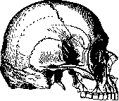
Figure 7. Side view of the skull with facial buttresses. «connected with the more superficially located m. orbicularis oculi, as a result of which the septum is extremely difficult to detect if one prepares it from the anterior surface, while from the orbit side, removing the fat mass with tweezers, one can with the smallest number of muscles of all the openings of the face and appears the least movable (see Nose.) The nose (nasus externus) opens outwardly by two openings of the nostrils (nares), which possess greater or lesser ability to narrow or widen,
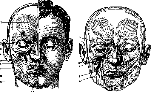
Figure 8.
Figure 9. m. frontalis; 2 - m. orbicularis oculi; Figure 8. Mimic musculature (superficial layer): 1 - m. ..., 2 - m. ..., 3 and 4 - caput angulare et caput infraorbit. m. quadrati labii superioris; 5 - m. caninus; 6 - m. zygomat.; 7 - m. risorius; 8 - platysma; 9 - m. triangul.; 10 - m. quadratus labii inferioris. Figure 9. Mimic musculature (deep layer): 1 - m. m. procerus; 2 - m. nasalis; 3 - m. caninus; 4 - m. buccinator; 5 - m. masseter. _________________
(According to Sicher-Tandler.) it is easy to open the posterior surface of the upper and lower eyelid ligaments. Lig. palpebrale mediale et laterale - the lateral medial and lateral ligaments of the tarsal plates of the eyelid, having various bends and a peculiar configuration that gives the face characteristic racial and individual differences. The tarsal plates, consisting not of cartilage, but of densely interwoven bundles of connective tissue, in the medial corner of the eye are connected by the so-called lig. palpebrale mediale, which extends from the designated corner to the frontal segment of the maxilla, lies immediately under the skin, in front of the blind end of the lacrimal sac and can be easily palpated with a closed eye. The lateral palpebral ligament (lig. palpebr. laterale) is usually absent in man. The commissures of the medial and lateral edges of the eyelids, more often the inner corner of the palpebral fissure, are designated by the term «canthus», well developed only in humans having an almond-shaped slit of the palpebral fissure. In animals, as is known, the palpebral fissure is round and corresponds in size to the circumference of the cornea, which is why the sclera is visible only at extreme degrees of rotation of the eyeball and the commissures of the eyelid corners are smoothed out almost completely. - Muscles surrounding the nasal opening. The nasal opening has the ability to widen. The muscular apparatus of the cartilaginous wings of the nose is represented partly by intrinsic muscles, but mainly by the muscles of the corners of the mouth. In the description of the nasal muscles in most authors, discrepancies can be seen. It is customary to describe five nasal muscles. 1) The procerus muscle, already described above. 2) The muscle that raises the wing of the nose (m. levator alae nasi) and represents part of the muscle that raises the upper eyelid; it begins at the edge of the orbit from the edges of the nasal process of the maxilla and attaches to the skin and cartilage of the wing of the nose. 3) The compressor nasi muscle (m. compressor nasi), begins from the jugum alveolare of the incisor teeth and, ascending, attaches to the edge of the cartilage of the wing of the nose along the outer surface of the wings of the nose to its back, where, having expanded, it merges with a tendinous bridge with the muscle of the opposite side, thanks to which a tendinous aponeurosis of the dorsum of the nose is formed. 4) The depressor of the nasal septum (m. depressor septi mobilis narium), represents as it were a detached bundle of the orbicularis oris muscle, which weaves into the mobile part of the cartilaginous nasal septum. The physiological action of the nasal muscles is characterized by the names of the muscles. Hyrtl also attributes to the muscle that raises the wing of the nose the ability to expand the nasal opening. 277 The muscles surrounding the oral opening represent the most numerous group of mimic musculature; they consist of two anatomical and physiological muscle groupings (Figure 11). The first group includes muscles that widen the mouth, diverging radially and located in the mass of the subcutaneous layer at various depths; the second group includes sphincters with a circular course of muscle fibers.
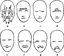
Figure 10. a - direction of traction of the radial musculature: 1 - m. front.; 2 - m. corrugat. supercil.; 3 - m. risorius; 4 - m. zygomat.; 5 - m. quadratus labii superioris; 6 - m. triangul.; 7 - quadr. labii inferioris; b - attention; c - reflection; d - pain; e - smile; f - laughter; g - crying; h - disgust.
Closer to the skin, at the border with the nasal region, are located: 1) M. lev. alae nasi et lab. super. 2) The proper muscle lifting the upper lip (m. levator labii proprius), which has a quadrilateral shape; it originates from the facial surface of the body of the maxilla, below the infraorbital margin, and, running downwards, forwards, and inwards, is lost in the skin in the middle of the extent of the nasolabial fold. 3) The zygomaticus minor muscle (m. zygomaticus minor, s. caput zygomaticum m. quadrati) has the shape of a narrow muscular ribbon, originating from the outer convex surface of the zygomatic bone, whence it runs obliquely downwards and forwards and is attached to the skin of the angle of the mouth, somewhat outwards and backwards from the preceding one. All three described muscles are united by Henle and Henke into a single muscle, which is given the name of the quadratus labii superioris muscle (m. quadratus labii superioris), in which three heads are distinguished: 1) the angular head (caput angulare), described above under the name of the muscle lifting the upper lip and the wing of the nose; 2) the infraorbital head (caput infraorbitale), described as the proper muscle lifting the upper lip; and finally 3) the zygomatic head (caput zygomaticum), corresponding to the zygomaticus minor muscle (see above). Outwards and somewhat more superficially lies 4) the zygomaticus major muscle (m. zygomaticus major), which originates from the margin of the zygomatic arch and runs forwards to the angle of the mouth, where it terminates on the nasolabial fold. Deeper than the quadratus muscle, in the mass of the underlying adipose tissue, is located 5) the levator anguli oris muscle (m. levator anguli oris), also called the triangularis superior muscle (m. triangularis superior), or the canine muscle (m. caninus); it originates with a broad base from the bottom of the canine fossa; the fibers run downwards and inwards to the angle of the mouth, where part of them terminates; the greater part, however, decussating, passes into the thickness of the lower lip, taking part in the formation of the superficial layer of the circular muscle that compresses the mouth. The remaining part of the muscles of the first group represents a group of antagonists to the described ones and is located in the thickness of the lower lip and chin. Superficial to all lies 6) the depressor anguli oris muscle (m. depressor anguli oris, s. triangularis); it has a triangular shape and originates with a broad base along the margin of the mandible, from the chin to the second premolar, whence the fibers, narrowing, run upwards to the angle of the mouth, where, just as in the preceding one, one portion is attached to the skin of the angles of the mouth, while the other, decussating, passes into the thickness of the upper lip and takes part in the formation of the superficial portion of the orbicularis oris muscle. Immediately deeper than the triangular, partly covering it, lies 7) the quadratus labii inferioris muscle (m. quadratus labii inferioris, s. depressor labii inferioris); it originates from the margin of the mandible throughout the extent from the mental foramen to the very angle of the mandible and terminates in the skin of the lower lip. 8) The mentalis muscle (m. levator menti) occupies the triangular space between the two quadratus muscles on
11,3 the anterior surface \il of the mandible. The muscle 1'.; fibers originate from the surface of the alveoli of the incisor teeth, whence they run downwards and are attached to the skin of the chin along its entire extent. 9) The risorius muscle (m. risorius, Santorini), a thin fan-shaped muscle, lies so
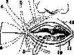
Figure 11. Diagram of the m. orbicularis oris: 1 and 13 - m. levat. nasi et labii sup. prof. et superficial.; 2 - quite superficially m. caninus; 3 and 4 - m. L
[. , zveromat. mm. et ma- above all muscles and with a broad base begins from -mm. incisivi labii inferioris et superioris. zygomat. min. et major; 5-m. buccinator; 6-m. risorius; 7-plat-ysma; 8-m. triangul.; TTT;rT, Trr.t,r4„DQTrilTTClW«,0 9-m. quadratus labii inferioris; 10-m. orbicul. oris; 11 and 12-the parotid gland; narrowing, its apex attaches to the corners of the mouth. The middle buccolingual section of the face, in addition to the mimic musculature, possesses muscles belonging to the visceral system. There are three such muscles. 1) The buccinator muscle (m. buccinator); forms the soft lateral walls of the oral cavity; on the inside it is covered by the mucous membrane, while on the outside it comes into contact with the adipose tissue forming Bichat's fat pad (corpus adiposum Bichati), and is pierced by the excretory duct of the parotid gland. The muscle fibers begin from both jaws at the level of the bottom of the molars and from there proceed to the corners of the mouth; the major part enters into the composition of the deep portion of the m. orbicul. oris. A fairly compact bundle of fibers originating from the bucco-pharyngeal fascia (fascia bucco-pharyngea) joins the crossed bundles (Fig. 11). 2) The orbicularis oris muscle (m. orbicularis oris); represents an individually variously developed muscular ring forming the thickness of the lips and pierced by dense fibrous bundles running vertically from the skin to the mucous membrane. 3) The incisive muscles (mm. incisivi); small muscle bundles beginning from the surface of the jaw bones and weaving into the composition of the orbicularis oris muscle.—Physiol. role of the circular and radial muscles of the oral orifice, despite the complexity of their anatomical differentiation, appears extremely simple. In no animal, including the highest anthropoid apes, does the oral orifice possess such significant and richly differentiated musculature as in man. Therefore, animal mimicry is considerably poorer than in man, and almost the entire play of the lips is limited to grasping food and baring the teeth. The orbicularis oris muscle of man is ascribed the ability to keep the oral fissure closed, and with strong contraction to pull the lips forward, which we do during eating, talking, whistling, kissing, etc. The m. quadr. labii sup. in addition to raising the wing of the nose and lip together with the m. corrug. superc. participates in the mimicry of crying and discontent. The m. zygom., raising the corner of the mouth, together with the m. orbitalis participates in the mimicry of joy. The m. caninus, pulling the corners of the mouth upward and baring the teeth, together with the m. quadr. lab. inf. participates in the mimicry of anger and discontent. The m. risorius, pulling back the corners of the mouth and widening the oral fissure, participates in the mimicry of discontent and daring; the m. triang. pulls the corners of the mouth downward and participates in the mimicry of dejection and discontent (see Mimicry). The fasciae of the face are two: temporal and buccal. The temporal fascia (fascia temporalis) consists of two leaves: superficial and deep. The superficial leaf represents as it were a continuation of the head aponeurotic helmet (galea aponeurotica), which thins out in the downward direction and, losing the properties of an aponeurosis, ends at the level of the zygomatic arch. The described superficial leaf is separated by a thin layer of loose cellular tissue from the deep leaf of the temporal fascia (fascia temporalis propria, s. profunda), which represents a continuation of the periosteum of the cranial bones. It begins from both temporal lines and, going down, splits into two leaves, of which the superficial one attaches to the upper edge of the zygomatic arch, and the deep one to the inner edge; the triangular space formed between them is filled with a small amount of adipose tissue (see Temporal region).—The buccal fascia (fascia buccalis) also breaks down into two plates—superficial and deep. The superficial fascia, called the parotid-masseteric (fascia parotideo-masseterica), extends downward from the zygomatic arch and, splitting, clothes the parotid gland and the buccinator muscle. Posteriorly it is connected with the mastoid process and the cartilage of the ear, downward it passes into the superficial leaf of the cervical fascia; in the forward direction it continues into the deep or bucco-pharyngeal fascia.—The bucco-pharyngeal fascia (fascia bucco-pharyngea) covers the outer surface of the buccal muscle and at its posterior edge merges with the pterygomandibular raphe (raphe pterygo-mandibularis) and from here continues along the outer surface of the superior pharyngeal constrictor, representing the superficial pharyngeal fascia (fascia pharyngea superficialis), or the so-called tunica adventitia pharyngis. Between both plates at the anterior edge of the buccinator muscle there remains a space filled with a lump of adipose tissue (corpus adiposum Bichati), which imparts roundness to the cheeks. With general emaciation the fat pad disappears, and the skin of the cheeks sinks in, giving a peculiar appearance to a thin face. Vessels and nerves of the face. The arterial vessels of the face are branches of the external carotid artery and partly of the ophthalmic. Among the numerous branches, the most important are: 1. The superficial temporal artery (a. temporalis superficialis), which alongside the internal maxillary artery (a. maxillaris interna) represents one of the terminal branches of the external carotid artery (see Carotis art.), divides into 2 large branches: anterior—frontal and posterior—temporal. Somewhat earlier, the temporal artery gives off a number of branches to the face running parallel to the zygomatic arch and supplying the muscles of the face: the transverse facial artery (a. transversa faciei), running below the zygomatic arch, between it and Stensen's duct, and the zygomatic artery (a. zygomatica), running parallel to the first, but above the zygomatic arch, at the level of the angle of the palpebral fissure. 2. The external maxillary artery (a. maxillaris externa) departs from the trunk of the external carotid artery at the level of the angle of the lower jaw; it is directed forward and upward along the inner surface of the submandibular salivary gland [along the outer surface descends the anterior facial vein (v. facialis anterior)] to the anterior edge of the masseter muscle, where the artery bends over the edge of the lower jaw and can be easily compressed with a finger during bleeding. On the face it runs, lying rather deep under the superficial layer of the mimic musculature, to the corners of the mouth, where it breaks down into three terminal branches: the superior and inferior labial coronary arteries (aa. coronariae labii super. et infer.) and the angular artery of the nose (a. angularis nasi), which ascends upward to the wing of the nose and anastomoses here with one of the branches of the ophthalmic artery. The coronary arteries, running in the thickness of the lips, form a circular arterial ring located under the mucosa in the vestibule of the oral cavity. Vascularization of the upper and lower lips occurs due to the labial arteries (arteriae labiales superiores et inferiores), which are tiny, breaking up into a dense capillary network and imparting the characteristic coloration to the lip mucosa, being the terminal branches of the labial coronary arteries (aa. coronariae labii superioris et inferioris). The soft parts of the buccal region are vascularized by the buccal artery (a. buccinatoria), departing from the trunk of the a. maxil. int., from which the second large branch of this region also departs—the infraorbital artery (a. infraorbitalis). The latter emerges onto the face through the foramen of the same name and breaks down into a whole network of branches nourishing the soft tissues in the middle region of the face and the anterior teeth. 0.5 cm higher than the lower edge of the lower jaw on its anterior surface from the foramen of the same name emerges a small mental artery (a. mentalis), which takes part in the nutrition of the soft tissues of the chin and anastomoses with the submental artery (a. submentalis), running from the external maxillary artery forward, where it bends onto the chin. Blood from the middle part of the face corresponding to the buccal region outflows through the buccal vein (v. buccinatoria) into the pterygoid venous plexus located in the region of the pterygopalatine fossa, and partly into the system of the anterior facial vein (v. facial. ant.). Lymphatic vessels of the face. The superficial vessels of the face draining the lymph of the skin and subcutaneous tissue can be divided into three groups: external, internal, and lower (see art. 165, fig. 20). The external group takes its origin in the skin of the outer half of the upper and lower eyelids and the zygomatic region. Their course corresponds to the zygomaticoorbital and transverse facial arteries (aa. zygomatico-orbitalis et transversa faciei), and their centers are the anterior auricular and superficial parotid glands. More
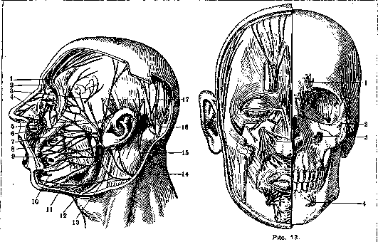
Figure 12.
Figure 12. Superficial branches of the trigeminal and facial nerves: 1 - supraorbital nerve; 2 - frontal branch of the frontal nerve; 3 - zygomaticotemporal nerve; 4 - external nasal branch of the anterior ethmoidal nerve; 5 - infraorbital nerve; 6 - zygomatic branches of the facial nerve; 7 - zygomatic and buccal branches of the facial nerve; 8 - buccal branches of the facial nerve; 9 - buccal nerve; 10 - mental nerve; 11 - marginal mandibular branch of the facial nerve; 12 - cervical branch of the facial nerve; 13 - transverse cervical nerve; 14 - great auricular nerve; 15 - lesser occipital nerve; 16 - greater occipital nerve; 17 - auriculotemporal nerve (After Sicher and Tandler). Figure 13. Exit points of the branches of the trigeminal nerve: 1 - supraorbital nerve; 2 - zygomaticofacial nerve; 3 - infraorbital nerve; 4 - mental nerve. The venous network fully corresponds to the arterial network and appears extremely rich; it envelops the arteries and nerves of the face. There are two main veins: the temporal and the anterior facial. The anterior facial vein (v. facialis anterior) corresponds to the branches of the external maxillary artery, and like it, at the edges of the orbit anastomoses with the dorsal nasal vein from the orbital vein system, and at the root of the nose and temple forms a rich anastomotic network with the superficial veins of the face and head. The main trunk is formed by the union of the two labial coronary veins at the corners of the mouth, whence the vein descends onto the neck along the anterior surface of the submandibular salivary gland and, beneath the angle of the mandible, joining with the posterior facial vein, forms the trunk of the common facial vein (v. facialis communis). The numerous internal group of vascular veins originates from the cutaneous networks of the glabella, nose, and its vestibule, the inner half of the upper and lower eyelids, the cheek, the upper and partly lower lips, and the angle of the mouth. The course of these vessels corresponds to the direction of the external maxillary artery, and their centers of convergence are the submandibular glands lying along the course of this artery. A third, less numerous group of superficial facial vessels originates in the skin of the lower lip and chin. This group follows an insignificant path, flowing into: 1) the submental glands lying on the diaphragm of the mouth at the inner margin of the anterior belly of the digastric muscle, and 2) the anterior submandibular glands lying at the outer margin of the same muscle. To the indicated three groups of superficial lymphatic vessels of the face correspond three groups of deep lymphatic vessels of the face, originating from deeper organs: the conjunctiva of the eye, the facial muscles, the mucous membrane of the cheek and lips with their salivary glands, as well as the outer gums, the mucous membrane and periosteum of the bones of the oral and nasal vestibules, and the periosteum of the outer surface of the bones forming the facial skeleton. Nerves of the face. Motor branches to the facial musculature of the face come from the seventh pair of cranial nerves (n. facialis), which forms under the skin the so-called greater goosefoot (pes anserinus major) (see Facialis nervus). The motor branches of the masticatory musculature represent branches of the motor part of the third pair of the trigeminal nerve (n. crotapnitico-buccinatorius). Sensory branches supplying the skin of the face are cutaneous branches of all three branches of the trigeminal nerve, distributed in strictly delimited corresponding regions of the face (Figs. 12, 13, and 14). The nerves of the upper lip are: sensory—branches of the infraorbital nerve (n. infraorbitalis), the second branch of the trigeminal nerve, and motor—branches of the facial nerve from the group of buccal branches (rami buccales). The nerves of the lower lip: sensory—originate from the mental nerve (n. mentalis)—the third pair of the trigeminal nerve, and motor—from the corresponding branches of the facial nerve. (See Trigeminus nervus.) Face and the doctrine of constitutions. In the face, individual, sexual, racial, and age characteristics are expressed most strongly; hence the structure of the face finds reflection in the modern doctrine of human constitutional types. In the digestive type, the facial skeleton, sitting on a short and thick neck, is strongly developed, especially in the lower division, corresponding to the masticatory apparatus, as a result of which the face takes the form of a truncated pyramid with a wide base pointing downwards. In the respiratory type, the facial division of the head is strongly developed in the middle part corresponding to the vestibule of the respiratory tract and is characterized by a long and large nose and sharply protruding zygomatic arches. The jaw apparatus is weakly developed. The facial musculature is best expressed in the same middle division and especially on the wings of the nose. In the muscular type, the face appears harmoniously developed; the contour of the head has the shape of a rectangle. The forehead is straight, and the superciliary arches are weakly developed. In the cerebral type, the head has the shape of a pyramid with its base pointing upwards, due to the weak development of the middle-respiratory and lower-digestive divisions and, conversely, the strong development of the upper cerebral division. A high, steep forehead is combined with weak development of the jaws and masticatory musculature. The facial musculature is especially well developed in the frontal division and around the orbital openings, with a generally good development of all facial musculature, which is distinguished by variety and richness of mimicry. In asthenics, the face is usually long, thin, and lean; the skin on the face is thin, due to which the subcutaneous vessels are clearly visible. Due to the weak development of the lower division of the face, the latter en face has the shape of a shortened egg. The athletic type, characterized by a strong development of the bone skeleton and subcutaneous fat layer, is provided with a high skull with a well-developed facial division. The attachment sites of the masticatory and occipital muscles are sharply expressed. The lower mandible is well developed and sharply protrudes against the background of the face; the superciliary and zygomatic arches sharply project, which gives the face the shape of an egg, sometimes a shield-like outline. In the pyknic type, the head, due to the shift of the shoulder girdle toward the midline, appears to sit on a short and thick neck and seems pushed between the raised shoulders. The skull is large, round, with significant circumference dimensions, and a mostly flat vertex; the height of the skull is not large, and the facial skeleton is well developed. The face repeats the features of the trunk: it is wide, soft, and rounded due to the abundance of the subcutaneous fat layer. The skin of the face is thin, and the vessels are strongly developed and visible through it. The nose and cheeks show a tendency toward a pink coloration, and the cheeks toward a large accumulation of fat, especially in the lower divisions, at the angles of the jaw and at the chin, due to which the pyknic face usually possesses a double chin. Cephalometry and craniometry as applied to the face. Due to the imperfection of the method of simple description, in which subjectivity plays a significant role in the evaluation of certain features, there is an inevitable aspiration to establish an objective, exact basis for studying the structural features of the face. In addition to the method of measurement following Baltz, another method of drawing outlines is used, which received its completed development in the works of Sarasin ("The Veddas of Ceylon") (Fig. 15). The shape of the facial part of the skull and head, as is known, is determined by its height and width, which is mainly determined by the width of the cheekbones, which can be measured not only on the skeleton, but also on a living person. The width of the face thus largely depends on the development of the zygomatic bones. Examining skulls at some distance from above, one can note the existence of two types of zygomatic arches, with which the notion of extreme forms of facial width is associated. They closely adjoin the facial skeleton or sharply protrude outward. According to Martin, the width of the zygomatic arches ranges from 116 to 158 mm in the living, and from 100 to 155 mm on the facial skeleton. Of greatest importance for characterizing the facial part of the skull are two indices determining the ratio of the maximum width of the zygomatic arch to the total height of the face and to the height of the upper part of the face. In humans
^W
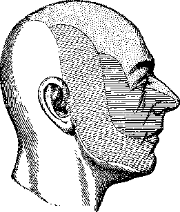
Figure 14. Diagram of the innervation zones of the branches of the trigeminal nerve on the face: dots — 1st branch; solid lines — 2nd branch; dashed lines — 3rd branch. cavity and nose, and the periosteum of the outer surface of the bones forming the facial skeleton. Nerves of the face. Motor branches to the facial musculature of the face come from the seventh pair of cranial nerves (n. facialis), forming under the skin the so-called greater goosefoot (pes anserinus major) (see Facialis nervus). The motor branches of the masticatory musculature represent branches of the motor part of the third pair of the trigeminal nerve (n. crotapnitico-buccinatorius). Sensory branches supplying the skin of the face are cutaneous branches of all three branches of the trigeminal nerve, distributed in strictly delimited corresponding regions of the face (Figs. 12, 13, and 14). The nerves of the upper lip are: sensory—branches of the infraorbital nerve (n. infraorbitalis), the II branch of the trigeminal nerve, and motor—branches of the facial nerve from the group of buccal branches (rami buccales). The nerves of the lower lip: sensory—originate from the mental nerve (n. mentalis)—the III pair of the trigeminal nerve, and motor—from the corresponding branches of the facial nerve. (See Trigeminus nervus.) Face and the doctrine of constitutions. In the face, individual, sexual, racial, and age characteristics are expressed most strongly; hence the structure of the face finds reflection in the modern doctrine of human constitutional types. In the digestive type, the facial skeleton, sitting on a short and thick neck, is strongly developed, especially in the lower division, corresponding to the masticatory apparatus, as a result of which the face takes the form of a truncated pyramid with a wide base pointing downwards. In the respiratory type, the facial division of the head is strongly developed in the middle part corresponding to the vestibule of the respiratory tract and is characterized by a long and large nose and sharply protruding zygomatic arches. The jaw apparatus is weakly developed. The facial musculature is best expressed in the same middle division and especially on the wings of the nose. In the muscular type, the face appears harmoniously developed; the contour of the head has the shape of a rectangle. The forehead is straight, and the superciliary arches are weakly developed. In the cerebral type, the head has the shape of a pyramid with its base pointing upwards, due to the weak development of the middle-respiratory and lower-digestive divisions and, conversely, the strong development of the upper cerebral division. A high, steep forehead is combined with weak development of the jaws and masticatory musculature. The facial musculature is especially well developed in the frontal division and around the orbital openings with a generally good development of all facial musculature of the face, distinguished by variety and richness of mimicry. In asthenics, the face is usually long, thin, and lean; the skin on the face is thin, due to which the subcutaneous vessels are clearly visible. Due to the weak development of the lower division of the face, the latter en face has the shape of a shortened egg. The athletic type, characterized by strong development of the bone skeleton and subcutaneous fat layer, is provided with a high skull with a well-developed facial division. The attachment sites of the masticatory and occipital muscles are sharply expressed. The lower jaw is well developed and sharply protrudes against the background of the face; the superciliary and zygomatic arches sharply project, which gives the face the shape of an egg, sometimes a shield-like outline. In the pyknic type, the head, due to the shift of the shoulder girdle to the midline, appears sitting on a short and thick neck and as if pushed between the raised shoulders. The skull is large, round, with significant circumference dimensions, more often a flat vertex; the height of the skull is not large, the facial skeleton is well developed. The face repeats the features of the trunk: it is wide, soft, rounded due to the abundance of the subcutaneous fat layer. The skin of the face is thin, vessels are strongly developed and visible through it. The nose and cheeks show a tendency to a pink coloration, and the cheeks to a large accumulation of fat, especially in the lower divisions, at the angles of the jaw and at the chin, due to which the pyknic face usually possesses a double chin. Cephalometry and craniometry as applied to the face. Due to the imperfection of the method of simple description, in which subjectivity plays a considerable role in the evaluation of certain features, there is inevitably an aspiration to bring an objective exact basis to the study of the features of the structure of the face. In addition to the method of measurement following Baltz, another method of drawing outlines is applied, which received its completed development in the works of Sarasin, "On the Veddas of Ceylon" (Fig. 15). The shape of the facial part of the skull and head, as is known, is caused by its height and width, which is mainly determined by the width of the cheekbones, which can be measured not only on the skeleton, but also on a living person. The width of the face thus largely depends on the development of the zygomatic bones. Examining skulls at some distance from above, one can note the existence of two types of zygomatic arches, with which the notion of extreme forms of facial width is connected. They closely adjoin the facial skeleton or sharply protrude outward. According to Martin, the width of the zygomatic arches fluctuates in the living from 116 to 158 mm, on the facial skeleton from 100 to 155 mm. The greatest significance for characterizing the facial part of the skull is held by two indices determining the ratio of the maximum width of the zygomatic arch to the height of the whole face and to the height of the upper part of the face. In man
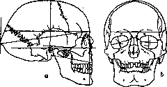
Figure 1;
Measurements on the skull: a — from the side; b — from the front. The height of the face ranges from 90 to 145 mm, and the height of the upper part of the face — from 52 to 91 mm (without the lower jaw). The shape and size of the face at all ages present extreme variety and can be reduced to two main morphological types — long-faced and broad-faced. Typical features are formed already in childhood, and in the process of ontogenesis the shape of the face and head as a whole remains quite stable. At the same time, it can be noted that various forms of the face are combined with certain morphological features of the head. Thus, long-facedness (leptoprosopia) is more often associated with long-headedness (dolichocephaly) and is considered an attribute of northern races. Broad-facedness (euryprosopia and mesoprosopia) is combined with mesocephalic and especially brachycephalic skulls, which is characteristic of Mongolian races. Topographic anatomy of the face. The following regions are distinguished: nasal, infraorbital, zygomatic, parotid, masticatory, oral, buccal, and mental. The location and boundaries of the enumerated regions are understood from their names (Fig. 16). 1. Nasal region (regio nasalis) occupies the region of the external nose and is separated from the infraorbital and buccal regions by means of the nasolabial sulcus (sulcus...)
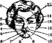
Figure 16. Natural folds and parts of the human face: 1 - glabella; 2 - nasus ext.; 3 - alae nasi; 4 - labium sup.; 5 - philtrum; 6 and 9 - lab. inf.; 7 - sulcus mento-labial.; 8 - mentum; 10 - tuberculum lab. sup.; 11 - sulcus naso-lab.; 12 - bucca; 13 - isthmus naso-buccalis; 14 - supercilium; 15 - radix nasi. [The text continues describing the anatomical regions of the face in layers: nasal, infraorbital, zygomatic, masseteric, buccal, labial, mental, and parotid regions, followed by a section on the clinical aspects of facial diseases, noting that the fully formed face results from the fusion of a system of clefts, recesses, and processes, and discussing developmental arrests and anomalies in embryogenesis]. N. Melik-Pashaev.

Figure 17. Transverse cleft of the face.—main) was one of the first to question the generally accepted mechanical explanation of facial malformations, admitting it only for a minority of cases in which the cause of the malformation is amniotic bands that impede facial development. Besides amniogenic causes, mechanical causes include skull malformations, an excessive enlargement of which can also delay facial development (hydrocephalus; Hentze). Furthermore, an obstacle to facial development may be the entrapment of a mispositioned embryo's hand in the embryonic cleft, the thumb of which may press on the face. A reduction in the cavity of the pregnant uterus (a uterine tumor or a growing embryo), by hindering fetal growth, can also serve as a cause of facial malformation. The most frequent facial malformation is hare-lip (see), which is often combined with cleft palate (see). Among the rarer malformations, lateral oblique cleft (meloschisis) (Fig. 17) deserves attention, occurring as a result of incomplete fusion between the lateral frontal and maxillary processes (the upper and lower processes of the first branchial arch). The oblique cleft usually begins from the free edge of the upper lip or from the angle of the mouth, runs obliquely toward the eye and ends near the lower eyelid or extends further along the temporal region to the hairline. Very rare cases of bilateral oblique facial cleft have been described. The oblique cleft is often found in combination with a transverse cleft, characterized by an enlargement of the mouth opening (macrostomia), which is also observed as an independent malformation. It depends on non-fusion between the first branchial arch and the maxillary process. It occurs either on one or on both sides. The length of the oral fissure in this case can be variable; in severe cases, the mouth reaches the external auditory meatus.—Besides malformations in the region of the lips and cheeks, a number of malformations occur in the nose region. Of these, the so-called bulldog nose (Doggennase) (Fig. 18) is of interest. The extreme degree of facial malformation, occurring always in combination with underdevelopment of the brain and a whole series of other pathological signs, is cyclopia, in which there is one eye, or rather one double eye. Often, together with cyclopia, arhinencephaly is encountered—abnormal development of the nose, instead of which a proboscis-like protrusion is sometimes observed. Such rare malformations include aprosopia (see), the congenital absence of the face. Eyelid malformations include ablepharia (see). Other malformations of the nose, jaws, and cerebral hernia—see the respective terms. The inconveniences caused by facial malformations are very different in character and severity. In newborns, alongside facial disfigurements, which sometimes reach significant degrees in complex forms, severe breathing and swallowing disorders occur that can become fatal (see. Cleft palate and Hare-lip). The prognosis depends on the prevalence and character of the malformation. While newborns suffering from cleft palate have a mortality rate (without surgery) of from 30% to 40%, newborns with more severe forms of malformations perish almost all, and while for the former one can argue regarding the choice of timing for surgical treatment, the latter should be operated on in the first days after their birth, without placing high hopes on a favorable outcome of this treatment. The principle of operation
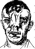
Figure 18. Median nasal cleft with displacement of the scalp.
with scars on it as traces of amniotic adhesions. The oblique fold of the cheek on the right with a coloboma of the lower eyelid generally corresponds to the rules applied in the operation for harelip: freshening of the edges of the clefts, their mobilization, and placement of sutures to approximate them. Hasselbach obtained excellent results with repeated operations for oblique facial cleft.-Atrophic (he-miatrophia faciei progressiva) (see Hemiatrophia) and hypertrophic processes, most frequently affecting one half of the face, also lead to its disfigurement. Facial hypertrophy is a congenital disease, which appears to be a particular manifestation of gigantism (see), and is frequently accompanied by excessive enlargement of other parts of the body (most often the extremities). To treat facial hypertrophy, ligation of the external carotid artery was proposed with the aim of stopping tissue growth. Pagenstecher and Werner, who performed this operation, obtained no results and proposed removing the hypertrophied tissues surgically. Completely standing apart are facial asymmetry in torticollis (see) and the characteristic enlargement of the face in acromegaly (see). As an exposed part of the body subject to the harmful action of the environment, the skin of the face is easily subject to various inflammatory processes, which often take a very severe course. Almost all acute inflammatory processes on the face are characterized from the very beginning by the fact that they rapidly lead to edema of a part or the whole face. This depends partly on the abundant blood supply of the skin of the face (especially in the region of the lips and cheeks), and partly on the nature of the subcutaneous tissue, which is distinguished by great looseness, especially in the region of the eyelids. Therefore, the appearance of such patients is peculiar and very similar regardless of the different nature and starting points of the inflammatory process: "the lips swell, often projecting in a proboscis-like manner, the cheeks are sharply edematous, the eyelids protrude in the form of wide rolls, between which only narrow slits remain, allowing light rays to pass" (de Quervain). Among the inflammatory processes, the most frequent are furuncles, carbuncles, and erysipelas. Not differing in their pathogenesis from similar processes in other areas of the human body, they can differ sharply from them in the severity of their course. This applies especially to carbuncles and furuncles located on the upper lip, the wings of the nose, and the region of the eyebrow arches, i.e., in the region of the branches of the anterior facial vein. Such cases sometimes end rapidly in death as a consequence of inflammatory thrombosis of the vein with subsequent sinus thrombosis and meningitis. Such so-called "malignant carbuncles" (of German authors) are not frequently encountered. Their prognosis is severe. Kuznetsov observed 5 deaths out of 6 cases, and Yakovlev 2 deaths out of 8. There is no unified opinion on the treatment of these processes on the face, and while some authors advise making a wide incision of the inflammatory focus in the early period, others adhere to an expectant course of action, using an incision only when softening appears in the center of the infiltrate; others (Lawen) propose exclusively conservative treatment in the form of local and general autohemotherapy, autovaccinotherapy, Besredka filtrates, X-ray therapy, quartz lamp irradiation, etc. (see Carbuncle). Clinical experience and literature data (Lexer, Kuznetsov) allow us to say that in cases having a severe septic character from the very beginning, an early incision through the entire infiltrate followed by cauterization of the wound surface with Paquelin is more rational. In the absence of symptoms of a general disease and a clear tendency toward localization of the process and softening of the inflammatory infiltrate, conservative methods are indicated. Sometimes confused with a carbuncle is the malignant pustule [pustula maligna, anthrax, anthrax (see)], which is also frequently localized on the face. According to W. Koch's statistics, out of 1,077 cases, the anthrax pustule was observed on the head and face in 490 cases. It is predominantly observed in individuals dealing with skins and hair of animals susceptible to anthrax. The entry gates are small skin cracks or insect bites (for clinic and treatment, see Anthrax).-Actinomycosis (see) of the face is also not a rare disease. Sometimes primary infection of the skin with actinomycosis is encountered; more often, however, the skin is infected secondarily from more deeply located foci (jaws, oral cavity, etc.).-The rarest of the specific infectious diseases of the face is glanders (see), appearing mainly on the conjunctiva, less often on the nasal mucosa and on the skin of the face.-Upon infection of wounds of the head and face with tetanus bacilli, so-called head tetanus arises. The disease, in contrast to the known picture of general tetanus rapidly encompassing the entire organism of the patient, is restricted to the region of the branches of the 12 pairs of cranial nerves. Characteristic spasms of the masticatory muscles, resembling the picture of rabies, gave Rose reason to call this form tetanus hydrophobicus (see Tetanus). Mention must be made of leprosy (see), the tuberous form of which leads to characteristic facial changes even in the early stages. Phlegmons and abscesses are frequent complications of various minor inflammatory processes on the face. They are treated according to general rules. Due to the peculiarities of the vascular network on the face, these processes can also take a very severe course. Standing apart are phlegmons originating from the lacrimal sac (see Dacryocystitis). Encountered on the face, especially near the corners of the mouth, the wings of the nose, and the corners of the eyes, sometimes barely noticeable cracks and excoriations can serve as entry gates for erysipelas (see), which is encountered on the face about ten times (Klapp) more often than in other areas of the human body. Sometimes erysipelas joins furuncles or carbuncles, being a severe complication of them. Most frequently encountered are the erythematous, less frequently the bullous, phlegmonous, and necrotic forms. The latter two forms, engulfing the eyelids as they spread, frequently lead to their necrosis with subsequent scar changes (ectropion of the eyelids), requiring various plastic operations (see Blepharoplasty). Recurrent erysipelas frequently leads to the development of elephantiasis (see Elephantiasis), which particularly readily and symmetrically affects the eyelids and the skin above the zygomatic arches.-The skin of the face is frequently affected by a tuberculous process (lupus), leaving extensive scars upon healing, the elimination of which requires various plastic operations.-Syphilis of the face is observed in all three stages. Practically the most important is its primary stage (primary affect on the lips) and the gummous stage, which affects the bony skeleton of the face particularly readily. Disfigurement of the face as a result of past syphilis is one of the main areas of application of numerous methods of plastic surgery. The skin of the face, especially in the mustache and beard region in men, is affected by sycosis, seborrheic, acute and chronic eczema, and other disease processes of both endo- and exogenous origin, causing temporary and sometimes permanent disfigurement of the face. The skin of the face is the site of most frequent localization of acne vulgaris, acne rosacea, acne necrotica, and other skin changes associated with diseases of the sebaceous glands. A great variety is presented by the clinical picture and especially the consequences of facial burns, which are observed in all 3 degrees. Among the causes of burns, burns by boiling liquids, caustic chemicals, direct action of flame, explosion of gunpowder, illuminating gas, firedamp in mines, etc., are described. For the most part, the eyes at the moment of the burn are protected by the closure of the eyelids from the direct action of the burn, so that usually only the eyelashes and eyebrows are burned, but cases of severe ulceration of the cornea and complete blindness due to deep damage to the eyeball are not uncommon. With burns by acids and alkalis, the bones of the facial skeleton can also become involved in the process. Remote results of facial burns are extensive scar disfigurements of the face, contractures of the lower jaw, etc., requiring various plastic operations.-Due to good vascularization of the face, extensive frostbite of it is observed comparatively rarely: only prominent parts of the face (nose, ears) suffer from low temperatures. In professions associated with prolonged constant stay in the frost (cab drivers, tram conductors, chauffeurs, militia officers, etc.), chronic ulcers often develop, appearing every winter and particularly frequently affecting the margins of the auricles. The latter are ultimately deformed in such a way that their upper edges appear as if nibbled away. For mild degrees of chronic frostbite, brown coloration of the skin of the nose is characteristic.-In individuals exhausted as a result of past severe diseases (especially infectious ones), mainly in children aged 2 to 12 years, a peculiar disease of the face is observed - water cancer (noma, see), expressed in extensive necroses of the cheeks, lips, gums, and in rare cases the jaw and palate; 75% of patients with noma die (Lexer).
In survivors, extensive cicatricial changes of the face, contracture of the lower jaw, etc., develop, which require repeated, often quite numerous plastic operations. Close in its pathogenesis and clinical course are cases of gangrene of the soft tissues and bony skeleton of the face, observed as complications of typhus (Hesse, Lwow). The pathogenesis and clinical features of diseases of the facial skeletal bones (mainly the jaw) are discussed under the entries Jaw, Frontal bone, Zygomatic bone, etc. Injuries to the soft tissues of the face occur as a result of a fall on the face, a blow, or are inflicted by a saber, knife, dagger, etc. Animal bites of the face are not uncommon, and human bites are rarer. Particularly severe injuries are caused by gunshot wounds, especially those inflicted at close range (in suicides, for example, when shooting into the mouth or from a rifle filled with water). Extensive destruction of the face due to bullet wounds, grenade fragments, or various kinds of explosions is observed in wartime. At the site of the destroyed tissues of the face in such cases, there is a large, ragged, heavily bleeding wound. Such wounded individuals often die from blood loss, blood poisoning, or aspiration pneumonia. Respiratory and swallowing disorders, depending on simultaneous injury to the tongue or pharynx, often require immediate tracheotomy. To combat hemorrhage in such extensive injuries, tamponade is used. The ligation of individual bleeding vessels may be difficult due to the deep position of their main trunks, which is why Pirogov in severe cases at the time preferred the ligation of the common carotid artery, which later gave way to the ligation of the external carotid artery. The healing of such wounds usually proceeds through the formation of extensive scars leading to contractures of the lower jaw, for the elimination of which numerous plastic operations must be used. Deep incised wounds of the face may be accompanied by the transection of branches or the main trunk of the facial nerve with all its consequences, the transection of Stensen's duct with the formation of a salivary fistula, etc. Damaged large arterial trunks can cause life-threatening hemorrhages; sometimes aneurysms develop on the basis of these injuries. Already older authors noted the good healing of facial injuries, especially its external integuments. This was explained by the abundance of blood and lymph supply. Katzenstein asserts that the soft tissues of the face, especially near the corners of the mouth, possess increased immunity against infection. These features of the face make it possible to use primary suturing in the treatment of its injuries, despite even obvious contamination of the wound. Experience shows that such wounds, of course after their careful toilet, can heal by primary intention. It goes without saying that the further treatment of such wounds must be conducted under the supervision of a physician. At the first sign of growing infection, such primarily sutured wounds are subject to immediate wide opening, since otherwise the infection can spread through the richly developed vascular network and lead to severe both local and general complications (sepsis). Tumors encountered on the face are very diverse. From the connective tissue series, there are numerous varieties of fibromas: fibroma molluscum, neurofibroma cirsoides, described by Bruns under the name elephantiasis congenita nervorum, and others. At the site of former ulcers of all kinds, keloid scars frequently develop, which are very difficult to treat. Lipomas are comparatively rare, while varieties of angiomas (haemangioma simplex, haemangioma cavernosum, angioma arteriale racemosum), lymphangiomas, and finally sarcomas, most often originating from the facial bones or salivary glands, are significantly more common. Pigmentary spots frequently encountered on the face, the so-called birthmarks, sometimes lead to malignant melanomas (melanosarcoma). Myomas are rarely encountered. Cancerous neoplasms of the skin, frequently encountered on the face, are of great practical importance. According to Gurlt, facial cancers occupy a seventh part of all cancerous neoplasms in general. Most often, the lower lip is affected, followed in decreasing frequency by the nose, eyelids, cheeks, forehead, auricles, temporal region, upper lip, and finally the chin. The frequency of cancerous neoplasms of the face is facilitated by special congenital and acquired skin anomalies: benign fibro-epithelial proliferations (warts, papillomas, cutaneous horns), hypertrophies of the sebaceous and sweat glands, their adenomas, atheromas, dermoids, etc., recurring inflammatory processes frequently encountered on the face (chronic eczemas, erysipelas, ulcers of a tuberculous or syphilitic character), etc. Squamous cell carcinoma is most often observed, the ulcus rodens of older authors, clinically appearing for the most part benign. In addition to squamous cell carcinoma, more malignant forms are encountered on the face, histologically corresponding to the structure of cancroids, affecting predominantly the lips and rapidly giving metastases to the regional lymph glands. Among cystic formations on the face, atheromas and dermoids are common, and teratomas are very rare. Due to the abundance of nerve branches and blood vessels, operations on the face require a precise knowledge of the topography of these structures, the injury of which, especially the branches of the facial nerve, can lead to irreparable consequences (paralysis). There are a number of schemes proposed by various authors for incisions on the face. Of these, Bockenheimer's scheme is practically the most useful (Figure 19).
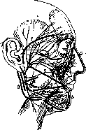
Figure 19. Direction of incisions on the face: 1—temporal; 2—brow; 3—zygomatic; 4—paranasal; 5—median nasal; 6 and 7—upper and lower buccal; 8—parotid; 9—cervical; 10—mandibular (after Bockenheimer).
Dataset segment.
N. Blumenthal. The face in diagnostics, or rather the changes in the structure and expression of the face in diseases of the nervous system, endocrine glands, and internal organs, as well as in infectious diseases and others, present such characteristic features that they acquire the significance of an important diagnostic symptom. Changes in facial expression in acute infectious diseases were already the subject of study of the Hippocratic school (facies hippocratica, s. abdominalis, f. cholerica, etc.) and until the middle of the 19th century, i.e., until the period of the heyday of modern diagnostics, this symptom was one of the main ones determining the recognition of such diseases (Krankenphysiognomik of Baumgartner; 1899). At that time, the semiotics of the face and the use of facial expression for diagnostic purposes were brought by individual physicians to a high degree of perfection (Curschmann, Klinische Abbildungen). Under physiological conditions, the expression of the face is determined by a whole series of anatomical and functional factors of both purely local and general character; in its emergence, along with a huge number of genotypic traits, an infinite multitude of paratypic influences are summed up. Therefore, the analysis of the mechanism of various facial changes sometimes appears extremely difficult, which, however, by no means diminishes the significance of a particular characteristic facies as a diagnostic sign. Among the factors determining the structure and expression of the face, it is necessary to single out the anatomical structure of the facial bones, the state of the facial musculature and the motor and sensory nerves innervating it, the turgor of the skin and subcutaneous tissue, in particular the water content in them, blood filling of the vessels (pallor, redness, cyanosis), skin color, dryness or moisture of the skin, as well as the features of the eyes (e.g., gloss), nose, position of the teeth, hair growth, etc. Under pathological conditions, the alteration of a number of the listed factors gives the face an expression of pain, fear, anxiety, laughter, excitement, indifference, etc. All these phenomena, including subtler changes in the function of the mimic musculature, constitute the subject of special study (see Mimics). In diagnostic terms, complex expressions of facial changes characterizing acute diseases of the internal organs and nervous system are particularly important. In acute diffuse peritonitis and advanced ileus (strangulated), as a rule, the face acquires an expression known as facies abdominalis, s. hippocratica; the face expresses severe suffering, it becomes senile, wrinkles become sharper; the chin and the tip of the nose appear pointed, the eyeballs sink, a cold, sticky sweat appears on the face; the complexion is pale or slightly cyanotic. A clinical picture of this kind, even in its pronounced form, can develop extremely rapidly, sometimes within a few hours, and serves as one of the main diagnostic signs of the transition of the inflammatory process from the abdominal cavity organs to the peritoneum. In the algid stage of cholera, typical changes of the face (facies cholerica) are also observed, characterized by extreme pallor (the "pallor of a dead man") of the facial skin, sunken and tense cheeks, a sharply demarcated nasolabial fold, sunken eyes and their rolling upwards. The main pathogenetic mechanism of the origin of the latter form, as well as of facies abdominalis, is apparently the loss of huge amounts of water by the organism and the rush of the bulk of the blood into the region innervated by the splanchnic nerves. In typhoidal infections, facial expression also plays a well-known, albeit lesser, differential diagnostic role, since upon comparative observation a sharp difference strikes the eye: the indifferent, tired, pale, sometimes "thoughtful" face of the typhoid patient and the excited, mobile, often red face of the typhus patient. However, the mentioned changes in facies typhosa cannot be recognized as pathognomonic, since they are also observed in other infectious diseases. Among acute diseases of the nervous system, very typical changes in facial expression are noted in tetanus, meningitis, and chorea. A tearful facial expression or a sharply forced expansion of the mouth with extreme tension of all facial musculature are characteristic of tetanus; the face thus appears to a certain extent smiling (risus sardonicus). The picture is often complicated by typical trismus caused by spasms of the masseter muscle. In chorea, involuntary twitchings of the striated musculature also pass to the face, which creates the impression of extremely diverse grimaces (expression of pain, fear, contempt), by no means related to the corresponding emotion of the patient. Finally, in meningitis, in connection with the tension of all motor musculature, the face acquires an exclusively tense, mask-like expression. Well-known and very typical are no less characteristic features of the face in many chronic diseases: the face of a cretin, a myxedematous, a Basedow's disease patient, an acromegalic, a consumptive, pregnant women, blind people; the same applies to the expression of the face in parkinsonism and a whole range of mental disorders (for details see the description of the symptomatology of the corresponding diseases). A bright pink coloring of the cheeks with clearly visible capillaries is characteristic of individuals suffering from heart defects (mitral stenosis), M. Vovsi.
Related articles
Cite this page
“Facial Nerve (FACIAL NERVE, see Facialis).” Soviet Medical Encyclopedia. English translation of Bolshaya Meditsinskaya Entsiklopediya, 1st ed. (Moscow, 1928–1936), ed. N. A. Semashko. https://sovietmedicalencyclopedia.pages.dev/article/facial-nerve/