Erysipelas
Historical document, translated for reference. It reflects medical knowledge of the 1920s–30s and is not medical advice.
Summary
This article from the 1928–1936 Soviet Great Medical Encyclopedia discusses the history, etiology, and pathogenesis of erysipelas, an acute inflammation of the skin and mucous membranes caused primarily by streptococci. It explores infection routes, including ectogenic, lymphogenic, and hematogenic pathways, and debunks the traditional view of high contagion.
Encyclopedia article (1928–1936)
ERYSIPELAS, erysipelas (from the Greek erythros - red and pella - skin), a progressive inflammation of the skin and mucous membranes, particularly of the finest lymphatic pathways of the skin (capillary lymphangitis of the skin of Dieulafoy), caused by streptococci, in other words - an acute parasitic dermatitis. History. Erysipelas was well known even to ancient Hindu physicians and was accurately described by Hippocrates. Galen was the first to separate erysipelas from a number of purely local purulent phlegmonous skin lesions. Sydenham in the 17th century noted features of similarity that bring erysipelas close to general acute exanthems. In the 19th century, a number of scientists in various countries—Hunter and Gregory in England, Velpeau and Trousseau in France, Volkmann in Germany, and others—spoke in favor of the contagiousness of erysipelas, and these assumptions received real justification in the works of Bouchard, Lukomsky, and others, who discovered the presence of microorganisms in tissues and tissue fluid affected by erysipelas. Finally, Orth and others managed to prove the possibility of artificial infection with erysipelas (by inoculation) in healthy animals. When verifying these experiments, it turned out that the inoculations resulted sometimes in erysipelas and sometimes in phlegmon. The tendency of bacteriologists to look for the cause of the disease in the multiplication of specific pathogens led to the fact that Fehleisen (1881–1883) also isolated such a microbe for erysipelas. Etiology and pathogenesis. Erysipelas is indeed caused in the vast majority of cases by a streptococcus. Inoculations of this microbe or the fluid of erysipelatous blisters containing streptococci cause true erysipelas in animals and humans. For a long time, these microbes were considered under the name of erysipelas streptococci (Streptococcus erysipelatis Fehleiseni) as a special species of chain-forming cocci, differing from the streptococci of pus (Streptococcus pyogenes), streptococci of mucous membranes, or of other origins. But clinical observations and especially the experiments and studies of Petruschky clarified that there is no special species of streptococci causing only erysipelas. It has been established that the erysipelas streptococcus can also cause suppuration, and the causative agent of ordinary suppuration—the pyogenic streptococcus—can cause erysipelas. On the surface of our body, for example, streptococci are found in the form of simple skin saprophytes, completely harmless under normal conditions; but a lapse in asepsis during surgery, or sometimes an insignificant wound or scratch, is enough for streptococci to penetrate deep into the skin, where they find conditions so favorable for their development that, thanks to increased virulence, they can already cause erysipelas (erysipelas traumaticum, s. spurium), suppuration, or even severe general infection. In the nasal and oral cavities, streptococci are also found as harmless inhabitants, but it is enough to increase their virulence in some way, and these microbes acquire all the pathogenic properties of fully virulent streptococci: they can cause erysipelas, suppuration, sepsis, ulcerative endocarditis in animals and humans (Widal, Besançon). Such erysipelas, arising seemingly spontaneously, bore the name of spontaneous erysipelas (erysipelas idiopathicum, s. spontaneum, s. verum) in contrast to the aforementioned traumatic, "false" erysipelas. At present, these two types of erysipelas are not separated from each other, since in the case of so-called spontaneous erysipelas, we are apparently dealing with minimal trauma, invisible to the naked eye, damage to the epidermis or the epithelium of the mucous membrane. Infection with erysipelas can occur not only from an erysipelas patient, but also in connection with postpartum sepsis, certain lymphangitis cases, and abscesses. Sometimes erysipelas develops after the opening of a phlegmon or puncture of a joint empyema, since the streptococci contained in the pus can penetrate into the opened lymphatic clefts of the skin. It also becomes understandable why erysipelas frequently arises after puerperal sepsis, which is mostly a general streptococcal infection. Often, a newborn during the mother's childbed fever is infected by her through the umbilical wound and falls ill with erysipelas, or the latter develops in a physician who had some injury on his finger while rendering assistance to a parturient woman. On the other hand, puerperal women may experience the development of puerperal sepsis after assistance by a midwife who was in contact with an erysipelas patient or was herself suffering from erysipelas. In view of the wide distribution and persistent viability of streptococci outside the human body, erysipelatous epidemics also become understandable, which were especially common in surgical hospitals in the pre-antiseptic era. According to a number of authors, erysipelas can also result from staphylococci. According to Pfahler, it also arises from diplococci, and according to Rheiner, from typhoid bacilli. In animals (rabbits), erysipelas can be easily induced by pneumococci and Bact. coli (Neufeld, Petruschky). Often we are dealing with a mixed infection. According to Catrin, the combination of streptococci with typhoid bacilli is particularly dangerous. In short, bacteriologically, erysipelas cannot be considered a strictly specific disease. Infection of the skin and mucous membranes can occur via three pathways: ectogenic, lymphogenic, and hematogenic. The starting point of the most frequently encountered ectogenic form is accidental injuries or surgical wounds (wound erysipelas—erysipelas traumaticum), as well as all kinds of other defects of the skin epithelium or mucous membrane: cracks, ulcers, old wounds (often already healed or covered with granulations), as well as skin damage from burns and wound surfaces resulting from them. Ectogenic erysipelas is most often located on the face, as can be seen from Table 1 (G. I. Kovarsky, Report on the work of the Erysipelas Ward of the Semashko Hospital in Moscow, 1930). Table 1. Area of lesion: Men, Women, Total. Head, face: 719, 1097, 1816. Neck: 24, 62, 86. Genitalia, perineum: 2, 10, 12. Upper limbs: 10, 62, 72. Lower limbs: 34, 163, 197. Total: 789, 1394, 2183. This is explained by the fact that on the face, especially near the nostrils and on the lips, cracks, eczemas, and abrasions are more common, through which the infectious agent can penetrate; furthermore, infection is possible via nasal discharge, which often contains streptococci (especially during influenza), or as a result of frequent touching of the face and rubbing it with dirty fingers. In addition, erysipelas of the mucous membrane of the nose and throat very often spreads to the face through the nostrils, nasolacrimal duct, Eustachian tube, and external auditory meatus. Ranking second in the frequency of ectogenic erysipelas involvement are the limbs, especially the lower ones (same table), the skin of which frequently suffers from bruises and all kinds of trauma. In men, who are more engaged in physical labor than women, the upper limbs are affected by erysipelas twice as often as in women. Lymphogenic erysipelas develops through the medium of lymphatic pathways over underlying inflammation (streptococcal lymphangitis, streptococcal subcutaneous phlegmon), sometimes at the site of a ruptured abscess of a bone, joint, or lymph node beneath the skin. The less frequently encountered hematogenic erysipelas is observed during metastasizing streptococcal sepsis, usually together with streptococcal phlegmon, joint suppuration, etc. (erysipelas in turn can be the cause of such a metastasizing form of sepsis and general bodily intoxication in general). The assumption that streptococci are contained in skin scales shedding from erysipelas patients during skin peeling and are carried by them through the air was refuted by Respinger and others. And since streptococci generally nest not on the skin, but in the thickness of the skin itself, in its smallest lymphatic vessels, it is clear that the patient and his skin generally cannot serve as a source of infection unless the integrity of the skin is broken and there are wounds with purulent discharge on it. If such wounds are present, then such a patient is no more dangerous to others than any purulent-surgical patient: an ordinary aseptic bandage completely protects persons surrounding the patient from the possibility of infection. The doctrine of the contagiousness of erysipelas must therefore be archived at present. Observations by G. I. Kovarsky in the Erysipelas Ward of the Semashko Hospital in Moscow, as well as observations by Markuze, Gulyaev, Ivashentsev (Botkin Hospital in Leningrad), and other authors fully confirm the view of the non-contagiousness of erysipelas; the staff and non-erysipelas purulent-surgical patients do not contract erysipelas.
G. Kovarsky. Statistics. Erysipelas is subject to compulsory registration only in some countries. Among European countries, its compulsory registration has been introduced in England and Wales, Free and Northern Ireland, and Scotland, in Bulgaria, Hungary, Gibraltar, Greece, Denmark, Iceland, Latvia, Malta, Norway, Poland, Switzerland (in some cantons), and Yugoslavia. According to official data, in 1930 the number of registered cases of erysipelas was: in England over 18,000, in Scotland about 4,000, in Bulgaria about 2,000, in Denmark over 3,000, in Norway about 2,500, in Poland about 5,000, and in Yugoslavia about 2,000. Table 2 shows the incidence rates of erysipelas in certain countries for 1925–30. Table 2. Number of registered erysipelas patients in certain countries (per 10,000 population). Countries: England and Wales... 3.95 3.7 3.7 4.2 4.5 4.6, Bulgaria... 0.3 0.5 1.1 1.5 3.2 3.4, Denmark.... 8.8 9.2 8.4 8.3 8.3 9.4, Ireland: Northern... 1.4 1.8 1.9 1.6 1.7, Free... 0.5 0.4 0.5 0.4 0.4, Latvia.... 0.3 1.3 1.3 2.3 3.3, Norway... 6.8 7.1 6.9 8.7 10.1 8.6, Poland... 0.8 0.8 1.1 1.5 1.4 1.6, Scotland... 6.9 6.9 6.9 6.7 8.0 8.3, Yugoslavia... 0.9 1.0 1.2 1.5 1.4 1.6. Attention is drawn to the high figures of erysipelas in Denmark, Norway, and Scotland; in England and Wales and in Scotland, erysipelas is registered significantly higher than in Ireland. The higher incidence rates of erysipelas in some countries may perhaps be explained by more complete
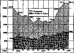
Figure 1. Number of deaths from erysipelas in England and Germany for 1919–30 (absolute figures).
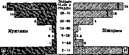
registration of patients. The figures of those dying annually from erysipelas in some countries are relatively very high. Thus, in England and Wales in recent years, over 1,000 people die annually from erysipelas, in Germany about 3,000, in Hungary about 500, in Czechoslovakia about 650, and in Mexico over 2,000. At the same time, over a number of years, not only is there no decrease in mortality (in absolute and relative figures), but, on the contrary, in many countries, there is a definite tendency toward its increase (Fig. 1). Table 3 gives the mortality rates from erysipelas for recent years in certain countries. Table 3. Mortality from erysipelas in certain countries for 1925–30 (per 100,000 population). Countries: England and Wales... 2.2 2.1 2.1 2.3 2.6 2.6, Bulgaria... 0.2 0.2 0.3 0.8 1.5 1.9, Hungary... - 4.5 5.2 5.0 6.0 4.8, Germany... 2.6 2.7 2.8 3.5 4.4 4.4, Denmark.... 2.2 1.8 2.0 1.8 2.5, Ireland | Free... - 1.1 1.2 1.0 1.6, Ireland Northern... 1.3 3.5 3.3 1.8 2.7 2.0, Norway... 0.8 1.9 2.4 3.4 4.8 -, Switzerland... 1.6 1.2 1.8 2.4 2.9 2.0, Scotland... 3.1 3.0 2.5 3.3 3.9 4.0, Czechoslovakia... 2.7 3.1 3.1 3.6 4.3 4.7, Estonia... - - 3.7 4.0 3.9 -, Yugoslavia... 0.4 0.3 0.6 0.7 0.8 0.7, Mexico... 9.1 7.0 10.9 11.2 12.5 14.1. The mortality rates from erysipelas in some of the cited countries, e.g., in England and Wales, Scotland, Germany, Denmark, Norway, are higher than the mortality rates from typhoid fever for the same years. An idea of the nature of erysipelas incidence by age groups and sex is given in Table 4, which shows the incidence rates of the disease by Moscow Governorate and the city of Moscow. Table 4. Incidence of erysipelas by age and sex (per 10,000 persons of each group). Moscow Governorate: Moscow Age 1903–08 m. f. m. f. Up to 1 yr..... 25.9 29.3, 1–4 yrs. 8.1 9.2, 5–9 » ... 4.6 6.2, 10–14 » ... 6.7 14.6, 15–19 » ... 11.6 31.4, 20–29 » ... 12.9 27.9, 30–39 » ... 17.1 35.7, 40–49 » ... 24.8 55.6, 50–59 » ... 27.7 63.3, 60 yrs. and older 21.1 45.6, Total... 14.7 31.7. The incidence rate for the population of both sexes in 1926 was 28 according to Moscow Governorate and 29 according to Moscow per 10,000. The incidence among women is higher than among men. Erysipelas is registered most highly in infants and at the age over 40 years. The nature of mortality by age and sex is illustrated by Table 5 and Fig. 2. A similar pattern of mortality is noted according to the materials of Moscow and Leningrad for a number of years, i.e., the highest mortality from erysipelas is observed in infants and in old age. In infancy, mortality from erysipelas in many countries in recent years occupies Table 5. Mortality from erysipelas by age and sex (per 100,000 persons of each group). Age Germany Age Switzerland 1901–20 m. f. m. f. Up to 1 yr... 24 22, 1–5 yrs... 1 1, 5–15 » ... 0 0, 15–30 » ... 1 1, 30–60 » ... 3 2, 60–70 » ... 9 7, 70 yrs. and older 24 47. Up to 1 yr... 30.8 30.6, 1–20 yrs... 0.2 0.4, 20–40 » ... 0.9 1.1, 40–60 » ... 3.6 2.5, 60 yrs. and older 11.5 11.2. Total Total: 2.8 2.7
a high place. Out of 1,000 children who died under the age of 1 year in 1927–28, deaths from erysipelas accounted for: in England and Wales—2.2, in Hungary—3.5, in Germany—4.0, in Holland
Figure 2. Mortality from erysipelas in Germany in 1930 by sex and age. 4.1, in Norway—4.0, in Poland (Warsaw and Lodz)—5.1, in Czechoslovakia—3.8, in Sweden—7.9, in Scotland—3.0, in the USA—6.2, in New Zealand—7.7 (1927), in Moscow (1926–27)—13.1, in Leningrad—7.0 (1926–27). Case fatality from erysipelas (per 100 patients) in hospitals in Germany for 1902–22 is given in Table 6. Table 6. Years Men Women 1902–07 8.4 5.8 1908–13 8.2 5.5 1914–19 11.1 6.6 1920–22 8.6 5.5. For 1924–29, the case fatality in hospitals in Germany was 7.6%. Of the Russian authors, Ivashentsev (Leningrad) and Kireyev (Moscow) estimate the case fatality in erysipelas at 2–5%; according to Stefansky (Odessa), it rarely exceeds 5% in adults, while in surgical patients the percentage of fatal outcomes is significantly higher—about 12–15. It is even higher in infants: according to data from the Odessa hospital for 1926–28, the case fatality among them was Table 7. Vienna Denmark Moscow Gov. Moscow Months 1891–1900 1923–27...
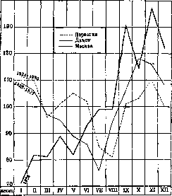
284 26%. The monthly distribution of erysipelas cases (the ratio of the daily average for a given month to the daily average for the corresponding years) is given in Table 7 and Fig. 3.
Figure 3. Monthly distribution of erysipelas cases.
In Moscow and Moscow Governorate, an increase in the incidence of erysipelas was noted in the autumn months, and in Vienna and Denmark—in the autumn-winter months.
I. Dobreytzer. Epidemiology. In the older literature, it was constantly pointed out that there were times when the widespread occurrence of erysipelas was a cruel scourge for medical institutions. In monographs devoted to erysipelas, descriptions of epidemic outbreaks of erysipelas in surgical and gynecological departments and maternity hospitals were constantly cited. However, the causes determining the occurrence of endemic outbreaks of erysipelas still remain unclear. With the development of antiseptics and especially asepsis, these outbreaks became less and less frequent. During the World War, the incidence curve of erysipelas rose sharply again (Izvolskaya). In the last two years, the number of erysipelatous diseases in the large cities of the USSR has been growing noticeably, and in the reports of the erysipelas departments of large cities, the large number of diseases among day laborers, housewives, domestic servants, cab drivers, seasonal workers, homeless people, etc., catches the eye—that is, precisely those strata of the population who, by virtue of their occupation or living conditions, find it most difficult to maintain cleanliness. Cold apparently also plays a role as a factor contributing to the onset of erysipelas: in November, December, and January, the number of cases of erysipelas and its relapses almost doubles. In summer, even in prolonged hot weather, erysipelatous diseases still do not disappear completely: the influence of the aforementioned factors of a domestic, social, and occupational nature obviously persists. According to some observations (Nekrasov), the number of erysipelatous diseases increases with rising atmospheric humidity. The increase in the number of erysipelatous diseases often coincides with epidemics of scarlet fever. Outbreaks of influenza epidemics can also play a significant role in increasing the number of erysipelatous diseases: in the anamnesis of very many erysipelatous patients, there is influenza, followed by a sudden outbreak of erysipelas, with the redness in such cases appearing earliest around the nose (its wings)—that is, at the site of prolonged skin irritation by the nasal discharge inevitable in influenza. It is well known that erysipelatous inflammation frequently complicates wound healing after all kinds of burns. Predisposition to erysipelatous inflammation undoubtedly also depends on sex and age. According to most statistics, women fall ill more often than men. Out of 5,602 patients with erysipelas who passed through the Botkin Hospital in Leningrad in 1924–1929, there were 2,305 men and 3,297 women. A number of authors note a hereditarily transmitted tendency to contract erysipelas and the concentration of cases within families without a direct connection between one disease and another (in time, opportunity for contact, etc.). Immunity. Once contracted, erysipelas not only does not protect against a new disease, but on the contrary, tends to recur. Menstruation and the postpartum period favor recurrences of erysipelas. The above-indicated factors of domestic, social, and occupational properties also favor recurrences, as does the cold season of the year. But in the presence of all these factors, erysipelas may be absent in people with certain constitutional features and increased resistance to all kinds of purulent infection, even in the presence of all kinds of skin damage, which undoubtedly gives the right to assume the existence of natural immunity to erysipelas. One often has to deal with phenomena of the opposite property: subjects who have once suffered from erysipelas fall ill with it again many times (up to 20 or even 30 times), despite all kinds of precautionary measures and efforts and the possibility of eliminating all factors favoring the onset of erysipelas (the so-called "habitual erysipelas"). However, the role of constitution in the etiology of erysipelatous inflammation is still almost unstudied at the present time, and this question is only just being put on the agenda. Only one thing is indisputable: with certain inherited or acquired constitutional defects (probably depending on one or another metabolic disturbance), allergic states of the organism in general are observed, in particular of its skin integuments, where one can speak of an increased state of readiness (Bereitschaft) for irritation of the epidermis or blood vessels (see Allergy). Pathological Anatomy. On the corpse, erysipelatous redness is barely noticeable. Upon incision, the skin appears thickened and fused with the subcutaneous tissue. Microscopic examination shows that the skin, especially in the depths, at the border with the subcutaneous tissue, is permeated with leukocytes, and its blood vessels are dilated. The subcutaneous tissue also takes part in the inflammatory process; in addition, lymphangitis is often, although not always, observed. Streptococci are found in the perivascular lymphatic spaces, in the lymphatic vessels of the base of the papillae, in the lymphatic clefts of the skin proper, as well as in the sheaths of the hair follicles (Cornil, Babes). Lukomsky and others found them in dilated capillaries, which was rejected by Fehleisen. Some of the streptococci may be phagocytosed. Among the leukocytes are rare specimens of large wandering cells. In the erysipelatous plaque, Fehleisen described three zones: in the zone located outside the ridge, or the so-called peripheral zone, the skin appears almost normal, but contains a multitude of streptococci. In the marginal zone of the plaque, the process is in full swing: here a significant accumulation of leukocytes and edema are found. In the central zone of the erysipelatous plaque, the process is in a state of regression, and there are no streptococci at all in the skin proper, or at least very few of them. It is easy to understand that the three zones described are essentially phases of the development of erysipelatous inflammation as it spreads. The epidermis also exhibits a number of changes. The cells comprising its various layers are partly separated and degenerated; in places, the epidermis is lifted in a sheet under the pressure of edematous fluid, so that a blister is formed, usually filled with a light straw-yellow fluid [see separate plate (to the article Sarcoma), fig. 3]. Sometimes, with further development, the process leads to the formation of abscesses, phlegmon, and even necrosis. These complications cannot always be explained; it is possible that they depend, on the one hand, on the nature of the infection (usually mixed in such cases), and on the other hand, on the reduced resistance of the organism of the given individual, caused by debilitating diseases, intoxications, alcoholism, and also age. In blood vessels, streptococci are found only in the hematogenous form or with an associated general infection (sepsis). In erythematous erysipelas, the tissue is destroyed insignificantly, so that after the disappearance of inflammatory phenomena, no traces remain except desquamation and hair loss. The latter depends on exudation into the sheaths of the hair roots; however, the hair soon grows back again. A sharp disturbance of skin nutrition can lead to its necrosis and gangrene. Such necrosis usually occurs in purulent-phlegmonous forms, while in the usual "erythematous" form, it occurs only in places where the skin is poorly stretchable due to a lack of adipose tissue (the edge of the tibia, patella, olecranon, zygomatic bone) or where an abundant exudate can form due to the loose structure of the skin (eyelids, scrotum). Course and Symptoms. Erysipelas of the face usually begins with prodromal symptoms: chills, malaise, headache, often vomiting, and a rise in temperature up to 39.0°–40.0°. These phenomena occur amidst complete health and usually precede the appearance of erysipelatous redness by a few hours or 1–2 days; sometimes patients note painful swelling of the submandibular lymph nodes even before the appearance of redness. Erysipelatous redness arises in the form of a red, sensitive plaque around the nostrils, in the corner of the eye, or on the auricle. If there is any excoriation (eczema, herpes, scratch) on the skin of the face or scalp, erysipelas usually begins from the excoriated part. The erysipelatous plaque soon becomes darker, shiny, and painful, and the skin in its vicinity is hot to the touch, tense, swollen, dry, and dense. Upon pressure, the redness does not completely disappear, as happens with erythema. The redness gradually increases and spreads unevenly across the skin. Its edges are raised in a ridge-like manner and often have a festooned, jagged, tongue-like appearance, so that it is sharply demarcated from the healthy parts of the skin. The speed of spread of erysipelatous redness across the skin varies greatly, sometimes up to 2–3 cm per hour. As it spreads, the redness disappears in the originally affected places and often stops where the skin is more firmly connected to the underlying parts (on the chin, at the border of the scalp). Simultaneously with the redness, severe edema appears around the affected areas, especially on the eyelids (due to the looseness of the subcutaneous tissue). The inflammatory process often captures the entire face, which in such cases becomes unrecognizable (hence the name erysipelas): red and edematous eyelids close the eyes, swollen and disfigured nostrils are almost plugged, the cheeks swell, and the auricles are red, shiny, and greatly increased in volume. Often the erysipelatous process also spreads to the forehead, head, back of the head, and neck. On the scalp, the erysipelatous redness is less intense than on the face, but the pain is greater. Such is the picture of ordinary erythematous erysipelas.
Sometimes the epidermis is raised by a serous exudate that accumulates in the form of larger or smaller vesicles—the vesicular or bullous form (erysipelas bullosum). The contents of the vesicles may become turbid, purulent, and bloody. Sometimes hemorrhages may occur spontaneously on individual areas of the erysipelatous spot (erysipelas haemorrhagicum). Pus may accumulate in the form of superficial abscesses of greater or lesser magnitude (pustular form), after the bursting of which thick yellowish crusts appear in their place (erysipelas squamosum, s. crustosum). When the roller-like elevation of the skin disappears and the redness pales, it can be said that erysipelas is coming to an end. Upon termination of the process, the skin desquamates in flakes. In erysipelas of the scalp, hair loss is commonly observed (from the accumulation of exudate in the hair follicles); complete baldness does not occur, but the hair can sometimes become very thin. Loss of eyebrows can be final. The disease lasts on average 4–10 days. The temperature reaches high figures in the evenings (up to 41° with tenths), while in the mornings it presents more or less pronounced remissions. In some cases it drops gradually, in others immediately, within a single night, critically, which is accompanied by heavy sweating (usually on the 7th–8th day, but sometimes as early as the 3rd–4th day). Not infrequently, erysipelas that seemed to have already ended flares up again, the temperature rises again, redness reappears, and the disease continues for 15–20 days. Such relapses of the disease sometimes occur 3–4 times during the patient's stay in a hospital institution. At the very beginning of the disease, patients lose their appetite, insomnia and constipation appear, urine is passed in small amounts and may contain protein. The content of urea and chlorides is generally reduced. The blood picture in erysipelas changes rather sharply (P. Y. Kaminer): the red blood cell count decreases, leukocytosis is usually observed, the degree of which corresponds to the severity of the disease form: 12,000–18,000 in moderately severe cases, up to 30,000 and higher in severe cases. The neutrophil shift (reaching up to 90%) also corresponds to the severity of intoxication. The number of neutrophils decreases by the time the temperature drops and is replaced by an increase in lymphocytosis. Eosinophils disappear at the height of the process, appearing by the beginning of recovery. Even before their appearance, before the temperature drop, the number of monocytes increases. With a combination of sharp leukocytosis with a shift to the left, the prognosis becomes unfavorable. According to Roger, prolonged polynucleosis or its resumption in the recovery period portends a relapse. Headaches can be very intense and in some patients constitute the main complaint. Headache should not be confused with sharp hyperesthesia of the affected skin areas (which is very painful even upon the lightest touch with a finger), which is also very distressing for patients. Not infrequently, nervous phenomena also reach a high degree (excitation, delirium), especially with involvement of the scalp and in alcoholics. Roger notes a frequent coincidence of delirium with albuminuria in men; the liver is often hypertrophied, the spleen enlarged. Suppuration may not be limited to the skin surface, but spread deeper into the subcutaneous tissue—phlegmonous erysipelas. In these cases, the detachment of the skin and the accumulation of pus can be very significant, and the absence of inflammatory phenomena on the skin in the area of the pus accumulation is often striking: the latter forms by the type of cold cold-abscess-like boils. After the evacuation of such abscesses, a large amount of necrotic tissue in the area of the entire scalp is often discharged. In the presence of great tension of the soft tissues, phlegmonous erysipelas, and even more often vesicular (bullous) erysipelas, in weakened or elderly subjects can pass into a necrotic, gangrenous form. On the face, the eyelids are most often subjected to the process of necrosis. In phlegmonous and gangrenous forms, the general condition appears even more severe. Sepsis is often joined. Erysipelas proceeds in a similar manner on other parts of the body. All differences depend on the anatomical and topographical features of the given body area. There are apparently several types of the course of this disease on different parts of the body, although essentially in each specific case we have only a definite stage of the same disease with the predominance of certain symptoms depending on the virulence of the streptococcus, secondary infection by other microbes, the anatomical and topographical features of the given case, and the reaction of the organism—its constitution, and not infrequently the severity of the course of individual cases depends on the allergic reaction of the given organism in the sense of its particularly increased sensitivity to the given infection. Returning to facial erysipelas, it should be noted that here it proceeds favorably in the vast majority of cases, contrary to the bad reputation of this disease established in textbooks precisely when it is localized on the face: bullous forms (not to mention erythematous ones) always result in complete recovery, and phlegmonous and gangrenous forms give only an insignificant mortality percentage, as can be seen from Table 8 when comparing it with Table 1—the total number of diseases on various parts of the body, and also fully corresponds to the observations of other authors (Ivanov). The edema gradually subsides, the crusts fall off, desquamation ends, and the face assumes its former appearance. Abscesses often form in the thickness of the eyelids, which must be emptied in a timely manner. In rare cases, an incision in the presence of clear fluctuation yields only the outflow of serous fluid, without pus, but this also produces the desired effect: the edema passes. This edema is very burdensome for patients, as it deprives them of vision for many days, but essentially it is completely harmless. Phlegmonous forms of erysipelas of the head and face sometimes lead to the accumulation of colossal amounts of pus under the skin of the scalp with its complete detachment from the underlying tissues over the entire skull. The condition of the patients in such cases is very severe, and the course of the disease is very protracted. Still, with the timely production of incisions with counter-openings, the phenomena subside, and complete recovery ensues with insignificant (scar) disfigurement of the cranial integuments. In gangrene of the eyelids, the course is severe; it usually occurs in depleted and elderly subjects, therefore the prognosis is always serious. On the neck, primary erysipelas is rare; more often the process passes here from neighboring areas, mainly from the face and head. The usual form is erythematous, sometimes turning into phlegmonous (which depends on the abundance of tissue on the anterior surface of the neck between the fascial layers, in the region of large vessels). On the trunk, the process most often passes from neighboring parts, and here erysipelas (usually erythematous) often bears the character of the so-called wandering erysipelas (erysipelas migrans, s. erraticum). The inflammatory process gradually bypasses the skin of the entire trunk and even the extremities, and a return of the process along the already traveled path is often observed. The course of the disease in these cases is very severe and, if the patient does not die in the coming days from general intoxication, protracted. The severity of the disease depends not only on poisoning by a large amount of bacterial toxins, but also on the shutdown of the respiratory functions of the skin over a large extent. Neither mountain sun treatment nor serum treatment, so effective in other cases of erysipelas, has any noticeable favorable influence on the course of the process here. On the female mammary gland, erysipelas often complicates the course of mastitis. Gangrenous forms with subsequent loss of tissue and scarring, often leading to the loss of organ function, are also encountered here. Erysipelas of the scrotum and penis often arises primarily and often passes into a gangrenous form, and this is preceded by a stage of colossal edematous infiltration of the integuments of the scrotum and penis, which can make it necessary to withdraw urine with a catheter and even leave it a demeure, since the opening of the urethra in the edematous prepuce is found with difficulty. Edema of the scrotum can in individual cases reach such colossal sizes that confusion with a strangulated hernia is possible (in the presence of any abdominal symptoms). The intensity of edema phenomena with subsequent frequent necrosis of the scrotum integuments depends apparently on the anatomical features of this organ: the thinness of some of its membranes, isolated blood circulation with insufficiently developed collectors for outflow and a highly developed network of lymphatic vessels that are easily compressed in the presence of inflammatory phenomena. After the shedding of the necrotic walls of the scrotum, the testicles turn out to be completely exposed, but quickly become covered with granulations. (Necrosis never spreads to the testicular tissue: independent blood and lymphatic systems protect it from this.) Subsequent scarring in such cases leads to one degree or another of testicular atrophy.
The course of gangrenous erysipelas of the scrotum is severe only until the rejection of the necrotic areas, and thereafter it is merely very prolonged. On the female genitalia and perineum, erythematous forms with fairly sharply pronounced edema are most frequently observed. On the extremities, even erythematous and bullous forms run a severe course. Bullous forms frequently turn into phlegmon, and less commonly result in necrosis with a high mortality rate of 4%. Even with correct surgical treatment, long-persisting edemas and more or less persistent functional impairments (especially of the hand) remain. It is necessary to emphasize another circumstance, namely the enormity of purulent accumulations in certain phlegmonous forms, and these accumulations appear in the most unexpected places, at a distance from the initial focus of skin necrosis, without preceding tissue infiltration and without inflammatory phenomena on the skin, i.e., by the type of cold abscesses. It is important to open such accumulations in a timely manner so as not to exhaust the patient's strength: such accumulations can easily be overlooked during insufficiently attentive examinations of the patient at dressings. The swelling of regional (inguinal and axillary) lymph nodes often even precedes the appearance of redness. Erysipelas of mucous membranes more often arises primarily, but sometimes, conversely, the process spreads to the mucous membranes from neighboring parts. The mucosal changes in this case are analogous to the described skin changes in erythematous erysipelas: bright, well-demarcated redness, swelling or edematousness, significant pain, and frequently the formation of small, rapidly rupturing vesicles and the formation of superficial defects. The oral cavity and tongue are affected rarely or at least only slightly involved by erysipelas, whereas the pharynx is one of the most frequent localizations of the disease, with this being primary. Erysipelas of the pharynx (Lasegue) is characterized by intensive glossy redness and sharply expressed phenomena of angina; sometimes it is accompanied by the formation of blisters, necrosis of the mucous membrane (in exhausted and elderly patients), or a retropharyngeal abscess. From the pharynx, erysipelas can spread to the face by various pathways—along the oral mucosa, through the nasal cavities, and through the lacrimal ducts (Peter). The nasal cavities are frequently the primary site of the appearance of erysipelas; such erysipelatous rhinitis gives strong pains and is accompanied by a high temperature and swelling of the cervical glands. Frequently, erysipelatous rhinitis spreads to the tympanic cavity, and from there to the cells of the mastoid process and the meninges. The larynx is usually affected by erysipelas secondarily; however, cases of primary erysipelatous laryngitis have been described. When erysipelas is localized in the larynx, there is often rapidly increasing edematous swelling of the glossoepiglottic and arytenoepiglottic folds and dyspnea; in such cases, emergency tracheotomy may be required. Erysipelatous bronchopneumonia was observed by Straus, Dieulafoy, and others, with the process penetrating the bronchial mucosa and pulmonary alveoli already at the end of the disease, when the temperature in patients was already falling. Such cases can lead to a fatal outcome; upon bacteriological examination of the pneumonia exudate, streptococci are usually discovered, but frequently bronchopneumonia is caused by a secondary diplococcal infection. Erysipelas of the mucous membrane of the genital tract in women (vulva) gives strong edemas of the labia majora and minora. It occurs not infrequently in puerperal women. Age-related features of the course of erysipelas: In infants, erysipelas runs a very heavy course and gives an enormous mortality rate. The creeping form predominates. Facial erysipelas at this age is frequently complicated by fatal meningitis; with creeping erysipelas of the abdomen, death from peritonitis is frequent. In older children (from one year to 10 years old), erysipelas proceeds on the whole satisfactorily, localizing mainly on the face and perineum. In adolescence and young age (up to 30 or even up to 40 years), erysipelas proceeds comparatively well and gives a minimal mortality rate (slightly more than 1%). Elderly subjects endure erysipelas very poorly—up to 12.5% mortality; phlegmonous, gangrenous forms, and sepsis are frequent. Complications of erysipelas occur as a result of the further spread and metastasization of the inflammatory process or the addition of a secondary infection. With the spread of inflammation into the depth, parotitis, suppurations of tendon sheaths, synovial bursae, muscles, and joints (so-called infectious false rheumatism) may develop. Thrombophlebitis on the extremities is not infrequently observed, and especially frequently in the system of the anterior facial vein, where this process passes to the veins of the orbit and further to the veins of the base of the brain, and ends almost always in fatal basal meningitis. Acute erysipelatous nephritis with albuminuria, casts, and sometimes streptococci in the urine is observed not infrequently; it usually passes, but can serve as an impetus for the development of chronic nephritis. Pericarditis is rarely encountered; streptococci may be found in the exudate (Denuce). Endocarditis is observed more often; for the most part, it is very benign and transient, but sometimes ulcerative, malignant endocarditis develops with all possible consequences in such cases. In such instances, one is dealing with sepsis that has already developed as a complication. Streptococci have been found in the vegetations of the endocardium (Achalme). Pleurisy is rare. Of the complications from the eyes in erysipelas, conjunctivitis and edema of the conjunctiva are most often observed, and less commonly iritis. Suppurations and gangrene of the eyelids have already been mentioned. Sometimes phlegmons of the eyelids can spread to the orbital cellular tissue and cause inflammation of the optic nerve, vascular thrombosis, and even panophthalmitis. Sometimes inflammatory processes in the orbit develop even without preceding necrosis or phlegmon of the eyelids. Pointing symptoms for timely diagnosis in these cases are complaints of pain, a sensation of tension, and exophthalmus. Lesion of the optic nerve can lead to a decrease in vision or even its complete loss as a result of optic atrophy. The latter may occur as a result of blockage of the central artery or even just from compression of the nerve and disruption of its nutrition by virtue of sharply pronounced prolonged edema. Complications from the ear have already been mentioned. Inflammations of the meninges are rare (0.1%) and, aside from the aforementioned pathways, can arise metastasically as a result of erysipelatous sepsis. Gas gangrene is observed as a very rare complication (Marcuse observed 4 cases with a 50% mortality rate). Delirium in erysipelas patients is rarely caused by meningitis; it undoubtedly depends on streptococcal toxins (toxic infectious psychosis) or on alcoholism. It is also frequently observed in mentally overworked subjects, responsible workers, research workers, and educators. In alcoholics, this delirium bears the character of delirium with sharply pronounced motor agitation: jumping out of bed, attempts to dress and go home, and even run or jump out of a window. It is interesting to note that the agitation happens in such patients not only at a high temperature, but also at the beginning of its drop. Since in such cases one is dealing most often with strongly built men, the agitated state of these patients causes not a little trouble for the medical personnel and requires from the latter maximum vigilance and skill, especially since any attempts at any coercive measures (tying to the bed, throwing on a straitjacket) drive the patients into a frenzy. Upon the termination of the delirious state, patients usually remember nothing about what happened or recall it very vaguely. In non-alcoholics, delusions of grandeur are frequently observed. In mentally ill patients, erysipelas often provokes a new exacerbation of the underlying disease. In tuberculous patients, an exacerbation of the process often sets in under the influence of erysipelas. Premature labor can occur in pregnant women. Menstruation also appears a few days earlier, usually simultaneously with the onset of the disease. The diagnosis of erysipelas is usually not difficult, since rapidly spreading, sharply demarcated redness and swelling of the skin and chills, replaced by a sharply elevated temperature, usually go hand in hand. If the characteristic redness is masked by hair, sunburn, or ointments applied for the treatment of the disease (especially ichthyol), the nature of the disease is recognized by the sharply expressed hyperesthesia of the skin, so characteristic precisely of erysipelas. Erysipelas is more difficult to diagnose in cases of the absence of a temperature reaction—in old people, debilitated subjects, and generally individuals reacting little to any kind of intoxication (according to Baryshev, normal temperature is observed in 15.6% of cases of primary erysipelas and in 31.5% of cases of recurrent erysipelas), but the general weakness, characteristic skin changes, and the course of the disease here too make it possible to make a correct diagnosis. The differential diagnosis between erysipelas and all kinds of erythemas and dermatitides is made on the basis of the following signs: in erysipelas, the anamnesis is very characteristic—a sudden onset with chills and subsequent rise in temperature, sometimes vomiting, following which patients discover redness and swelling somewhere; severe general malaise.
The erysipelatous plaque differs by specific features: it is glossy, painful, dense, the redness at its site is not interrupted by patches of normal skin and does not disappear upon pressure, unlike the redness in erythemas, and finally, what is especially important, the edges of the erysipelatous redness are sharply demarcated from the healthy skin and raised in the form of a ridge, which also does not occur in erythemas. In addition, the spots in infectious erythema are painless and as a rule appear on the extensor surfaces of the extremities. The elevation of temperature in them is insignificant or entirely absent. Eczema rubrum—acute eczema accompanied by redness of the face—resembles erysipelas at first glance, but here the process from the very beginning affects the entire face, while the redness passes imperceptibly, without sharp boundaries, into healthy skin; almost one can always find several small, strongly itching vesicles. The elevation of temperature is absent or very insignificant. Sometimes it is very difficult to distinguish Erysipelas from phlegmon, especially when erysipelas is localized on the extremities (on the shin). The differences stem from the pathogenesis of both diseases: in Erysipelas, the inflammatory process is nested in the skin and is most intense at the periphery of the affected areas, whereas in phlegmon the process is nested in the subcutaneous tissue and concentrated at one definite place, with the redness from reactive hyperemia of the skin being brightest in the center, of a somewhat purplish hue, and gradually fading to none. The maximum pain in phlegmon is in the center of the lesion; instead of skin edema while preserving its mobility over the underlying tissues and organs, there is a sharp induration of the subcutaneous tissue, fixing the skin and giving a woody feel to the touch (Ivashintsev). In doubtful cases, observation of the course of the process usually clarifies the diagnosis. To the actual erysipelatous process on the skin there can of course (see above) be added a phlegmonous inflammation of the underlying tissue and even intermuscular phlegmon. Then the symptoms of both diseases naturally merge, but the erysipelatous process on the skin does not lose its characteristic features. A confusion of Erysipelas with primary non-erysipelatous lymphangitis is also possible, but in the latter the red stripes or fine red net correspond to the course of the subcutaneous lymphatic vessels. In erysipelatous lymphangitis, such stripes run from the skin area primarily affected by erysipelatous redness, as if being its branches.—Similar in its external manifestation to Erysipelas is the process formerly known under the name of chron[ic] Erysipelas or wandering erythema and named by Rosenbach erysipeloid. Most often the starting point of the process is small injuries on the fingers of the hands, but it can also be observed on the nose, cheeks, and neck. Without an increase in temperature and without any disturbance of the general state, there appears a slightly burning or rather itching, sometimes painful swelling and strong redness or purplish coloration of the skin, which has sharp boundaries and spreads from the finger towards the hand, while the old places pale. In approximately 10% of cases, persistent lymphangitis on the upper extremity is observed. The disease usually lasts about a week, less often stretching for 3–4 weeks. Erysipeloid is related to dead animal substances that begin to undergo decomposition, which is why housewives, butchers, tanners, dealers in game or fish often fall ill with it. Almost always, some damage can be found on the skin of patients through which the infectious principle penetrated. In 1887, Rosenbach isolated in a pure culture from the skin areas affected by erysipeloid a microorganism (resembling Cladothrix in appearance), inoculation of which into skin wounds caused the same disease. Its pathogenic significance was confirmed in 1904 by the studies of Ohlemann. The classification of this microbe, which has an irregular rounded shape and forms threads in old cultures, is difficult. The prognosis in Erysipelas depends on the age, the form of the disease, and sometimes on constitutional features. The erythematous form of erysipelas of the face and head gives the best prognosis; it is less favorable in erysipelas of the trunk and extremities. Most dangerous (even the erythematous) is Erysipelas in alcoholics, in persons weakened by other diseases, in the elderly, and also in newborns, especially in those cases where in the enumerated categories of patients it takes on the character of wandering erysipelas. The prognosis is extremely unfavorable in erysipelas joining severe forms of tuberculosis, typhoid fever, diphtheria, or puerperal sepsis. Purulent, phlegmonous forms often lead to sepsis, which is most often the cause of death, along with cardiac weakness, meningitis, and pneumonia. The prognosis is equally serious in the gangrenous form in elderly subjects. In young people, after the rejection of dead skin and tissue, the wound is cleansed and complete recovery often occurs.—The prognosis of E[rysipelas] quoad sanationem is overshadowed by the constant threat of a recurrence of the disease. There is no way to foresee whether erysipelas will recur in a given patient or not. Even in stubborn recidivists, it is impossible to say which relapse will finally be the last. The percentage of relapses according to literature data ranges from 6 (Soerensen) to 39.4 (Frickhinger). According to Gulyayev and Markuze—18.24%, according to Baryshev—17% (of which 31% with normal temperature), in Kovarsky's department in the Semashko hospital, where treatment with ultraviolet rays is the method of choice—about 4% (Gorev). The tendency to relapses decreases with time, the maximum of relapses falls on the year of the first disease. The cause of relapses, according to the majority of authors, is a latent infection that flares up with renewed vigor in the presence of all kinds of trauma and factors of a domestic, social, and occupational nature, the cold season of the year, and especially in the presence of an allergic state of the organism (see above). Repeated relapses of E[rysipelas] in the same place on the body (especially on the extremities) lead, due to the degeneration of the lymphatic vessels, to the phenomena of so-called elephantiasis of the skin.—Treatment. In ancient Egypt, the treatment of E[rysipelas] was performed by rubbing a mixture of human milk and donkey dung (Kovner), but even until recently the treatment continues to remain purely empirical and, it must be confessed, little successful, as evidenced by up to 40 different methods of treatment proposed for it over the past 30 years (Alisov and Morozkin). The local application of various agents proposed to stop the further spread of erysipelatous inflammation over the skin—iodine tincture, lapis solution according to Gaugele, etc.—proved completely ineffective, since cauterization with these agents of the surrounding healthy skin apparently did not lead to the closure of lymphatic clefts, just as neither did pressing the skin with adhesive plaster (according to Wölfler) or collodion. Injection of disinfecting substances into the inflamed skin likewise gave no results; even less benefit was noted from antiseptic lotions (with corrosive sublimate, boric acid, lysoform) with or without preliminary incisions of the skin. Until now, even in large centers, Nussbaum's ichthyoal ointment is still willingly used, believing in its antiseptic properties penetrating deep into the skin. But it works no better than any other ointments, and its dark color masks the redness, thus preventing one from following the course of the process on the skin, and delays ultraviolet rays, which have such a beneficial effect in erysipelatous inflammation (see below).—The same shortcomings are characteristic of methylene blue, a 5% solution of which Nobécourt proposed to lubricate the skin with in erysipelas. Bandages with non-irritating ointments (vaseline, zinc ointment, 10% naphthalane ointment, camphor oil) definitely act as painkillers and sharply reduce the sensation of itching and burning, but apparently do not affect the course of the process itself; they are nevertheless willingly used as symptomatic agents in the absence of the possibility of using other, more rational and effective methods of treatment (mountain sun, serotherapy, autohemotherapy). By the use in erysipelas of collargol, electrargol (10–20 cm3 of a 2% solution intravenously), trypaflavin, rivanol (30–40 cm3 of a 0.1% solution intravenously each time), an attempt is made to achieve disinfection through the blood. Among such disinfecting methods, one can also include the treatment with calomel recommended by old authors (0.5 for a single dose). Treatment with injections of antistreptococcal serum was proposed by Marmorek back in 1895. Such a correctly prepared and sufficiently aged (3–4 weeks after the last inoculation) horse serum is completely harmless and possesses prophylactic and healing properties. In the treatment of E[rysipelas] with injections of antistreptococcal serum (10, 15, or 20 cm3 subcutaneously), mortality, excluding cases that cannot be taken into account (complications in other organs, addition of other diseases, etc.), amounts to only 1–2%, while in the treatment of erysipelas by old methods it reaches 5% and more (Chantemesse). In addition, the duration of the disease is noticeably reduced, and in many cases the development of local and general phenomena is clearly halted. In some patients, after the serum injection, the temperature drops critically and the erysipelas breaks off.
At present, polyvalent antistreptococcal or antitoxic scarlet fever serum (observations of G. I. Kovarsky from the erysipelas ward at the Semashko Hospital in Moscow), as well as antierysipelas serum (Birkhaug) prepared at the Mechnikov Institute in Moscow, are used particularly willingly in the treatment of erysipelas. In the majority of cases, after subcutaneous injection (observing all antiseptic precautions) of immediately 9, 12, 15, or even 20 thousand units (depending on the weight of the patient), the morbid phenomena rapidly subside: a few hours after the injection, the patient feels better, pain, unpleasant itching, and tension decrease; not infrequently, 6-8 hours after the injection, the temperature begins to drop and reaches normal within a day. Erysipelatous redness and tissue tension significantly weaken within 12-24 hours, and sometimes desquamation sets in as early as 4-5 hours after the injection. In patients who received the injection from the very beginning of erysipelas, albuminuria is almost never observed, and sometimes even pre-existing albuminuria disappeared. The number of suppurations, according to some observations, also sharply decreases in patients treated with serum injections. In cases where a single injection proves insufficient, a second or third injection in increased doses is given a day later or on subsequent days, which ultimately leads to the desired result. Normal horse serum also exerts an extremely favorable influence on the course of the erysipelatous process (Rose). In patients treated with sera, erythemas with or without an increase in temperature sometimes arise, occasionally in the form of purpura. These complications have no serious consequences and are nothing other than a manifestation of anaphylaxis (see). By first injecting a small dose subcutaneously, one can achieve desensitization of the organism. Infiltrates may arise at the site of the serum injection, sometimes rather difficult to resolve. In an erysipelatous patient, such an infiltrate can easily become infected with streptococcus and suppurate; then it has to be opened. Whether serum injections act prophylactically in individuals with a tendency to relapses of erysipelas is difficult to say: there is yet no convincing data to this effect. A major disadvantage of this method of treatment is its comparative expensiveness (each injection costs no less than 3-4 rubles); therefore, other cheaper methods of a biological character successfully compete with it, among which autohemotherapy (see) occupies not the last place. The beginning of its use can be referred to the end of the 19th century and the beginning of the 20th century (Nourney); recently (1923) it was especially popularized by Lawen. The method of application is very simple: blood in an amount of 5-10 cm3 is taken with a syringe from the cubital vein of the patient and injected into him either subcutaneously (of the abdomen) or intramuscularly. In erysipelas, injection around the focus is also recommended for reasons concerning the barrier role of the injected blood. A number of authors have used autohemotherapy with great success in erysipelas. 6-12 hours after the blood injection, the inflammation phenomena sharply decrease or even cease altogether: general well-being improves, temperature drops (often critically), pain, edema, enlargement of glands disappear, redness pales and takes on a bluish hue. Leukocytosis sharply rises at first, but gradually returns to normal. In rare cases, the injection has to be repeated a day later (in a slightly larger dose) or even injected a 3rd time. Only in wandering erysipelas is the result often unsatisfactory (Khomenko). The method is absolutely harmless, ideally simple, and requires no expenses. The use of bacterial filtrates according to Besredka has also found a place in the treatment of erysipelatous inflammation. Such a filtrate contains the corresponding antivirus; its action when applied to the skin is expressed in the fact that it desensitizes the skin in relation to the virus. The use of this method for treating erysipelas in the hands of a whole series of authors has yielded very favorable results: a critical or accelerated lytic drop in temperature, improvement in general well-being, localization of the process, a threefold reduction in the percentage of complications, and a twofold reduction in mortality (Baryshev). The filtrate is used in the form of warming compresses or ointment dressings on the affected area, as well as in the form of infiltration using a thin intradermal needle at a distance of 8-10 cm from the affected area (intervals of 1 cm). During the period of enthusiasm for lactotherapy, this method also found application for the treatment of erysipelas (Skorodumov), supposedly with fairly good results. All these methods are left far behind in simplicity of application and rapidity of the obtained effect by the method of treating erysipelatous inflammation with ultraviolet rays - artificial mountain sun. Ultraviolet rays began to be used for the treatment of erysipelas only recently. The first reports on this are found in Carl, Becker (1927). The local action of ultraviolet rays depends to some extent on their bactericidal properties (Buchner, Finsen) (truth be told, manifesting only on the surface), but mainly on the inflammatory reaction that arises in the area of their application not only on the skin, but also in deeper-lying tissues. For the treatment of erysipelas with ultraviolet rays, almost exclusively local irradiation of the affected skin at the border with healthy skin ("border irradiation") is used using a Bach quartz lamp. The dose of rays varies depending on the age, weight of the patient, extent and intensity of the process, as well as the individual skin sensitivity of the patient. The effective dose is the so-called "erythemal" dose, i.e., one that causes the appearance of skin erythema (which is why the method is called erythemotherapy by many). Becker considers the most effective to be the "one-and-a-half erythemal" dose, which he obtained upon irradiation for 9-12 minutes at a distance of 100 cm (Melin). In the erysipelas ward of the Botkin Hospital in Moscow, supraperythemal doses are also used. In the erysipelas ward of the Semashko Hospital in Moscow, they are limited to erythemal doses. In many cases, cure occurs already after 1-2 irradiations: temperature drops critically, general well-being sharply improves, redness pales. The best result is obtained in fresh cases—on the 2nd or 3rd day of the disease, whereas in neglected cases (admitted for treatment on the 7th day and later), repeated irradiation often yields no success. Sometimes no effect is obtained in wandering erysipelas: despite the irradiation of freshly affected areas, the process spreads further and further. In bullous forms, some caution in dosage is required; in gangrenous forms, irradiation is not used. In incipient suppurations, irradiation either accelerates the formation of an abscess or even leads to the resolution of the infiltrate. In wound erysipelas, erythemotherapy is also a stimulating factor (Minakova). Repeated repetition of irradiation should be avoided, since it indisputably increases the intensity of edema even in the first 24 hours (thrombosis of the smallest skin vessels), makes the edema rather persistent, and increases the erythematous coloration of the skin, needlessly frightening patients ("the erysipelas has not passed yet!") and misleading insufficiently experienced doctors. The authors' observations on the treatment of erysipelas with ultraviolet rays lead them to the following conclusions: 1) treatment with ultraviolet rays halves the duration of treatment of erysipelas and thus leads to a significant decrease in the figure of the average stay of the patient in a medical institution; 2) a major advantage of the method compared to the treatment of erysipelas with ointments is its beneficial effect on the entire organism; 3) the use of this method is significantly cheaper than other generally accepted methods of treating erysipelatous inflammation; 4) an important advantage of the method is the cleanliness, simplicity, and convenience of its application; 5) it is very probable that the use of the method significantly reduces the number of relapses and even in the most stubborn cases not amenable to treatment by other methods (Minakova's case with relapses for 30 years) sometimes yields a complete cure. The reason for failures is insufficient irradiation intensity. In stubborn cases, wonderful results are frequently obtained from combined treatment with ultraviolet rays and antitoxic serum. The method of treatment with ultraviolet rays is contraindicated in alcoholics, in whom irradiation often provokes excitation and delirious ravings by disrupting the so-called blood-brain barrier (Mogilnitsky, Markuze). This method is also contraindicated in elderly subjects with sharply expressed phenomena of sclerosis and decompensation. In infants, the method is used (with an appropriate reduction in dosage), but without noticeable success. Good results (Mikhailovsky) have been described from the use of X-ray therapy in chronic recurrent erysipelas. Some other methods of treating erysipelas proposed in our country and abroad in the very last years are based on the enhancement of the local inflammatory reaction.
Hüttin in 1925 proposed freezing with ethyl chloride, by which he sought to induce artificial inflammation in healthy tissues and thereby weaken the virulence of bacteria that had reached the areas subjected to cooling. According to the author, the redness, swelling, rise in temperature, and sensation of tension in the skin after the application of ethyl chloride persist for up to 24 hours. The technique of the method consists in freezing the skin until blisters form, at a distance of 11/2 cm from the edge of the erysipelatous redness; although the process is not aborted, it does not spread beyond the zone subjected to the action of ethyl chloride. The treatment of erysipelas with ethereal compresses proposed by Brusser is based on the same principle. Ritter introduced treatment with active hyperemia induced by hot air. In erysipelas of the extremities, the latter are placed in Bier boxes; in facial erysipelas, a stream of air heated by an alcohol lamp is directed onto the face through a tube, the opening of which is as close as the patient can bear. Treatment sessions last for 1/2 to 1 hour 2 to 3 times a day. In severe cases of erysipelas with a septic state, great assistance is rendered by infusions of physiological sodium chloride solution (sometimes together with serum), and in patients who have starved for a long time, by a 5% glucose solution (up to 500.0). In complicated surgical erysipelas, surgical treatment comes into its own: incisions of phlegmons and abscesses, bandaging of the resulting wounds, and sometimes amputation of extremities or parts thereof. Suppurating wounds, especially in the presence of dead tissue and abundant discharge, are recommended to be bandaged (wet dressings) with hypertonic solutions of 20% magnesium sulfate or 10% sodium chloride. In erysipelatous patients, it is desirable not to undertake operations that can be postponed until the patient recovers from erysipelas. However, operations undertaken for complications of erysipelas usually lead to the subsidence of the erysipelatous process, if such was present in the patient at the time of the operation. The use of the above-described methods of treatment for erysipelas shortens the number of days the patient stays in a medical institution to 13 (formerly 20), while in uncomplicated cases the patient occupies a bed for only 6-7 days (on average). After the discharge of an erysipelatous patient from a medical institution, it is necessary to grant him leave for some time depending on his profession, since erysipelatous inflammation, even after the final drop in temperature, almost always leaves behind severe weakness. The presence of minor edema is not a contraindication to the discharge of the patient, since this edema can persist for weeks and depends not only on the erysipelatous inflammation as such, but also on the changes in the tissues (thrombosis of the smallest vessels) that occur as a result of the action of ultraviolet rays on them. Nor should erythema from irradiation with ultraviolet rays (photoerythema) be confused with erysipelatous redness. Such erythema persists quite stably, but has no relation to erysipelatous inflammation. The characteristic carmine hue of the skin makes it possible to distinguish this erythema from erysipelatous redness (which usually has a somewhat brick-red hue). Prophylaxis. It was pointed out above that erysipelas is not a contagious disease, that a specific causative agent of erysipelas does not exist, that streptococci are present on the skin and mucous membranes in every person, but erysipelas is caused by this microorganism only in the presence of: a) a predisposition to this disease (constitutional factor), b) a violation of the integrity of the skin or mucous membranes, c) a number of socio-domestic and occupational factors, and d) factors weakening the resistance of the given individual (all kinds of diseases, surgical interventions, severe trauma, burns, etc.). Based on the study of the corresponding data, erysipelatous inflammation must be considered predominantly a disease of working people. To reduce morbidity and prevent epidemic outbreaks, it is necessary to improve the working and living conditions of workers and employees, and first of all: a) reducing the possibility of traumatization, i.e., combating industrial, transport, and domestic traumatism; b) improving the sanitary and hygienic conditions of work (especially in those shops where the skin is easily soiled due to working conditions) and housing of the least provided groups of the population, i.e., the struggle for the sanitary minimum; c) combating influenza epidemics and their complications; d) improving the organization of work in surgical, rhinological, dental, and maternity departments of hospitals so as to exclude the possibility of outbreaks of nosocomial epidemics of postoperative erysipelas; e) strengthening sanitary and educational work among workers, employees, and the unorganized population. Further study of the influence of all the above-mentioned factors on the occurrence of erysipelatous inflammation is necessary, as well as further study of its causative agent with the aim of finding methods of specific treatment of the disease and the possibility of producing mass prophylactic vaccinations. Birkhaug proposed using erysipelas toxin for active immunization of patients suffering from relapses of erysipelas. An erysipelatous patient does not require isolation. With proper care and sufficient qualification of the attending personnel, the patient is not dangerous to surgical patients, in any case no more dangerous than any pyogenic-surgical patient, e.g., with phlegmon, mastitis, furunculosis. The possibility of infection of those living with the patient in the same room is negligible, therefore hospitalization is necessary only in more severe cases requiring constant medical supervision, careful care, and the use of hospital treatment methods (quartz lamp) and clinical and laboratory research methods. The construction of portable (more compact) quartz units and the widespread use of serum treatment methods (cheaper), autohemotherapy, etc., would greatly reduce the number of those in need of hospitalization: only cases of complicated erysipelas requiring surgical intervention and any more complex interventions in general would end up in hospitals. Outpatient treatment of erysipelas (e.g., with ultraviolet rays) is technically entirely feasible, but is hardly indicated in view of the presence in erysipelas of a severe general reaction—high temperature, weakness, and phenomena of intoxication so characteristic of this disease. After the disease has been suffered, disinfection of the patient alone is required; disinfection of the premises is not required unless the objects surrounding the patient, the bed, or the floor were soiled with wound discharge; in the latter case, wet disinfection is performed by any method. Individual prophylaxis, especially for persons who have already suffered from erysipelas, boils down to the following measures: a) not rubbing itching areas of the skin; b) not washing the face with cold water; c) not shaving the face for 2 weeks after suffering facial erysipelas, limiting oneself to clipping with a hair clipper; d) lubricating flaking and itching areas with boric petroleum jelly; e) lubricating the skin around the nostrils with petroleum jelly in the presence of a runny nose; f) in the presence of a suddenly appeared high temperature, chills, and the appearance of burning red spots, immediately consulting a doctor.
G. Kovarsky. Erysipelas in children. There are indications in the literature that erysipelas can be transmitted to the fetus from the mother, and that the fetus can fall ill with erysipelas in the womb; there are observations of the birth of children with erysipelas or with traces of erysipelatous inflammation from a mother who is ill or has just been ill. It should be assumed that the mother evidently had septic erysipelas, i.e., septicemia, which also affects the placenta. Erysipelas of newborns is due in most cases to the introduction of infection into the umbilical wound via the hands of the nursing personnel or dressing material, or in the case of contamination of the infant's tissues with the infectious agent during its passage through the mother's birth canal. Cases of erysipelas in infants are known which are sometimes associated with inflammatory processes in the mother's breast, and conversely, practice shows that the suckling of an erysipelatous infant threatens the mother with infection of the breast with erysipelas, and is especially easy in the presence of obvious nipple fissures. The primary erysipelatous focus of the newborn, not always with all its characteristic features, develops in the skin surrounding the umbilicus, but with the same umbilical portals, the primary localization of the focus is very often found in the pubic region. With a normal temperature or a slight rise of it on the first day of the disease, the child shows obvious weakness, becomes restless, is capricious, but may still continue to take the breast as before. Regional lymph nodes are not enlarged. After 24-36 hours, the process usually spreads to the genitals and then begins to crawl from there along the lower extremities, passes to the buttocks, back, abdomen, affects the entire trunk and often the upper extremities over the course of 5-7 days, fading on the earlier foci of the outbreak (where desquamation may even occur) and developing brightly on new ones. The period of spread of the erysipelatous inflammation of the skin is accompanied by a rising temperature up to 41° and an increase in general symptoms—the child stops sleeping, cries a lot, does not take the breast; diarrhea and often vomiting appear, general weakness increases, the pulse is small and very frequent, there are phenomena of nephroso-nephritis in the urine, and an increased leukocyte count in the blood, although not always. In atrophic children, erysipelas proceeds with very weak phenomena on the skin; cases of "white erysipelas" without redness, but only in the form of edema, have been described (Filatov). Erysipelas of the newborn proceeds almost always with sharp phenomena of general septico-toxicosis and in the form of "wandering erysipelas" with a great propensity for severe complications; this course of wandering erysipelas of the newborn differs from its course in an adult, and such a course is independent of localization. Lesage explains the rapid transition of the local process of erysipelas in children into general septicemia by the fact that in the subcutaneous tissue and lymphatic pathways there is an insignificant leukocytic reaction, which is why, in his opinion, there is no suppuration of the glands. By the 6th or 7th day of the disease, or sometimes earlier or a few days later, complications may occur, among which peritonitis, preceded or accompanied by inflammation of the umbilical or portal vein, and sometimes purulent pleurisy, occupies first place in newborn children. Such complications, along with general sepsis, usually serve as the cause of the rapid death of newborns, but cases are frequent when an autopsy, apart from skin changes, yields nothing except signs of general sepsis. One can often observe during the course of the disease the development of gangrenous areas in the affected region, especially the scrotum and labia; the ensuing clearly expressed edemas of the skin of the penis can make urination very painful and difficult. Cases of a protracted course of erysipelas of newborns are observed—up to 20 odd days, and the process can often recur again in those places where it had already faded many days ago. In these cases, one can most often encounter the appearance of phlegmonous foci with the formation of abscesses. Trousseau noted that the formation of phlegmonous foci and abscesses at the site of erysipelatous lesions in such cases is, as it were, a clash of the disease with the body's mechanisms (stronger leukocytic reaction) and serves in protracted cases as a crisis that can resolve in a favorable sense—the process is limited; this has also been confirmed on the material of the erysipelas barracks in the Botkin Hospital in Moscow (Anikin). Very often, the erysipelatous process on the skin is only an expression of sepsis, appearing simultaneously in different places and combined with rashes of a septic character (Speransky). According Reich's opinion, puerperal erysipelas can also arise through the penetration of infection through the genitals; then the primary focus develops on them. Erysipelas in a child can be a complication of smallpox vaccination, and in previous years, with insufficient asepsis in smallpox vaccination and in the preparation of the vaccine material, it was a frequent occurrence; it occurs either in the first days after smallpox vaccination—early vaccinal erysipelas—or arises upon the maturation of pustules—late vaccinal erysipelas (Bonn, Gerhardt). Here too, in rare cases, it remains a local process; almost always it proceeds in the form of migrating erysipelas with high temperature and often with a fatal outcome due to septicemia. Erysipelas of infants can also arise on the face, starting from the nose, or it can have a focus of primary localization on the neck and other places where there is diaper rash and skin maceration in skin folds. The form of erysipelas in children of all ages can be erythematous, bullous (especially in young children), phlegmonous, gangrenous; on the material from the Botkin Hospital, cited below, hemorrhagic erysipelas was never noted in children. In the Botkin Hospital in Moscow, in the erysipelas ward in 1931-32, 107 children aged up to 10 years passed through the quartz room out of 3,500 cases of other ages. Of the 107 indicated children, 86 recovered, 21 died (about 20%), of them 12 children died under 1 year of age (39 were treated), and 9 people died aged from 1 to 6 years. Most authors indicate a high mortality rate from erysipelas in infancy with all modern methods of treatment used (Schlippe—70%). "When erysipelas appears during the first 15-20 days of life, it always ends in death. The physician remains unarmed against this formidable disease, resisting all our efforts," writes Trousseau in his lectures; such was the pessimistic view of the course of erysipelas in newborns in the 19th century. Erysipelas in children after the age of one begins to approach the course of erysipelas in adults in its form, and from 3 years of age its course does not differ from that in an adult. For the treatment of childhood erysipelas, authors used all the methods indicated for adults. Especially noteworthy is the method of ultraviolet irradiation used in the Botkin Hospital in Moscow; the comparatively low mortality rate on the material of the Botkin Hospital serves as an indication of the effectiveness of irradiation in childhood erysipelas, just as has been established for adults. The methodology of irradiation of children is as follows: if the process is limited to a small area of the skin, then this area and the boundary zone of yet uninflamed skin around it to a width of 3-4 cm is irradiated with an erythemal dose of ultraviolet rays from a quartz burner of a Bach lamp for 3-5 minutes at a distance of 50 cm. Such erythemal irradiation in children is always better combined with irradiation of the entire body with a therapeutic dose (not erythemal) from 4 sides (from 1/2 to 1 minute at a burner distance of 50 cm). In cases of a wider spread of the process, i.e., with a clear tendency to wandering of the erysipelas or with already clear wandering erysipelas, it is necessary, together with general irradiation with therapeutic doses from 4 sides, to perform irradiation according to the following method: stepping back 4-5 centimeters from the edge of the erysipelatous redness, a strip of skin 4-5 cm wide is irradiated with an erythemal dose, and this strip of irradiation must encompass the entire circumference of the process or go around the trunk in a belt from above and below and in cuffs around the extremities. Such an irradiation method can stop the further spread of the process. General therapeutic irradiation is performed daily, and boundary erythemal irradiation is repeated if the redness has crawled beyond the border of the first erythemal strip, and the repetition of irradiation must again be carried out according to the indicated method, i.e., stepping back from the edge of the redness. It has been established that the earlier (i.e., on the first day and even hours) irradiation is started, the greater the chances of an abortive course of the disease.
M. Anikin.
Related articles
Mentioned in
Cite this page
“Erysipelas.” Soviet Medical Encyclopedia. English translation of Bolshaya Meditsinskaya Entsiklopediya, 1st ed. (Moscow, 1928–1936), ed. N. A. Semashko. https://sovietmedicalencyclopedia.pages.dev/article/erysipelas/