Dermatitis
Historical document, translated for reference. It reflects medical knowledge of the 1920s–30s and is not medical advice.
Summary
This article from the 1928–1936 Soviet Great Medical Encyclopedia discusses various forms of dermatitis, specifically focusing on skin inflammation caused by contact with plants and herbs. It details the clinical presentation and mechanisms of these phytogenic skin reactions.
Encyclopedia article (1928–1936)
694 Dermatitis caused by plants and herbs...
Definition and signs of dermatitis. Dermatitis (from Greek derma—skin), inflammation of the skin, to which, from a general pathological point of view, one could also attribute various infectious exanthems. However, from a dermatological point of view, the concept of dermatitis is more complex, especially if one sets the task of isolating dermatitis into a separate category of skin diseases from the vast and clinically diverse group of so-called dermatoses, which are also primarily characterized by a local inflammatory reaction of the skin. In this sense, the subject of a particularly lively discussion (which has not ended to this day) between various dermatological schools is the question of separating, in each individual case, dermatitis from the acute stage of eczema, since, according to the clinical picture, both these processes can be completely identical, and in practice, one mainly has to conduct a differential diagnosis between these two diseases. Many authors classify changes on the skin that clinically proceed in the form of acute eczema, but are directly caused by external irritants, as dermatitis. Others, on the contrary, based on clinical similarity, are inclined to consider dermatitis that has developed directly as a result of external irritation as acute eczema. One thing is certain: both artificially induced skin inflammation (depending on a specific external irritation) and acute eczema with a completely unknown etiology can proceed under the guise of dermatitis. Oppenheim and Pulay, as if uniting both these points of view, emphasize that not every dermatitis must necessarily be eczema, but every eczema is undoubtedly dermatitis. Every acute dermatitis arising depending on the most diverse external irritations (of a chemical, physical, mechanical, or other nature) is characterized by the following clinical signs: a sensation of heat or a feeling of burning and even pain, bright redness, swelling of the affected area, sometimes the formation of follicular nodules and weeping; in rare cases, destruction of various layers of the skin up to necrosis (e.g., in burns). During the regression of dermatitis, all inflammatory phenomena subside, peeling often appears, after which the normal skin cover is usually restored; sometimes hyperpigmentation persists for a short time, and only in rare cases does a scar form. To distinguish acute eczema from dermatitis, the following data are usually taken into account. 1. Itching, always more or less pronounced in eczema, is often absent in dermatitis, being replaced by a feeling of tingling, burning, and even pain. 2. The size of the affected area and the qualitative nature of the inflammatory changes in dermatitis are, as a rule, precisely limited and correspond to the place of application and the nature of the external irritation. In eczema, on the contrary, the intensity of the inflammatory reaction on the part of the skin usually far exceeds the nature of the irritating agent and has a tendency to spread per continuitatem to areas that have not been subjected to irritation, and even to arise reflexively in areas distant from the initial focus of the disease. 3. In dermatitis, the responsive inflammatory reaction occurs immediately after the application of the irritant. The time of its appearance is usually proportional to the strength of the irritating agent. In eczema, inflammatory changes can be detected only after a more or less long incubation period and, moreover, often depending on such irritants that would not cause any response in the majority of people. 4. In dermatitis, the skin, as the action of the external irritant ceases, tends to return to normal and restore its functions in the shortest possible time (often in a few days). In eczema, the inflammatory process drags on, continuing to exist even after the removal of the irritant, and usually shows a tendency to pass from an acute to a chronic stage. The listed differences between dermatitis and eczema clearly show that the reason for the differences lies not so much in the nature and duration of the action of external irritants, but mainly in the hereditary or acquired immuno-biological characteristics of the skin. In dermatitis, therefore, we are talking about the natural reaction of normal skin to strong external irritations; in eczema, about the peculiar reactive ability of the skin of certain people (potential eczematics), in whom a certain latent state of readiness for eczema (Ekzembereitschaft) can at any moment, under the influence of external agents, turn into an open disease. The latter circumstance has been brilliantly confirmed by the experiments of Bruno Bloch, who showed that the skin of eczematics exhibits seven times greater sensitivity to various irritants than the skin of healthy people. Some authors have tried to substantiate the difference between dermatitis and acute eczema, in addition to the data on the clinical picture and course cited above, also from the point of view of the pathological-anatomical essence. Thus, Samberger noted that in acute inflammatory states of the skin, two processes are particularly striking: hyperemia and hyperproduction of lymph. In this case, the acute stage of eczema is usually accompanied by increased lymph formation, and therefore, in it, there is a primary irritation of the lymph-secretory nerves of the skin. On the contrary, in dermatitis, phenomena of hyperemia, i.e., a disorder of the vasomotor nerves regulating blood circulation, come to the fore. Both these processes seem to compete with each other—and if hyperemia is in the foreground, then there is dermatitis; if hyperproduction of lymph, then eczema. Consequently, Samberger sees the specific feature of the eczematic's skin in the congenital or acquired sensitivity of its lymph-forming apparatus, i.e., the capillaries of the papillary layer. Thus, an irritant that causes the appearance of dermatitis in a healthy person will cause eczema in a potential eczematic with an increased reaction of the lymph-secretory apparatus. Based on all the data presented, it is not surprising that the majority of authors strive to exclude dermatitis caused by various external irritants from the vast group of eczema. This is all the more justified because, in addition to the general concept that has been given here, in dermatology, the name dermatitis is assigned to a special and rather extensive group of diseases that arise as a result of various external or internal influences, are characterized by a certain clinical picture, and in the basis of the pathological-anatomical essence of which phenomena of acute inflammation come to the fore on the part of the skin cover. Dermatitis of endogenous origin. Dermatitis symmetrica dysmenorrhoica (symmetric dysmenorrheic dermatitis), see Dermatoses. D. exfoliativa—a disease characterized by persistent inflammatory redness and peeling of the entire body with simultaneous involvement of the skin appendages (hair and nails). Primary and secondary exfoliative dermatitis are distinguished. The secondary one develops in subjects already suffering from certain dermatoses (psoriasis, eczema, lichen ruber), sometimes under the influence of irritating local treatment (mercury preparations, chrysarobin). Primary exfoliative dermatitis can be associated with a pathological process of an inflammatory-degenerative nature in the nervous system, with severe mental trauma, with leukemia, with intoxication from the intestine. Pathological-histological changes consist of round-cell infiltration of the papillary and subpapillary layer, vacuolar degeneration of the cells of the Malpighian layer, widespread parakeratosis, atrophy of hair and sebaceous glands. The disease usually begins acutely, with the appearance of large red spots that rapidly increase in size, merge, and soon cover the entire body. After 1-2 weeks, abundant, predominantly large-lamellar peeling appears. The amount of falling scales can reach 30-40 g per day. The affected skin becomes infiltrated and in places cracks. Sweating and sebum secretion decrease and even disappear, and the pilomotor reflex also disappears. Dermographism is red at the very beginning, is not elicited at the height of the process, then turns into white, and only upon recovery does a normal vascular reaction (a pale red strip) appear. The disease can be accompanied by hair and nail loss and simultaneous involvement of the mucous membranes (redness, swelling). Subjectively, a painful tightening of the skin, severe chilliness, and sometimes itching are felt, especially at the very beginning. The disease is accompanied by irregular fever, general weakness, increased reflexes, sensitivity to pressure on the sympathetic nodes and peripheral nerves, enlargement of lymph nodes, and persistent diarrhea or constipation. Recovery is possible both spontaneously and as a result of treatment. Death can also occur, more often as a result of complicating pneumonia or colitis. The duration of the process is from several weeks to many years. Widal and Raymond note hypoazoturia, to which they attribute great prognostic significance. The classification of exfoliative dermatitis and forms close to it, united under the general name of erythroderma, is still extremely difficult. Brocq has proposed the following classification. Secondary erythroderma: I. Acute generalized eruptions during other dermatoses. II.
Erythroderma in mycosis fungoides and leukemids. III. Terminal erythroderma in cachectic patients suffering from chronic persistent dermatoses (eczema, psoriasis, pemphigus). Primary erythroderma is divided into I. Exfoliative dermatitis; a) acute - desquamating scarlatiniform erythema and b) subacute and chronic - exfoliative dermatitis proper of Wilson-Brocq. II. Epidemic exfoliative dermatitis (Savill) and exfoliative dermatitis of the newborn. III. Pityriasis rubra: a) subacute benign form, b) chronic severe (Hebra type) and c) chronic benign form. Treatment must be adapted to the general condition of the patient and the stage of the disease. Indicated are strengthening agents, agents calming the nervous system, laxatives, agents promoting the restoration of disturbed metabolism, and vasodilators. Locally - initially skin-protecting agents, and then resolving agents (baths, massage, compresses, tar ointments). Dermatitis exfoliativa neonatorum, syn.: D. exfol. infantum, keratolysis neonatorum, keratolysis exfoliativa congenitalis, Ritter von Rittershain's disease, a disease of newborns, rapidly generalizing and proceeding in the form of universal erythema. The epidermis peels off in sheets without the prior formation of blisters, which are encountered only rarely (see separate plate, fig. 4). The mucous membranes are also affected. In 50% of cases, death occurs. The disease is associated with pemphigus neonatorum and pemphigus foliaceus. Treatment - protect against heat loss, warm baths, protective ointment dressings, and dusting powders. Dermatitis exfoliativa secundaria occurs as a complication of certain skin diseases, e.g., in eczema, psoriasis, lichen planus and lichen acuminatus, pemphigus, and epidermolysis bullosa hereditaria. Secondary dermatitis arises both as a result of irrational treatment of the primary affliction (for example, after the application of Wilkinson's ointment in acute eczema) and spontaneously. The clinical picture of secondary dermatitis does not differ in variety; more often they take on a universal character, then resembling exfoliative erythroderma (Wilson-Brocq disease). The diffusely red, shiny, tense skin peels in large scales and whole sheets; over the joints, the skin cracks during movement, forming deep, bleeding, painful fissures; in severe cases, hair falls out and nails are affected. Patients lie motionless with bent limbs and fingers. Almost always, secondary dermatitis is accompanied by fever, itching, severe tension of the skin, and insomnia. The rapid exhaustion occurring in the most severe cases can lead to death. The duration of secondary dermatitis varies from a few days to several months. Far from all cases is it possible to recognize in time the primary affliction of the secondary dermatitis; for this, one must find the primary elements of the latter (e.g., typical psoriatic elements or individual nodules of lichen planus). More often, one has to wait to make a final diagnosis until the acute phenomena of the dermatitis subside. The prognosis is always favorable in cases of dermatitis from irrational, strongly irritating therapy, while in cases of spontaneous development of dermatitis, the prognosis is somewhat worse, especially in psoriasis and pemphigus. It should, however, be borne in mind that in some cases (lichen planus, psoriasis), dermatitis has a favorable effect on the further course of the primary dermatosis. One can speak of prophylaxis only in relation to secondary medicamentous dermatitis. Careful, especially initial, local treatment of inflammatory dermatoses allows in some cases to avoid secondary dermatitis. Local treatment of the latter consists of the use of anti-inflammatory (cold compresses of aluminum acetate, lead water, etc., cooling ointments) and antipruritic agents. General treatment: bromine preparations, hypnotics, and in appropriate cases, cardiac and diuretic agents. D. scarlatiniformis recidivans (erytheme scarlatiniforme recidivant) - a relatively rare disease, representing a recurrent acute erythroderma affecting both children and adults. The etiology is not precisely determined. Along with assumptions about the infectious nature of the disease, the view of Besnier deserves attention, which consists in the fact that D. scarlatiniformis recidivans is a response reaction of the skin to various irritations in peculiarly sensitive individuals. Contagiousness is absent. Histologically - thickening of the epidermis and detachment of the horny layer in sheets, sometimes disappearance of the granular layer, an increase in the number of rows of cells of the Malpighian layer (acanthosis). In the upper part of the dermis proper - edema and perivascular lymphocytic infiltrate. In the deep layers of the skin - splitting of the connective tissue. Course. 2-3 days before the eruption, there are prodromal phenomena: headache, drowsiness, vomiting, chills, increase in temperature. The febrile state persists in the first days of the eruption as well. A rapidly spreading, itchy, macular scarlatiniform rash usually evenly covers the skin of the entire body (sometimes with the exception of the head). In the very first days of the existence of the rash, white desquamation is noted. Sometimes the oral mucosa, hair, and nails (transverse grooves on the latter) are involved in the process. The number of relapses varies: from 2-3 to several dozen. An individual attack lasts on average 15-18 days; intervals between relapses last in some cases for several years, more often from several months to a year. Over time, relapses become less frequent and proceed more mildly. In a differential diagnostic respect, it is sometimes difficult to distinguish this dermatitis from a true scarlatinal rash. Weak general phenomena, almost diffuse redness of the face, including parts of the face usually free in scarlet fever, absence of scarlatinal angina, changes in the nails, and often the anamnesis (relapses) allow in these cases to recognize D. scarlatiniformis recidivans. The prognosis is favorable. The course is always benign. Prophylaxis is absent; human-to-human transmission has not been observed. Local treatment is purely symptomatic, mainly antipruritic and anti-inflammatory agents (cold compresses of aluminum acetate, etc., cooling ointments). General treatment - agents strengthening the activity of the heart and kidneys. Dermatitis from vaccines and sera. After vaccinations with smallpox detritus, toxic exanthema (roseola vaccinica) often arises in children, especially in the hot season, in the form of generalized, small-spotted, bright red erythematous spots. Scarlatiniform and urticarial eruptions are often encountered. Vaccine rash can sometimes simulate polymorphic exudative erythema. The rash disappears spontaneously, usually causing insignificant disorders. In weakened children, there are sometimes fever and a sharp disturbance of the general condition. The culprit of vaccine erythema is apparently not the infectious agent itself, but the side pathological products of the vaccine. Therapy - symptomatic. After injection of antitoxic therapeutic sera (e.g., antidiphtheritic, antimeningococcal, etc.) in a subject with an idiosyncratic predisposition, various eruptions develop (in approximately 20% of cases), mainly in the form of urticarial wheals, scarlatinal and measles-like erythemas. Less often, a rash of a hemorrhagic character is observed. Simultaneously, edematous swelling of the face, hands, and genitals occurs, disappearing after a few days. Serum rash usually appears after a certain incubation period (8-12 days) and is initially limited to the area around the injection site, accompanied by swelling of the nearest regional lymph glands. In exceptional cases, fever, nervous symptoms, arthralgia, and even more rarely, lesions of the mucous membranes (bronchitis, laryngitis, bloody diarrhea) are observed. Injection of foreign serum gives rise to the so-called colloid-hemoclastic crises of Widal (lowering of blood pressure, anaphylactic shock, metabolic disturbance, leukocytosis, and with the development of serum phenomena - leukopenia, etc.). Upon repeated injection of serum (3-8 weeks later), dermatitis and phenomena of serum sickness develop more often (in 50%) and significantly faster, which depends not on the therapeutic antitoxins contained in the serum, but on the foreign, usually horse, serum itself (see Anaphylaxis). The prognosis is favorable. Treatment: calcium chloride (1-3 g); antianaphylaxis, or skeptophylaxis according to the Besredka method (i.e., initially 1 cm3 of serum subcutaneously, an hour later - 2 cm3, and then the full dose). Dermatitis in internal diseases. Various diseases of internal organs, such as: nephritis, pyelonephritis, functional insufficiency of the liver, cholemia, gastrointestinal dyspepsia, as well as constitutional anomalies, autointoxications, septicemia, uremia, dyscrasias or metabolic anomalies, gout, uric acid diathesis, diabetes, etc., can cause various eruptions on the skin, as an important excretory organ, as a result of the accumulation of toxic products in the body (erythema, urticarial wheals, purpura; eczema, acne, lichen, itching, prurigo, etc.).
In diabetes, due to blood toxicity (acetone, acetoacetic acid) and malnutrition, there occurs a disturbance in the vital activity of cells and changes in the vessels (endarteritis). Diabetic skin lesions include changes depending either on a disturbance of metabolism alone or arising under the influence of the irritating action of sugar contained in the urine, which, undergoing fermentation and oxidation, presents an excellent nutrient medium for bacterial flora and fungi (see Diabetids). In differential diagnosis, the presence of a "general disease" is of decisive importance. Treatment boils down to eliminating the underlying cause of the disease, hygienic measures, astringent and antiseptic agents, and an appropriate dietary regimen. Dermatitis from psychological influences, which occurs, apparently, extremely rarely, usually has an urticarial character and is accompanied by severe itching. Usually, these dermatites exist for a very short time (sometimes only a few hours), recurring each time with the corresponding "trauma" to the psyche. An example of dermatitis from psychological influences can be the experimentally confirmed case of Dufke, in which dermatitis developed each time the patient had thoughts about contact with silk or velvet. Dermatitis from food intake (derm. ab ingestis). After consuming various products or drinks, such as: stale fish, fatty types of fish, especially carp and dorada (swordfish), eel, herring, sardines, mackerel, crayfish and crabs, edible sea shellfish, pork, salt meat, stale eggs, spoiled cheese, mushrooms, certain fruits (especially acidic ones), cornelian cherry, strawberries, tomatoes, cucumbers, almonds, nuts, coffee, tea, alcohol, liqueurs, etc., diverse toxic acute alimentary eruptions often develop on the skin in the form of urticarial wheals, urticarial or scarlatinoid erythema, purpura, acne, phlyctenae, etc. The rash usually appears immediately or some time after eating (from a few minutes to 28 hours) and is often accompanied by a disturbance of the general condition (vomiting, nausea, diarrhea, fever, headaches, various nervous disorders). This also includes cases of recurrent or giant urticaria (Quincke's disease), which should undoubtedly be regarded as an expression of alimentary anaphylaxis. The cause is a special predisposition, an individual hypersensitivity [so-called colloidoclastic idiosyncrasy (see)] to a harmful food product or autointoxication. Treatment: energetic laxative, disinfectants (salol, benzonaphthol), strict diet and even an exclusively milk diet; removal of the corresponding harmful toxic substances from food, desensitization; use per os of antianaphylactic polyvalent agents such as peptalmin, consisting of meat and fish peptones, as well as ovalbumin and lactalbumin. Dermatitis medicamentosa. Dermatitis from antipyretics (dermatitis ex antipyreticis). After taking antipyrine, various skin phenomena can develop, most often large-spotted erythematous plaques, randomly scattered over the torso and limbs. In rare cases, the rash can simulate syphilitic roseola. Under the name of fixed erythematous-pigmentary antipyrine rash, Brocq first described skin changes characterized by large oval or round nummular, sharply defined disks, the size of a palm, of a mottled red color, sometimes slightly infiltrated, causing severe burning. Having existed for several days, and sometimes weeks, the redness disappears spontaneously, accompanied by lamellar or pityriasis-like desquamation and leaving a slowly disappearing dense brownish-brown or blackish pigmentation. Upon the reappearance of erythema after each subsequent intake of antipyrine (after a few hours or even 20 minutes), the same places are always affected (hence the name "fixed erythema"). Sometimes scarlatiniform and measles-like erythemas form. In rare cases, individual eruptions can acquire a hemorrhagic character. Less common are vesicular, herpetiform, and pemphigoid elements, which usually arise against a background of erythematous spots, sometimes resembling exudative erythema and usually located on the lips, hands, feet, and penis. Sometimes limited edema appears in predisposed individuals and, in very rare cases, fulminant gangrene. Favorite localization of edema: the circumference of the mouth, eyes, nose, and genital organs (scrotum and foreskin). The mucous membranes of the oral cavity and especially the conjunctiva can also swell; laryngeal edema is less common. More often there are minor general disorders of the body (headaches, malaise), less often fever, vomiting, etc. Skin irritation after antipyrine is noted in 10% of cases, especially in typhoid fever patients. Idiosyncrasy varies in duration and intensity. The prognosis is generally favorable. Treatment: cessation of antipyrine intake, indifferent powders, desensitization with negligible doses of antipyrine. In case of significant laryngeal edema, tracheotomy. Very similar skin changes are sometimes caused by salicylic preparations (aspirin, salipyrin), derivatives of antipyrine (melubrin, migrenin, and especially pyramidon), as well as a number of other aniline derivatives. After the use of salicylic preparations, spotted redness, urticaria, bullous dermatitis, hemorrhagic spots, and even gangrene are sometimes noted. Salicylic rash sometimes shows a tendency to serpiginous spread, forming sharply defined ring-shaped erythematous disks, despite the immediate cessation of the corresponding agent. Pyramidon sometimes causes large, elevated, solitary plaques of a bluish-red color with localization on the face, torso, neck, and hands. Undergoing slow regression and causing minor disorders, the disks disappear without a trace, sometimes leaving slight desquamation. Quinine and its derivatives (optochin, atophan) cause mainly a scarlatiniform erythema spread over the entire body, accompanied by itching and violent general phenomena (shaking chills, fever, nausea, vomiting), which usually disappear after one or several days. Quinine rash is accompanied by large-lamellar desquamation. Sometimes intense conjunctivitis occurs. On the limbs, isolated erythematous spots or nodules and large areas of skin free from lesions are often noticeable. Treatment is purely symptomatic; desensitization with very small doses of quinine; autohemotherapy.
Salvarsan dermatitis is a frequent complication following the intravenous or intramuscular administration of preparations of the salvarsan or neosalvarsan type. With the introduction into the therapy and prophylaxis of syphilis of a new method—the administration of arsenic preparations per os—dermatitis has also begun to be observed with this method (after stovarsol, spirocid, osarsol, etc.). Autopsy data of those who died from salvarsan dermatitis do not always provide indications of direct damage to any organs by salvarsan; death more often occurs from an associated secondary infection. Among the numerous hypotheses proposed to explain the pathogenesis of salvarsan dermatitis, the following deserve the greatest attention: the theory of primary functional damage to the liver, due to which the organism is flooded with insufficiently detoxified products of salvarsan decomposition (E. Hoffmann); the hypothesis of Stühmer regarding the conversion of salvarsan in the blood into so-called salvarsan oxide, which, by binding with blood proteins, acquires antigenic properties and sensitizes the organism to salvarsan; the view of Buschke and others regarding damage to the autonomic nervous system; the hypothesis of Milian regarding latent "microbiosis"; and finally, the view of Zieler, which is the most acceptable at the present time, regarding idiosyncrasy to salvarsan as the cause of dermatitis. This idiosyncrasy can manifest clinically in the form of dermatitis even after the first administration of salvarsan (rarely) or, remaining latent for a certain time, manifest only after subsequent administrations of the preparation. In both cases, the idiosyncrasy is apparently congenital, which is supported by cases of familial hypersensitivity to salvarsan. Biberstein, Fuhs, and G. Riehl succeeded in the passive transfer of hypersensitivity to salvarsan to individuals who had previously tolerated it perfectly. In some cases, the direct cause of dermatitis is an absolute or relative overdose, and sometimes the poor quality of the preparation. There is no characteristic histological picture; there are always greater or lesser inflammatory changes: intracellular and intercellular edema of the thinned epidermis; edema of the papillae; in the subpapillary layer of the dermis, a perivascular, mainly lymphocytic-leukocytic infiltrate; dilation of vessels; and significant loosening of the connective tissue fibers of the dermis. Salvarsan dermatitis begins, for the most part, 5-7 days after the last administration of salvarsan; in rare cases, it arises even after 1-2 months. Dermatitis appearing in the first hours and days after the administration of salvarsan belongs to the mildest salvarsan rashes; they are for the most part urticarial or erythematous in nature, are accompanied by a burning sensation and severe itching, and are often limited to a specific skin area—so-called fixed salvarsan dermatitis, described for the first time by Nägeli: with each new administration of salvarsan, a rash of the same character always appears in the same places. Fixed salvarsan dermatitis is characterized by a short duration. Generalized dermatitis appearing after the first infusions of salvarsan (so-called early salvarsan dermatitis) usually runs a milder course than dermatitis developing 2-6 weeks after the last administration of salvarsan (so-called late salvarsan dermatitis). In severe cases, the dermatitis is preceded by prodromal phenomena (disturbances of the general condition, fever, etc.). In some cases, the eruption is preceded by itching. Generalized dermatitis is mainly of a scarlatiniform, measles-like, or urticarial character; sometimes it resembles exudative erythema, rarely lichen planus, herpes zoster, and some other dermatoses. Salvarsan dermatitis begins with the appearance of either small, itchy red spots (measles-like form), which in some cases quickly take on a blister-like character, or small follicular nodules, the close arrangement of which bears a significant resemblance to a scarlet fever rash. Sometimes, having begun in the form of a scarlatiniform exanthema, the dermatitis takes on a measles-like character after 1-2 days. The initial preferred localization of salvarsan dermatitis is the extensor surfaces of the limbs in the area of the elbow and knee joints and the face. Subsequently, the most affected areas are usually the so-called seborrheic sites. In severe cases, the dermatitis takes on a "universal" character. Edematous, intensely red, in places weeping and profusely peeling skin of almost the entire body creates a picture of diffuse erythroderma [see separate table (col. 739-740, 2nd table), fig. 2]. Subjectively, there is itching, burning, and a feeling of tightness of the skin. The temperature usually remains high the whole time, sometimes taking on an intermittent character. In a number of cases, lymphadenitis (often painful), diarrhea, a decrease in the amount of urine, etc., are added to these phenomena. The stage of diffuse erythroderma can last for several weeks, after which the weeping gradually ceases, and large-lamellar peeling intensifies, the temperature falls, and the general condition improves. In this period of beginning recovery, various complications are not rare, delaying recovery for a long period and sometimes leading to death. These include complications from the lungs, kidneys (anuria), protracted pyodermas and furunculosis, phlegmons, and necroses with subsequent pyemia, etc. After an apparent complete recovery, spontaneous relapses of dermatitis sometimes occur. Differential diagnostic difficulties are rare. Scarlet fever and measles in doubtful cases are excluded both by the general course and by the absence of characteristic lesions of the mucous membranes and the initial appearance of the rash on the limbs and face. Furthermore, in childhood, salvarsan dermatitis is a great rarity. Much greater difficulties in diagnosis can be caused by salvarsan exanthemas simulating various other dermatoses, such as, for example, lichen planus or exudative erythema. When dermatitis develops after combined specific treatment, it is often difficult on the basis of the clinical picture alone to decide the question of which agent caused the dermatitis in the given case. Sometimes, skin tests with salvarsan and mercury can help in resolving this question, but at the same time, it should be kept in mind that in many cases after dermatitis caused, for example, by salvarsan, hypersensitivity remains to both or even to several medicinal agents; the idiosyncrasy becomes polyvalent. If one succeeds in protecting the patient from complications, the prognosis is for the most part favorable. Individuals of the so-called lymphatic constitution tolerate salvarsan dermatitis particularly poorly. Treatment with sodium thiosulfate significantly improves the prognosis even in severe cases. The opinion expressed by Buschke and Freimann regarding the favorable influence of salvarsan dermatitis on the course of syphilis has remained unconfirmed so far. Individual prophylaxis of salvarsan dermatitis is of great importance, since individual tolerance to salvarsan fluctuates within very wide limits. If even insignificant signs of poor tolerance are noted, the intervals between infusions should be lengthened, the dosage reduced, and, if possible, the preparation changed. One should be especially cautious when there is any disturbance on the part of the kidneys or liver (of a non-specific character), as well as in individuals who have suffered from eczema or have already once undergone any medicinal dermatitis, even if in a mild form. If a patient has already once undergone salvarsan dermatitis, it is best to completely refuse any new administration of salvarsan; if one nevertheless decides for some reason to continue salvarsan therapy, one must always start with minimal doses. For the purpose of prophylaxis of side effects of salvarsan in general, including dermatitis, a number of authors recommend "detoxifying" neosalvarsan preparations by replacing distilled water and physiological saline with various special solvents (glucose, calcium, gelatin, serum, etc.). Often, skin hypersensitivity to salvarsan persists for a long time after the complete healing of the dermatitis. Thus, Kerl observed the recurrence of dermatitis from a small dose after two years. Local treatment consists mainly in the application of anti-inflammatory agents (cold resorcinol, lead, or boric-borax compresses, cooling ointments). To protect against secondary skin infection, frequent baths with potassium permanganate are recommended. In the case of already arisen pyoderma—autovaccine. General treatment consists in the prescription of diuretic and cardiac agents, injections of adrenaline (1:1,000), the administration of calcium chloride intravenously or per os, a milk diet, and the application of the best remedy for salvarsan dermatitis—sodium thiosulfate (Natriumthiosulfat), first recommended for this purpose by Ravaut in 1920 and usually administered intravenously (administration per os or per rectum can be recommended only for mild dermatitis) in a 10-15% aqueous solution, daily or every other day, starting with 0.15-0.3 of the preparation and reaching 1.0-1.5. If one begins to administer sodium thiosulfate at the very beginning of the development of dermatitis, it is almost always possible to arrest the skin phenomena; in cases of later application, a more favorable course of the dermatitis is often achieved. Sodium thiosulfate has almost stopped mortality from salvarsan dermatitis. The mechanism of its action is not yet exactly known.
Dermatitis from arsenic upon internal use is rare; clinically, dermatitis manifests either as hyperemia and edema, predominantly of the palms and soles (sometimes with subsequent formation of keratoderma in these same areas), or as pemphigoid blisters. Dermatitis from bismuth develops after parenteral administration of bismuth preparations. The cause of the development of bismuth dermatitis is seen in the toxic effect of the metal on the sympathetic nervous system. A decisive role is apparently played by hypersensitivity to bismuth. The clinical picture of bismuth dermatitis is very diverse. Along with scarlatiniform and measles-like exanthems, there are eruptions of the nature of exfoliative dermatitis. In almost all cases, there is intense itching. Early and late dermatitis are distinguished. Early ones, developing after the very first injections or in the middle of treatment, have for the most part a scarlatiniform character; late ones proceed more often according to the type of generalized exfoliative dermatitis. Sometimes a more benign scarlatiniform or measles-like dermatitis subsequently turns into a more severe weeping exfoliative erythroderma. The prognosis in the majority of cases is favorable. Prophylaxis consists of cautious dosage, especially the initial one, and in careful observation of the tolerance to the bismuth preparation, especially the state of the kidneys, liver, and skin. General treatment consists of the use of agents that strengthen the work of the heart and kidneys. In some cases, intravenous injections of sodium thiosulfate are beneficial (for more details on this, see dermatitis from salvarsan). Local treatment consists of the use of anti-inflammatory and anti-pruritic agents. Dermatitis from gold develops after intravenous injections of preparations containing gold salts: chrysolgan, triphal, sanocrysin, aurophos, and others, used mainly for the treatment of tuberculosis of various organs. According to some data, dermatitis occurs in 10% of all cases treated with gold salts. Some authors see their cause in intoxication due to the accumulation of the administered gold salts, while others consider them to be phenomena of an anaphylactic nature. Histologically, one finds greater or lesser inflammatory phenomena in the epidermis, edema of the papillary and subpapillary layers of the dermis, and leukocytic infiltration of these same layers. The clinical picture is very diverse—ranging from relatively mild scarlatiniform and measles-like exanthems to very severe, generalized erythematous-exudative eruptions lasting for months. Sometimes the mucous membrane of the mouth is also affected. Dermatitis from gold is almost always accompanied by severe itching. Differential diagnostic difficulties occur in those rare cases when dermatitis simulates other dermatoses, for example, pityriasis rosea of Gibert, herpes zoster, etc. The prognosis in the majority of cases is favorable. Prophylaxis consists of cautious dosage and careful observation of the patient, especially his skin and kidneys. Galewsky recommends, in order to avoid severe side effects, to abandon permanent treatment with gold preparations and to conduct it in stages. For local treatment, cooling and anti-pruritic ointments and lotions are used; for general treatment, some authors recommend intravenous infusions of sodium thiosulfate (for details on this method, see dermatitis from salvarsan). Dermatitis from iodine, arising relatively often upon ingestion or parenteral administration of its preparations (more often potassium iodide), can clinically have a different character. Besides the most frequently occurring acne rash (see Acne) and iododerma, iodine preparations sometimes cause more or less intense scarlatiniform, nodular, vesicular, bullous, and hemorrhagic eruptions. Wolf observed a lethal case in which bullous dermatitis developed after the intake of 7 teaspoons of 3% potassium iodide. Dermatitis from iodine is more often localized on the back of the limbs, mainly the hands, on the face, and on the nape. In persons with increased sensitivity to iodine, dermatitis arises mainly due to irritation of vasomotor centers by iodine. For the purpose of prophylaxis of severe dermatitis, one should always begin treatment with iodine with small doses and inform the patient about the possibility of the appearance of a rash; in this case, the intake of the medicine should be stopped. Dermatitis from bromine can appear both upon ingestion of its preparations and upon their intravenous administration. Clinically, dermatitis from bromine more often proceeds in the form of so-called bromide acne (see Acne) and bromoderma (see), and only very rarely are there macular and bullous erythemas. Dermatitis from balsamic agents (dermatitis balsamica). After the use of some balsamic preparation (copaiba and Peruvian balsams, cubeb, sandalwood oil, turpentine), large and small macular, non-confluent erythematous plaques of rounded or irregular outline, of a characteristic cinnabar-red color, similar to syphilitic roseola, can arise, with frequent localization on the extensor sides of the limbs and on the trunk, especially in places subjected to mechanical influence—pressure of clothing, footwear, garters, etc. (see separate table, fig. 5). Not infrequently, the rash has an urticarial character. Sometimes, due to the crowding of wheals, annular ridges are formed under the influence of pressure. Papular, vesicular, bullous, and petechial eruptions are also observed. Sometimes there is intense burning and itching. The rash is distinguished by its ephemerality and usually disappears quite quickly with the cessation of the intake of the balsam, sometimes leaving behind very persistent pigmentations, especially in hemorrhagic forms. Dermatitis from narcotic and hypnotic agents is relatively rare. More often than others, rashes are caused by chloral hydrate, luminal, veronal, sulfonal, opium, morphine, and codeine. The eruptions are most diverse, and by the character of the rash, it is never possible to determine by which agent it was caused. There are erythematous (roseola-like, scarlatiniform, measles-like, rubella-like), maculopapular, urticarial, and finally the most severe eruptions of the nature of erythroderma, resembling salvarsan dermatitis; sometimes the mucous membrane of the mouth and pharynx is involved in the process. Regression is almost always accompanied by abundant desquamation. Almost always there is severe itching. Not infrequently, dermatitis is localized only on the limbs. The prognosis is favorable if only, immediately upon the appearance of the initial signs of dermatitis, the administration of the agent is stopped; otherwise, the matter can even reach a lethal outcome (as, for example, in the case of Geill, in which dermatitis from chloral hydrate was not recognized at all). Dermatitis of exogenous origin. Dermatitis actinica (actinic dermatitis)—acute or chronic inflammation of the skin caused by the action of X-rays, radium, ultraviolet rays, etc.—Roentgen dermatitis (dermatitis e radiis Roentgeni). The reaction arising under the influence of excessive exposure to X-rays varies, depending on the degree of intensity and duration of the action, from a reddish erythema, similar to a sunburn, to true gangrene. Inflammation of the skin from X-rays forms upon the use of a dose higher than the erythema dose. Exposures are completely painless. After the expiration of the corresponding latent period (about 3–21 days), inflammatory changes of the skin develop on the exposed areas, which, like burns, can be divided into 3–4 degrees.—Dermatitis of the first degree: after a single but intense exposure, the skin usually does not show any visible inflammatory phenomena; however, after a latent incubation period lasting about 2–3 weeks, ordinary redness of the skin with a bluish tint arises with phenomena of mild itching and burning, and a loosening of the hair and its temporary loss occurs due to atrophy of the hair papillae. If the affected skin (sycosis, lupus) was subjected to roentgenization, then already after the passage of one week, an exacerbation of the existing inflammatory process occurs. The mild erythematous redness disappears spontaneously within a week or 10 days, leaving no traces or causing insignificant pigmentation or superficial bran-like desquamation.—Dermatitis of the second degree (radiodermatitis hyperaemica sive erythematosa). With insufficient protection, after prolonged action of X-rays, after 2 weeks, hair loss occurs with phenomena of diffuse inflammatory hyperemia, swelling, and infiltration of the skin, accompanied by a sensation of heat, tingling, and itching. The duration of these phenomena is 3–6 weeks. The regression of reactive inflammatory changes of the skin is accompanied by abundant desquamation. Sometimes persistent hyperpigmentations are formed.—Dermatitis of the third degree (radiodermatitis bullosa sive excoriativa). In severe acute cases, under the influence of intense exposures, sharply expressed inflammatory changes of the skin arise, accompanied by severe pain, with the formation of vesicles, excoriations, blisters, and pustules, with partial destruction of the papillae of the skin, sweat and sebaceous glands [so-called bullous X-ray dermatitis (see separate table, fig. 3)]. The latent period lasts about 1 week. The course is 6–12 weeks, sometimes accompanied by prolonged hair loss.
However, after a possible second incubation period, several months or even years after visible recovery, unpleasant consequences begin to develop in the form of skin atrophy, delicate scars, numerous vascular ectasias, and pigmentation, which sometimes are persistent in nature. Dermatitis of the fourth degree (radiodermatitis necroticans sive ulcerosa). The most severe results are entailed by the gangrenous form of dermatitis, where, a few days after exposure (latent period of 2–8 days), the situation leads to the destruction of large areas of skin, to necrosis and more or less deep death of the skin and underlying tissue, which sloughs off after the onset of demarcation, to the formation of a torpid ulcer covered with a thick, hard, compact scab of a brown or grayish color. The ulcerative process causes unbearable neuritic pain, depriving the patient of sleep, and heals extremely slowly, and at times shows no tendency toward scarring, being distinguished by extraordinary resistance to various therapeutic measures and stubbornly failing to heal for many months and even years. The course is very long and indefinite. Consequences include persistent senile atrophy of the skin, permanent alopecia, the formation of dense hypertrophic, keloid, and taut scars up to complete disfigurement (ectropion of the eyelids, immobility of the fingers), hyperpigmentation, and sharply pronounced telangiectasias on the surface of the scars. On the basis of X-ray dermatitis, a cancerous neoplasm may develop (so-called X-ray carcinoma), such that one must resort to timely radical surgical intervention. The latter is extremely important in view of the fact that X-ray carcinoma does not show a rapid tendency toward metastases. Acute inflammatory changes either heal or transition into a chronic disease. The aforementioned severe forms of acute X-ray dermatitis have been encountered comparatively less frequently in recent years due to the fact that the therapeutic application of X-rays has reached a proper level to a certain extent. It goes without saying that in order to avoid distressing consequences, X-ray therapy must be in the hands of experienced radiologists who know how to properly handle and manipulate this agent, observing extreme caution. Late X-ray dermatitis is observed comparatively more frequently. The picture of chronic radiodermatitis (dermato-pathia radiogenica) differs sharply from the acute form. In chronic cases, arising as a result of frequent exposures to the harmful agent, even in such weak doses that are usually unable to cause erythema, the skin becomes dry, thin, wrinkled, atrophic, and hyperpigmented; tears and cracks easily form on it. Sometimes slight hyperemia and swelling precede this. Nails may thin; they become brittle, fragile, and marked with longitudinal grooves. Tactile sensation is significantly reduced. Long-standing dry seborrhea often gives rise to the development of hyperkeratoses, which subsequently may undergo cancerous degeneration. Pathological anatomical changes are in close dependence on the dose applied, the degree of damage, and individual sensitivity. Initial changes boil down to a violation of the integrity of the epidermal covering (hair loss, destruction of sweat and sebaceous glands), to dilation of vessels, degenerative changes in cells, and to a violation of the cells' ability to divide (shriveling of the nuclei of the cells of the basal and prickle-cell layers, vacuolization of the cellular protoplasm). In the skin proper, perivascular cellular infiltration, consisting almost exclusively of lymphocytes, and edema are expressed to a greater or lesser degree. Blood vessels show proliferation and degenerative changes of the intima, as a result of which narrowing or even complete obliteration of the vessel lumen occurs. The disintegration of the cells of the covering epithelium and connective tissue elements, inflammatory changes and damage to the walls of vessels, hemorrhages, thrombi, obliteration of vessels, degenerative changes in nerve trunks—all this leads to the development of an ulcerative process and tissue necrosis. Most characteristic for acute X-ray dermatitis are hypertrophy and hydropic swelling of cells and especially multinuclearity of fibroblasts, endothelial, and vascular-muscular cells. Etiology: the method of development of X-ray dermatitis is unclear. Increased susceptibility is undoubtedly an extremely important factor. Especially sensitive to the action of the harmful agent are individuals with a very meager amount of pigment in the skin. The choice of tubes (soft ones are more effective), the duration of exposure, and the number of sessions are also of enormous importance. Treatment: vasoconstrictive, astringent, disinfectant, and analgesic agents. For torpid ulcers, hot air and the Pfannenstiel method (constant irrigation of the ulcerated surface with hydrogen peroxide from a dropper with simultaneous oral intake of iodide alkali) sometimes have a remarkable effect. Often one has to resort to surgical intervention (excision with skin grafting). Superficial dermatitis, hyperkeratoses, wart-like formations, and epitheliomas are often cured under the influence of radium. Dermatitis from radium emanation. Inflammation of the skin from the emanation of radium and other radioactive bodies (mesothorium II, thorium X, doramad) is encountered less frequently; alpha- and beta-rays of radium cause extremely rapid superficial inflammatory changes of the skin, lasting about 2 weeks, followed by peeling. In general, burns from radium are clinically and histologically very similar to burns from X-rays, but are less painful and heal significantly faster. Dermatitis from solar rays (dermatitis Solaris). Under the influence of the prolonged action of direct solar rays, superficial inflammation of the skin arises in the form of slight swelling and diffuse, more or less strong erythematous redness, coupled with unpleasant subjective sensations (intense burning and itching), followed by peeling and the formation of brownish pigmentation. With the sudden and strong action of actinic rays, vesicles and blisters develop against the background of inflammatory redness. Brocq characterizes the initial changes of the skin in sunburns as "pigment-circulatory changes of the tissue, keratosis of the orifices of the sebaceous-hair follicles, and acneiform rash." Sometimes solar urticaria arises. The appearance of solar erythema was previously explained by the influence of heat rays, and the subsequent pigmentation by the same rays in combination with the action of atmospheric agents (movement of air masses, wind, dust). Hence arose the expressions "tan," "weathering," and "cold burn," used to designate that form of thermal dermatitis which appears not infrequently in the sun in the cold air of plateaus. Thanks to the experimental studies of Unna, Widmark, and Hammer, it became clear that erythematous redness and pigment spots are caused not by heat rays (i.e., red and infrared), but exclusively by the short-wave blue, violet, and ultraviolet rays of the spectrum, which in everyday life bear the not entirely correct name of "chemical" rays. Increased deposition of pigment in the skin under the influence of actinic rays is an expression of the skin's reaction to external irritants. The physiological role of pigment is the self-defense of the organism from the penetration of ultraviolet rays. By absorbing these rays, the pigment protects the deeper, delicate tissues from the harmful influence of light. Solar dermatitis, and especially glacier burns and even gangrene, observed on the slopes of high mountain ranges due to the reflection of blinding light from eternal snows, usually spare pigmented areas. Spotted and striped animals are subject to solar burns only on areas of skin devoid of pigment. However, not all individuals exposed to insolation react to the same degree to the actinic rays of the sun. Blondes, subjects with delicate skin, those not accustomed to prolonged stays in the open fresh air, and some neuropathic individuals are more prone to solar dermatitis. Sailors, travelers, soldiers, farmers, cattle breeders, mountain dwellers, guides, field workers, etc., who are exposed to the constant action of solar rays and sharp climatic fluctuations, are often afflicted with a severe form of dermatitis, which can entail the formation of persistent brown pigmentation, freckles, lentigo-like spots, atrophic scars, white sclerotic spots, wart-like growths, and epitheliomas of the spinocellular type (late pigmentary xeroderma, or ephelides graves). Great similarity to the skin lesion in sailors, described by Unna as Seemannshaut and Seemannscarcinom, is borne by the so-called "tropical skin" (bearing the name "farmer's skin" in America), which develops in young people aged 20–30 years. In essence, this is chronic solar dermatitis. The actinic rays of the sun undoubtedly play an enormous role in the pathogenesis of various skin diseases. Apparently, the "abnormal reactive capacity of the skin depends on a congenital or acquired insufficient resistance of the skin vessels to rays with a short wavelength, on a persistent, increased individual sensitivity of the skin to these rays."
Anatomically, active hyperemia of the vessels, caused by irritation, is noted. In the words of Dastre, actinic rays of the sun produce a "biotic" effect on living cells, causing dehydration and coagulation of the protoplasm. Treatment is generally symptomatic and boils down primarily to eliminating the harmful influence of ultraviolet rays and, in sensitive subjects, to the prophylactic use, for protection, of red or yellow veils, various ointments, solutions, and powders [10% solution of quinine in glycerin; Mattan sive Eumattan, i.e., a combination of lycopodium, fat, and water; Gletscher-mattan, or Lichtschutzmattan according to Unna, i.e., Curcuma 3.0, Boli alb., Glycerini, Dextrini, Aq. dest. aa 1.75; Ultrazeozon, Zeozoncreme, and Heliovertin (i.e., esculin ointment)]. Dermatitis from temperature influences. Dermatitis from heat (dermatitis calorica s. ex ambustione) develops on areas of the skin exposed to high temperatures (hot water, molten resin and metals, heating pads, etc.). In the first degree of burn, swelling and sharply defined redness appear on the corresponding areas, and local skin temperature rises; subjectively, there is pain and burning. Usually, after a short time (up to several days), the process regresses, almost always accompanied by peeling. Upon exposure to higher temperatures, second- and third-degree burns develop. In some cases, after prolonged and intensive application of heat in the form of heating pads, thermophores, hot wraps, or after long warming by a stove, red and then dark pigmented stripes, several millimeters wide, appear on the skin of the corresponding areas, which, intertwining with each other, form peculiar nets, sometimes having a tree-like, loop-shaped form (so-called dermatitis reticularis). Persons with thin and delicate skin (women and children) are especially predisposed to dermatitis from heat, as well as those suffering from general or local circulatory disorders (for example, dermatitis from heating pads on varicose shins in pregnant women is not uncommon). In Peru, a strongly itching erythematous-urticarial dermatitis is observed, which depends on the climate; it develops when moving from high cold places to low, warmer places. 2. Dermatitis from cold (dermatitis e frigore) develops on areas exposed to prolonged exposure to low temperatures, mainly on the fingers and toes, on the auricles, and on the tip of the nose. A bluish-red color and swelling of the affected areas, as well as greater or lesser soreness and often intense itching, characterize clinically the 1st degree of frostbite (chilblains—perniones). Anemic subjects and persons with weakened heart activity are especially prone to dermatitis from cold; insufficiently rapid blood circulation in the peripheral parts of the body creates a predisposition to chilblains and frostbite. Dermatitis from the action of electricity (dermatitis photoelectrica). Electric light is capable of causing erythema or intense dermatitis with phlyctenae, very similar to sunburn, in the exact same way. This form of dermatitis is often observed in persons working on
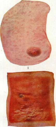
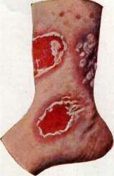
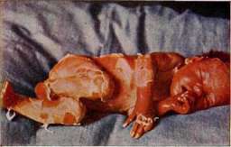
Figure 1. Toxicodendron mercurialis. Figure 2. Dermatitis dysmenorrhoica. Figure 3. Dermatitis roentgenologica.
Fig. 4. Dermatitis exfoliativa. Figure 5. Toxicodermia ex usu Balsami Copaivae. (Figs. 2, 3, and 4 are from Frieboes, Figs. 1 and 5 are from Jakoby.) Regarding dermatitis, see Dermatitis. ...when exposed to the light of arc lamps, in solderers of the voltaic arc who deal with high-voltage currents. Lightning strikes result in burns of the 1st and 2nd degree due to the impact of a huge amount of electrical energy, which heal quickly, leaving pigmentation. This also includes skin changes caused by Finsen phototherapy (light therapy with concentrated light) and the Kromayer quartz lamp, in which mercury vapor, rich in chemical ultraviolet rays, glows due to the passage of an electric current. Medicinal dermatitis (from external use). (Regarding dermatitis arising as a result of enteral or parenteral administration of drugs, see dermatitis of endogenous origin.) Dermatitis from arnica tincture (Tinctura Arnicae) is characterized by intense redness and a vesicular-bullous eruption at the sites of application of the arnica tincture. In rare cases, the dermatitis spreads beyond the boundaries of the arnica tincture application. Dermatitis from tar preparations occurs frequently and almost always has a follicular character. In some cases of special sensitivity, diffuse erythematous-vesicular dermatitis arises. Juniper tar (Oleum cadinum) and coal tar preparations (Liquor carbonis detergens, etc.) are less irritating than others. Prevention consists of cautious prescription of tar for inflammatory dermatoses, especially for eczema; one should avoid the use of tar preparations on skin abundantly covered with hair. Dermatitis from iodine usually develops only at the sites of its application; in cases of increased sensitivity to it, the external use of iodine preparations on limited areas sometimes causes a widespread measles-like or urticarial exanthema. Clinically, dermatitis at the sites of frequent smearing with iodine tincture or the use of ointments with potassium iodide, Jothion, and other preparations is characterized by redness, vesicular eruptions, abundant large-lamellar desquamation, and sometimes burning and itching. Dermatitis from iodoform (Jodoformium) arises in individuals particularly sensitive to it when applied to the skin; in some cases, dermatitis develops after a single application of iodoform, sometimes from a minimal amount of it. There are isolated observations of the appearance of dermatitis even upon contact of the preparation with a mucous membrane, for example, when introduced into the urinary bladder. Frei observed skin erythema even after subcutaneous injection of iodoform. Recent experimental work, mainly by Bruno Bloch and Jadassohn, has shown that idiosyncrasy to iodoform is entirely independent of idiosyncrasy to iodine; the methyl radical contained in it is responsible for the occurrence of dermatitis from iodoform. Dermatitis from iodoform usually proceeds violently, accompanied by significant subjective sensations. Eruptions are either of an erythematous-vesicular character or in the form of an erysipelas-like exanthema, sometimes accompanied by pseudo-phlegmonous edema; during regression, the epidermal covering is shed over significant areas. In most cases, the dermatitis is limited to the site of iodoform application, affecting only adjacent areas of the skin. In rarer generalized cases, there are scarlatiniform eruptions, often accompanied by more or less severe general symptoms (high temperature, delirium, prostration, etc.). The duration of the disease is 2-3 weeks. Bearing in mind the existence of idiosyncrasy in some individuals, for the purpose of prevention, one should limit oneself to small amounts of it during the first applications of iodoform. Local treatment consists of the use of anti-inflammatory agents (compresses of aluminum acetate or resorcinol, cooling ointments). Agents replacing iodoform—airol, aristol, iodol, europhen, etc.—cause dermatitis much less frequently than iodoform. Orthoform, on the contrary, often causes erysipelatous and gangrenous dermatitis. Dermatitis from pyrogallol (Pyrogallol, Acidum pyrogallicum) occurs relatively frequently when using preparations containing pyrogallol for the treatment of various dermatoses, mainly inflammatory processes of the skin of the face and scalp (psoriasis, lupus). Pyrogallol generally causes a yellowish-brown discoloration of the skin at the sites of its application very often. With an overdose or with special sensitivity to it, intense widespread dermatitis arises, accompanied by significant edema, an increase in body temperature, and severe pain in the affected areas. Such dermatitis lasts for several weeks, during which areas of skin that did not come into contact with pyrogallol are also involved in the process. During regression, bran-like desquamation occurs. Histologically: vacuolization of the cells of the prickle cell layer of the epidermis, formation of blisters in the latter, edema, and perivascular round-cell infiltrate in the skin proper. The prognosis is favorable in most cases. For the purpose of prevention, extremely cautious prescription of ointments with pyrogallol is recommended, especially for inflammatory processes. Treatment consists of thorough removal of ointment residues and the prescription of indifferent anti-inflammatory agents. Dermatitis from mercury (dermatitis mercurialis) can develop both with external use of the latter and with any method of introduction into the body, but more often with the former. Most researchers see the cause of mercurial dermatitis in the toxic effect of mercury on the sympathetic nervous system; as a result of the paralysis of the latter, vasodilation and the formation of transudate occur. In most cases, when dermatitis develops after ordinary doses of mercury, it is a matter of individual hypersensitivity to mercury; minimal doses in these cases cause skin changes. Some even speak of hypersensitivity to mercury of individual parts of the body or organs, with some (Almkvist) considering the cells of the corresponding sympathetic ganglia to be the site of localization of mercurial hypersensitivity, while others (Tomaszewski) look for hypersensitivity in the cells of the organs themselves. The causes of limited dermatitis arising from the introduction of mercury through the skin, mainly from inunctions of gray mercury ointment, are mechanical irritation during friction, chemical irritation from the penetration of the ointment oxidizing there into the follicles, and the introduction of pyogenic cocci into the follicular orifices. Some individuals are sensitive only to a specific method of introducing mercury, for example, dermatitis develops in them only after injections, while when mercury is introduced by other methods, dermatitis does not arise. The same is noted in relation to individual mercury preparations with the same method of its introduction; for example, a patient in whom dermatitis develops after an injection of calomel tolerates injections of mercury salicylate perfectly. Histologically: dilation of the upper vascular network, edema of the papillae, perivascular infiltrate of leukocytes and lymphocytes in the upper layers of the dermis; in the epidermis, infiltration with fluid and leukocytes, vacuolization of cells, parakeratosis, sometimes acanthosis. In severe cases, the infiltrate is more powerful, diffuse, and includes mast cells, plasma cells, and eosinophilic cells; often, significant accumulations of leukocytes are found in the upper layers of the epidermis, lifting the overlying layers of epidermal cells; in the stratum corneum, bacterial masses. Dermatitis can appear 1-40 days after the start of mercury treatment. Starting usually on the extremities, it soon spreads to the trunk and face, sometimes affecting the mucous membranes as well. When mercury is introduced through the skin, the dermatitis begins at the site of application of the mercury preparation, from where it sometimes (rarely) spreads further per continuitatem. Clinically, mercurial dermatitis is very diverse; scarlatiniform exanthema is more common, measles-like and urticarial are rarer, and rashes similar to pemphigus, exudative erythema, erysipelas, purpura, etc., are even rarer. Densely arranged violet- or blue-red (often with a yellowish tint) small spots or nodules confined to the follicles can create the impression of diffuse erythema (see separate table, Fig. 1). Simple and complicated mercurial dermatitis are distinguished (Almkvist). The former (exanthema mercuriale simplex) is characterized by an acute course (1-3 weeks), redness of the skin, formation of vesicles and hemorrhages, slight edema and infiltration of the skin, fever, and severe itching. Complicated mercurial dermatitis (exanthema mercuriale complicatum infectum) is characterized by a very slow course (several months) and frequent exacerbations. On the foul-smelling, heavily infiltrated, and edematous dirty-red skin, there are spots, papules, vesicles, purulent-bloody blisters, crusts, and large scales; abundant weeping, especially in the flexures and skin folds (sometimes a picture of severe exfoliative dermatitis develops). Prolonged and exhausting fever and accompanying severe changes in internal organs lead to death in some cases. Limited dermatitis, developing upon the introduction of mercury through the skin, is characterized in the developed state by redness, edema, the formation of purulent folliculitis, weeping, and sometimes significant pain. Very rarely, there are so-called fixed (always recurring in the same places) mercurial dermatitis.
Mercury dermatitis differs from a true scarlatiniform rash by a more bluish, and sometimes yellowish, shade of the rash, by starting in most cases on the extremities, by weaker fever, by the absence of scarlatinous angina, and often by the presence of mercury stomatitis. Sometimes mercury erythema may resemble a large-spotted roseola. Prognosis is doubtful in cases of long-lasting dermatitis complicated by surface infection. Prophylaxis consists of the caution with which treatment with mercury should be conducted (do not allow overdosing, use small initial doses). In case of poor tolerance, especially on the part of the kidneys, liver, and skin, it is recommended to change the preparation or the method of administration; in extreme cases, abandon mercury entirely. Special caution is required for individuals who have already suffered from mercury dermatitis or salvarsan dermatitis (since the latter often sensitizes the organism to mercury, and vice versa): in them, the smallest dose of mercury can cause very severe dermatitis. As for the prophylaxis of dermatitis during the external application of mercury, it should not be prescribed to individuals with delicate, thin skin, those with inflammatory dermatoses, individuals who have previously suffered from eczema or Duhring's dermatitis, and finally, those who are very hairy. Individuals with oily, hairy skin covered with nodules of lichen pilaris are especially prone to mercury dermatitis. Furthermore, during inunctions, to avoid dermatitis, it is recommended to change the sites of friction, prefer the flexor and posterior surfaces of the extremities for them, and powder the skin with talc after inunctions. General treatment consists of using agents that strengthen the activity of the heart and kidneys; in the initial stage, adrenaline. Locally, washings with oxidizing agents work best; daily washings with a 2% solution of KMnO4 often quickly cure mild forms of dermatitis. Dermatitis from sulfur usually arises in cases of its application in a highly concentrated form for various inflammatory dermatoses. In rare cases of special hypersensitivity to sulfur, dermatitis develops even after the application of sulfur preparations in weak concentration. Clinically, the dermatitis is characterized by redness and itching; sometimes vesicular eruptions with subsequent weeping are added. For individuals sensitive to sulfur, it is better to prescribe less skin-irritating preparations of colloidal sulfur (Sulfidal, Sulioform, etc.). Dermatitis from chrysarobin (Chrysarobinum) is a not infrequent complication during the treatment of various skin diseases with it, mainly psoriasis. Being dissolved on the skin by the alkali of sweat or soaps, chrysarobin almost always causes a brownish-violet (often the color of red wine) discoloration of the skin accompanied by itching, which usually continues to exist for several weeks even after the cessation of chrysarobin use. Characteristic is the so-called bronze chrysarobin erythema (more or less edematous skin of a dark red color). In cases of overdose, especially in individuals hypersensitive to chrysarobin (mainly in psoriatics), the latter sometimes causes severe weeping exfoliative dermatitis, accompanied by a febrile state and other disorders of the general condition. In some cases, dermatitis from chrysarobin in psoriatics gives an impetus to a widespread acute eruption of psoriasis. Not infrequently, the dermatitis is accompanied by conjunctivitis. Histological studies of dermatitis from chrysarobin, induced experimentally both in rabbits and in humans, have shown intense inflammation in the skin, accompanied by significant edema and the formation of epithelial microabscesses under the necrotized layers of the epidermis (Hodara, Racinowski). The prognosis is in most cases favorable. Prophylaxis consists of the cautious prescription of agents containing chrysarobin, especially in inflammatory diseases (psoriasis, eczema, neurodermatitis, etc.). The initial concentration of chrysarobin in such cases should be minimal (1/4-3%). Treatment. In cases of mild dermatitis, one can limit oneself to stopping the use of chrysarobin, thoroughly cleaning the skin of ointment residues, and using an indifferent powder (rice powder or talc). In more severe cases, anti-inflammatory agents, cooling ointments, etc. are used. Besides the above-listed medicinal agents that can cause dermatitis upon external use, there is a number of other medications that sometimes cause inflammatory processes in the skin; these include: novocaine, picric acid, salicylic acid, resorcinol (in strong concentrations), Veratrum album, Tinct. Capsici annui, Tinct. Cantharid., paraffin, formalin, turpentine, carbolic acid, etc. (regarding some of them, see Occupational Dermatoses). Dermatitis from chemical causes (occupational dermatitis—see Dermatoses). Dermatitis from acridine—a derivative of anthracene (a component of coal tar)—arises in some individuals on the auricles after regular, prolonged contact with the earpieces of a radio or telephone, in the manufacture of which acridine is used. Dermatitis from non-professional dyes is encountered relatively rarely. In the first place here are dermatitis from hair dyes, which usually proceed very violently. More often than others, dermatitis is caused by dyes containing para-phenylenediamine [C6H4(NH2)2], much less often by dyes with pyrogallic acid and silver nitrate. Usually, already a few hours after contact with the dye, edema and an itchy erythematous (later vesicular) rash appear on the skin of the face and eyelids, rapidly spreading further—to the neck, torso, and sometimes the extremities. In some cases, there is a significant disturbance of the general condition. The swelling and redness last for several weeks, and the peeling continues longer. Dermatitis from dyed fur develops when wearing fur items. Almost all authors consider the cause of this dermatitis to be poor washing of the fur after dyeing with derivatives of the same para-phenylenediamine. Mainly women fall ill; the dermatitis usually develops only after 5-6 weeks of wearing the fur, and in 98% of cases, it is localized on the neck. Dermatitis from poorly dyed stockings is observed rarely and is apparently caused not by the dye substance itself, but by the mordant used to fix the dye substance. In two cases by Porosz, such a mordant was some kind of iron salt (ferrous sulfate?). The dermatitis, localizing on the feet and shins, is accompanied by severe itching and can sometimes simulate erysipelas. Dermatitis "from matchboxes" was repeatedly noted during the years of the World War in Germany. Its cause was always the skin-irritating mass (phosphorus sesquisulfide) covering the matchboxes in these cases, which was introduced by some match factories during the war years due to a shortage of the usual composition that was indifferent to the skin. On the areas of skin that came into contact through the fabric of pockets with such matchboxes, an acute, weeping, sharply defined dermatitis developed; in some cases, a severe universal dermatitis developed. Dermatitis from chromic acid was also observed mainly in Germany during the war, when, instead of the usual leather inner linings of hats, canvas linings dyed brown with chromic acid were used. Clinically, the dermatitis manifested itself in the form of strongly itching, wide, hoop-like red stripes on the forehead, and in those with short-cropped hair—also on the head ("Hautledereczem"). Dermatitis caused by parasites. Dermatitis caused by vegetable parasites, or dermatophytes—see Dermatomycetes. Dermatitis caused by animal parasites. Animal parasites causing painful changes in the skin, or dermatozoonoses, can be divided into 3 groups depending on whether: a) the parasites are located on the surface of the skin and mucous membranes, in the hair, in clothing, accidentally getting in from the outside and causing itching and inflammation of the skin through bites (so-called epizoa, or ectoparasites), b) they settle in the skin permanently or temporarily for the purpose of feeding, finding favorable conditions for their development and reproduction and causing intense itching (so-called intradermal parasites, or true dermatozoa), c) they penetrate into the connective tissue layer of the skin and subcutaneous tissue, causing a general infection of the organism. Dermatitis caused by parasitic insects (derm. epizoogenes). Among the surface parasites, or epizoa, that can cause dermatitis are lice (see Pediculosis), fleas, bedbugs, mosquitoes (see), and sandflies. It is also necessary to note dermatitis from the bites of various species of poisonous flies (see). The sting of a bee (Apis mellifica), bumblebee (Bombus terrestris), wasp (Vespa vulgaris), hornet (Vespa crabro), scorpion, and the bites of spiders are accompanied by excruciating pain, the rapid development of inflammatory redness, edematous swelling, vesicles, and blisters due to the local action of the venom, as well as a special hypersensitivity to it. If the stings and bites are multiple, they often give rise to severe, violent phenomena (dizziness, fever, respiratory distress, fainting, cold sweat, convulsions) and even death (e.g., bites of the tongue or larynx). Treatment: often hygienic and disinfecting measures are sufficient.
Among other parasites, some worms (Oxyuris = Enterobius vermicularis) can cause very itchy rashes on the skin of the inner surface of the thighs, near the anus, on the mucous membrane of the vulva, and on the scrotum, resembling eczema. Sometimes, under the influence of scratching, eczematous phenomena of secondary origin arise. In the folds and grooves of the skin, there is a very foul-smelling creamy coating. - Treatment: anthelmintic agents, washings. - Dermatitis caused by actual skin parasites (Dermatozoa). This includes parasites that penetrate the epithelial cover, for example, itch mites (see Scabies), or into the sebaceous-hair follicles (Demodex folliculorum). This parasite of the hair follicles is not dangerous to humans and usually does not cause inflammatory changes. Sometimes it is found in large quantities on seborrheic skin of the face, but it plays no role in comedones and acne rosacea. In rare cases, it may perhaps be the culprit of local pigmentation (Dubreuilh), lichen spinulosus (Darier), epithelioma (Borrel). Among other parasites that can cause dermatitis, Dermanyssus and other mites (see) have been noted. - Dermatitis caused by animal parasites that can penetrate under the skin and deeper into the organism. This refers primarily to tropical or subtropical parasitic worms, such as: filariae (Filaria, Dracunculus-guinea worm), bladder worm, etc. - Under the name craw-craw (kro-kro), a peculiar itchy dermatosis in Negroes of West and Central Africa has been described (O'Neill), especially on the lower extremities, consisting of papulo-vesiculo-pustular elements and clinically proceeding in the form of eczema, scabies, and prurigo. The cause of the disease in some cases is the filaria Onchocerca volvulus, the larvae (microfilariae) of which are localized in the skin. Cutaneous ancylostomiasis (derm. uncinarialis, dochmiasis, panighao, gourme des mineurs), ground itch, water sore - inflammatory diseases of the skin caused by the penetration of larvae of small parasitic worms Ancylostoma duodenale or Necator americanus. Any surface of the skin can be affected, most often the extremities, especially the hands and feet (during work on damp ground). At the site of the parasite's penetration, a papulo-vesicular dermatitis arises, accompanied by agonizing itching and significant edema of the skin. Often dermatitis is complicated by secondary purulent infection (pustules, abscesses). Sometimes persistent ulcers form. Treatment: compresses of 3% salicylic acid solution in ethyl alcohol, turpentine, etc. Dermatitis caused by poisonous insects. The hairs of the caterpillars of the oak processionary moth (Thaumetopoea processionea) and the pine processionary moth (Thaumetopoea pinivora) cause, as a result of local irritation on uncovered parts of the body, urticarial wheals, and in children and sensitive subjects with delicate skin - reflexively generalized urticaria over the whole body. The poisonous hairs of the gold-tail moth caterpillar (Euproctis, Porthesia) also have an irritating effect on human skin. In collectors, entomologists, and gardeners, when handling the larvae of the silkworm caterpillar (Lasiocampa rubra), dermatitis and erythematous redness arise on the hands and face due to the penetration of brittle poisonous hairs into the skin. Sometimes skin irritation is also caused by the cocoons of Euproctis, the hairs of which, piercing the skin and releasing chemically irritating substances, produce intense dermatitis and urticarial wheals. - Histologically - necrosis of epithelial cells and vesicles in the vicinity of the penetrated hairs, as well as inflammation of the skin proper (see Caterpillars). Jellyfish, sea anemones, cochineal, Spanish fly (Lytta vesicatoria) cause dermatitis and various rashes upon contact with the skin. In workers extracting sponges from the sea bottom, inflammatory redness, blisters, and sometimes numerous abscesses and even gangrene (the so-called sponge diver's disease) often develop due to contact with the anemone (Sagartia rosea - a coelenterate living on sponges). In one case (Zervos), detachment of the skin on the penis and gaping of the cavernous bodies were observed. The prognosis is favorable if the lesion has not ended in gangrene. Dermatitis caused by pyogenic cocci (derm. coccogenes) [see Impetigo, Furuncle, Erysipelas, Folliculitis (folliculitis decalvans, perifolliculitis capitis suffodiens et abscedens), Acne (acne keloid)]. Vegetating pustular dermatitis - see Chronic pyoderma. Under the name chronic streptococcal epidermitis, Sabouraud described a skin lesion in the form of small erosive rashes, 3-5 mm wide, which quickly turn into papules and become covered with dry, delicate crusts, and then can undergo lichenification, resembling eczematized prurigo. The cause of the disease is the penetration of streptococci. - Dermatitis of staphylococcal and streptococcal origin. Around deep wounds or superficial ulcers, skin changes often arise in the form of sharply defined red, sometimes weeping or scaly plaques of scalloped or rounded outline with a delicate epidermal rim at the periphery. Sometimes the plaques reach large sizes and clinically resemble psoriasiform parakeratosis. The origin of the process is undoubtedly microbial. - Derm. glutealis syphiloides neonatorum (or ecthyma vacciniforme syphiloides), a rare skin lesion in very emaciated infants with localization on the buttocks, genitals, and in the anal region. It begins in the form of erythematous spots and small nodules with a vesicle or pustule at the apex, which often undergo ulceration, resembling a smallpox or vaccine rash. It is especially easy to confuse this dermatosis with a weeping, ulcerative papulo-hypertrophic syphilid. In essence, the disease is an epidermo-dermatitis of microbial origin. The prognosis is unfavorable, as children often die from bronchopneumonia or gastroenteritis. Treatment: hygienic measures, indifferent powders. Dermatitis caused by plants and herbs (dermatitis venenata phytogenes) arise mainly in predisposed individuals from contact with certain plants and herbs. They are most frequent in America, where a significant quantity of plants grows that can cause skin changes in humans. Dermatitis from plants and herbs have especially begun to attract the attention of dermatologists in recent years, when a number of researchers succeeded in experiments of sensitization and desensitization with some of them. In total, there are more than 100 species of plants and herbs that can cause dermatitis. More frequent than others and better studied are dermatitis from primrose, sumac, geranium, and asparagus. Dermatitis from primrose (Chinese primrose, botanical species Primulaceae) occurs especially often due to the wide distribution of this plant, which is used for decorative purposes. Nestler showed by experiments on himself that the skin-irritating substance is located in the glandular hairs with which all aerial parts of the primrose are covered; the yellowish-green secretion of the cells of the glandular hairs contains needle-like and prismatic crystals of the monoclinic system, which are, according to Nestler, the skin poison; the chemical composition of the latter has not yet been precisely established. In dermatitis from primrose, as in most other dermatitis from plants and herbs, hypersensitivity to the plant plays the main role. This hypersensitivity, not being congenital in the vast majority of cases, begins to manifest itself clinically mostly only after repeated contact with the plant. There are apparently no people who are completely immune to the skin poison of the primrose. Bloch and Steiner succeeded in sensitizing 100% of the subjects exposed to the experiment by inoculating a concentrated extract ("antigen") obtained from primrose leaves, i.e., everyone who had previously tolerated contact with the primrose well became, after this inoculation, idiosyncratic to this plant, and the sensitization lasted for a long time. Recently, Walthard succeeded by applying an extract from the leaves of Primula obconica to the skin of white mice in causing an inflammatory reaction in them that fully corresponded (clinically and histologically) to the picture of acute eczema in humans. Not every species of primrose can cause skin irritation in humans; poisonous in this respect are: Primula obconica Hance, Primula sinensis Lindl., Primula Sieboldii Morren, and Primula Arondsii. Primula officinalis and Primula Auricula never irritate the skin. After the last contact, an "incubation period" of from 7 hours to 14 days passes. Beginning at the sites of contact with the plant, mostly on the hands, the lesion often spreads due to being carried by the fingers to other places, mainly to the face, neck, and genital parts. The rash often very much resembles acute eczema; on the strongly reddened and edematous skin are located groups of small, pointed, shiny red papules and vesicles; at the site of the ruptured vesicles - yellowish crusts; during the regression of the rash - peeling. Subjectively, there is very intense, often agonizing itching and burning. There are isolated observations of severe stomatitis that developed in individuals who put primrose flowers in their mouths.
In some cases, the eruption proceeds in the form of urticaria, in others it simulates erysipelas, and in others it is accompanied by the formation of large blisters.
With frequent recurrences, a picture arises that is very reminiscent of chronic eczema, with each subsequent recurrence usually being more severe than the previous one.
Histological changes, located mainly in the epidermis (intracellular and intercellular edema), generally differ little from those in acute eczema.
In diagnosis, the anamnesis plays the main role; the disease usually begins after cutting wilted flowers (the drying of flowers does not rid them of their toxicity).
With the exclusive localization of eruptions on the back of the hands (the palms are rarely affected) and fingers and on the face, one should not forget about the possibility of dermatitis from plants, and mainly from primrose.
The prognosis is favorable, provided the patient manages to avoid contact with the plant.
For prophylactic purposes, some authors (Pinkus) recommend completely banning the use of primroses.
Others consider this irrational, since according to some data only 6% of people are sensitive to primrose (figures for sensitivity to primrose are contradictory), and at the same time there are species of the plant that never cause dermatitis.
Inanimate objects can apparently also be carriers of primrose poison.
Usually, a few days after ceasing contact with the plant, the regression of the eruptions begins.
For treatment, local hot baths with a solution of boric acid or aluminum acetate, indifferent boric-bismuth ointments are recommended; often an abundant dusting with talc proves sufficient; alcohol rubs are also recommended to remove skin fat, in which the primrose poison dissolves.
Dermatitis from poison sumac (poison ivy, Rhus venenata, Toxicodendron, etc., from the botanical family Anacardiaceae) is more common in North America.
Experiments on humans (animals are insensitive to sumac poison) with an alcoholic extract of the plant's leaves showed that 65% of people are sensitive to the skin poison of poison ivy, with children under 11/2 years old proving to be generally insensitive.
Natural immunity to sumac is rarely absolute.
In summer, during flowering, ivy poison acts more strongly.
Dried leaves can still cause dermatitis for several years (Mac Nair).
Data regarding the chemistry of the poison are contradictory: according to Simpson - a glycoside, according to Mac Nair - toxicodendrol oil and lobinol-polyhydrophenols with unsubstituted side chains.
The first skin phenomena usually arise 48 hours after contact with the plant ('incubation period' - from several hours to 5 days).
The clinical picture of the eruptions is extremely diverse: from relatively benign erythema or papulo-vesicular (sometimes urticarial) eczema of the hands, face, and genitals to severe widespread dermatitis with erysipelas-like redness and swelling of the face, vesicles, weeping, crusts, agonizing itching, and burning.
Dermatitis is often accompanied by fever, vomiting, albuminuria, enlargement of lymph nodes and spleen, and a severe disturbance of the general condition.
The duration of the disease is from 11/2 to 6 weeks.
There are reports of three fatal cases.
Histologically, the main changes in the epidermis are of the character of acute eczema; in the cutis, there is insignificant infiltration, mainly around the sebaceous and sweat glands.
According to Touton, the stronger the secretion of the sebaceous glands, the greater the solubility of the sumac skin poison, and because of this, the more severe the disease.
The eczematous form of dermatitis can be distinguished from true eczema by the dark red color of the affected areas, the monomorphism of the rash in each stage of the disease, and the frequent absence of a papular stage.
The prognosis is favorable in most cases.
Prophylactically, in view of the significant percentage of individuals hypersensitive to sumac, it is recommended to avoid contact with it.
Treatment: desensitization is in the first place, which succeeds in most cases (in contrast to dermatitis from primrose, in which it never succeeds).
Schamberg recommends for this Tinct. Rhois toxicodendri internally in increasing doses; Strickler successfully combines administration per os with subcutaneous injections of the plant toxin.
Local treatment is as for dermatitis from primrose.
Dermatitis from shore plants and grasses usually arises 24-48 hours after swimming followed by lying on the shore grass.
Siemens, who first noted this dermatitis, as well as other authors, consider sedge to be the cause of the eruptions.
The exanthema appears on the areas of contact with the grass, mainly on the buttocks, abdomen, back, knees, outer surfaces of the thighs, and other places.
Maculo-vesicular eruptions are always arranged in stripes (corresponding to the strip-like shape of the sedge leaves), resembling herpetiform rashes in places.
Histologically, a small accumulation of fluid and leukocytes is found between the cells of the epidermis, their vacuolization, edema of the papillae, dilation of vessels in the upper parts of the dermis, and a small perivascular lymphocytic infiltrate.
Indifferent local therapy quickly leads to the cure of this dermatitis.
Traumatic dermatitis (d. traumatica), or mechanical dermatitis, is a local inflammatory change of the skin arising as a result of the irritating action of various kinds of external agents directly in contact with the outer covering.
Some authors divide them into two groups depending on whether the eruptions originate from purely mechanical irritation (traumatic dermatitis in the proper sense of the word) or from chemical and toxic exposure (derm. venenata).
Depending on the type of causative agent, the strength of the impact, the duration, and the frequency of the irritant, the clinical picture of traumatic dermatitis can manifest in various forms.
Mild traumatic damage to the skin can cause slight swelling and erythematous redness, which, undergoing regression, usually leaves no traces.
However, with individual predisposition, it may lead to increased pigment deposition.
Similar phenomena are noted in pediculosis and scabies as a result of scratching.
Conversely, more or less intense traumatic impact, such as: strong friction during scratching, massages, prolonged pressure, a blow, etc., leads to the formation of redness, vesicles, blisters, and superficial desquamation of the epithelium with occasional exposure of the papillary layer and subsequent development of scales and crusts.
This includes all sorts of manipulations performed by patients on themselves (scratches, abrasions, excoriations, pressure), further skin changes as a result of the mechanical impact of tight shoes, clothing, friction of darned stockings and garters during intense walking, when handling tools (bullae mechanicae on the hands of soldiers, rowers, workers), as well as due to maceration of two surfaces in contact with each other (intertriginous eczema in obese subjects) and urticaria factitia.
Artificially reproduced dermatitis, observed more often in hysterical women, arises directly through intense mechanical friction with the fingers and is usually sharply demarcated.
For the purpose of simulation, they intentionally exacerbate an existing dermatosis by subjecting the skin to the irritating action of some harmful agent (e.g., caustic alkali or acid).
In subjects with individual irritability, strong and prolonged pressure on a certain area of the skin, constant irritation of the tissue leads to the formation of erythema, excoriations, phlyctenae, ulcerations, and even gangrene.
If the skin is simultaneously macerated by any secretions or excretions (leukorrhea, sweat), then, due to the violation of the integrity of the horny layer, the presence of inflammatory irritation of the skin, and exudation, the process can easily turn into eczema and be complicated by a secondary pyogenic infection (pus-forming microorganisms).
In this way, folliculitis and perifolliculitis, pustules, acne, etc., arise.
With constant irritation due to itching, scratching, and weeping, it often leads to the development of a persistent skin affliction.
Nervous itching, prurigo, scabies, pediculosis, circulatory disorders in the lower extremities (varicose veins, venous stasis), especially in predisposed subjects, often give rise, due to scratching, to the formation of traumatic eczema.
Around wounds, deep ulcers, and long-standing fistulas, especially during the war, so-called paratraumatic eczemas (eczema paratraumaticum Darier) developed, distinguished by sharply defined edges and a polycyclic configuration, which, due to maceration of the surrounding skin with iodine, hydrogen peroxide, various antiseptic agents, and local circulatory disorders, were often subjected to complication by secondary purulent infection and impetiginization.
Sometimes under the influence of intense itching and prolonged scratching, less often due to pressure (by a corset, bandage), persistent thickening of the skin (lichenification) develops.
Friction and mechanical pressure thicken the epidermal layer on the palms and soles, forming calluses and callous thickenings of the skin (callus, callositas, clavus), i.e., chronic traumatic dermatitis with hypertrophy of the dermis and hyperkeratosis.
Irritation of peripheral nerves due to pressure, compression, or rupture can often cause herpetic eruptions on the skin (herpes traumaticus). A decrease in the resistance of the skin and its particular vulnerability under the influence of the slightest trauma, jolts, blows, or pressure are observed in congenital dystrophy of the skin, such as pemphigus hereditarius traumaticus simplex (see Epidermolysis bullosa). Friction of the skin of the scalp against a headboard, especially in weakened children and hydrocephalics, or accidental or intentional (for the purpose of simulation) scratching due to itching often causes hair loss of traumatic origin (see Alopecia). According to clinical and pathohistological features, acute traumatic dermatitis essentially differs little from eczema. Diagnosis of traumatic dermatitis is not difficult. Treatment is symptomatic: elimination of the harmful mechanical factor. N. Efron, L. Mashkilleyson, P. Kozhevnikov, M. Per.
Related articles
Mentioned in
Cite this page
“Dermatitis.” Soviet Medical Encyclopedia. English translation of Bolshaya Meditsinskaya Entsiklopediya, 1st ed. (Moscow, 1928–1936), ed. N. A. Semashko. https://sovietmedicalencyclopedia.pages.dev/article/dermatitis/