Regression
Historical document, translated for reference. It reflects medical knowledge of the 1920s–30s and is not medical advice.
Summary
Regression refers to quantitative changes in one phenomenon that occur in connection with changes in another phenomenon that is in correlation with the first. The article explains how regression coefficients are calculated and used to determine theoretical values of one phenomenon based on changes in another.
Encyclopedia article (1928–1936)
REGRESSION, quantitative changes in one phenomenon that occur in connection with changes in another phenomenon that is in correlation with the first. Thus, successive changes in weight in different parts of a certain collective that is homogeneous in basic characteristics (by age, sex, social status, etc.) in connection with successive changes in height in the corresponding parts of this collective represent regression of weight with respect to height. Reverse changes represent regression of height with respect to weight. The degree of change (magnitude of regression) is determined by the regression coefficient: Rx=r·σy and Ry=r·σx. In both expressions: r is the correlation coefficient of the variation series being studied (see Correlation), and σx and σy are the standard deviations of the same series (see Variation Statistics). Thus, if the correlation coefficient between height and weight for a certain group of people is +0.5384, the standard deviation (σx) of the variation series representing the height of the same group is 6.7272 cm, and the standard deviation (σy) of the weight series is 8.2143 kg, then the regression coefficient of height with respect to weight is: +0.5384·8.2143/6.7272 = +0.4409 cm, and the regression coefficient of weight with respect to height is: +0.5384·6.7272/8.2143 = +0.6574 kg. The first number shows that an increase in weight by 1 kg is associated with an increase in height of approximately 0.5 cm; the second number indicates that an increase in height by 1 cm is associated with an increase in weight of 0.66 kg. It should be particularly emphasized that when determining the regression coefficient, each standard deviation is taken in its absolute value (taking into account the size of the intervals of the corresponding variation series). In addition to the application mentioned - in the analysis of phenomena - the regression coefficient is very useful in certain cases for finding theoretical values of one phenomenon depending on changes in another. For this purpose, equations of the following form are used: Xx = Mx - Ry·My/σy + Ry·Yx/σy and Yx = My - Rx·Mx/σx + Rx·Xx/σx, where in the first equation: Xx is the sought value of one phenomenon at the corresponding given value of the second phenomenon (Yx) and at the arithmetic means Mx and My of both variation series being studied. The sought value in the second equation is the value of the second phenomenon - Yx at a given size of the first phenomenon (Xx) and at the same arithmetic means. Substituting for the algebraic expressions the corresponding values from the above example about height and weight: My = 65.65 kg, Mx = 175.32 cm, Ry = +0.6574 kg and Rx = +0.4409 cm, we will have: Xx = 65.65 - 0.6574·175.32 + 0.6574 Yx, Yx = 175.32 - 0.4409·65.65 + 0.4409 Xx, from which: Xx = -49.61 + 0.6574 Yx and Yx = 146.13 + 0.4409 Xx. If we substitute certain given values of height (Yx = 157.5, Y2 = 163.5, Y3 = 169.5, etc.) into the first equation, we obtain the following theoretical values of weight: X1 = 53.9, X2 = 57.8, X3 = 61.8, etc. Substituting given values of weight (X1 = 48, X2 = 57, X3 = 66, etc.) into the second equation, we obtain the following theoretical values of height: Y1 = 167.3, Y2 = 171.3, Y3 = 175.2, etc. The presented relationships in the sizes of height and weight (and vice versa) would be observed in the mass of people being studied if the existing dependence between both phenomena were not disrupted by extraneous circumstances. The geometric mean of both regression coefficients equals the correlation coefficient: r = √Rx·Ry. The mean error of the regression coefficient is mRy = mr·σy and mRx = mr·σx where mr is the mean error of the correlation coefficient.
ax
y
ay
P. Kuvshinnikov. REGULATION (of functions), the ability of an organism to change functions in the form of adapting them to certain changed conditions. The result of such a change in functions is the maintenance of the body's basic functions at the level necessary for its existence. This special property of organisms to change functions in the presence of changed living conditions should be understood not as an exhaustion of the entire reactive capacity of organisms, but as a change in the direction of increasing or decreasing functions, and consequently also of reactive capacity under the influence of various irritants. R. is thus connected with "such a state of the organism, which is characterized primarily by the preservation of basic functions, allowing the organism to exist without harm. Understanding R. in this way, one should distinguish between physiological and pathological "R. In the first case, it is a matter of adaptation to ordinary, and in the second to special, going beyond physiological fluctuations, conditions. An example of physiological R. can be the fluctuations in the lumen of peripheral vessels under the influence of thermal conditions, protecting the organism from both cooling and overheating. This also includes: working hypertrophy of skeletal muscle in people engaged in physical labor or sports, increased blood flow to working organs, carried out through local acidosis as a result of the organ's activity, temporary changes in the functions of respiration and circulation when an individual transitions from a state of rest to movement, increased secretory activity of glands under the influence of various irritants, differences in the secretion of digestive enzymes depending on the type of food, and much more. All functions of the organism are subject to physiological R., and the latter occurs constantly and completely, since under ordinary conditions it is a matter of accommodation to such fluctuations in the effects of the external and internal environment to which the organism has already adapted phylogenetically. The presence of pathological conditions places R. in special conditions, mainly in the form of a change in the reactive sensitivity of tissues to ordinary irritants. As examples of pathological R., one could point to the fact of a sharp increase in intracardiac pressure in the cavity of the right ventricle of the heart, marking a significant increase in the work of the latter in an experiment with artificial gradual narrowing of the lumen of the pulmonary artery, or a change in the frequency and magnitude of respiratory excursions in an experiment with artificial pneumothorax, with the replacement of negative pressure in the pleural cavity with positive, or finally a similar change in the function of respiration with artificial restriction of air access to the trachea. In all three of the mentioned cases, the conditions are unusual and go beyond physiological fluctuations, nevertheless, thanks to pathological R., the affected basic functions of the organism remain for a certain time and within certain limits at their previous level: in the first experiment, thanks to the increased work of the right ventricle, the heart manages in a unit of time to push the necessary amount of blood through the narrowed opening, and gas exchange in the last two experiments does not decrease. Pathological R. differs from physiological R. only in the unusualness of the conditions of its origin, often in the degree of its manifestation, and sometimes in the qualitative change of functions, but in any case, no new functions arise in this process. Thus, the heart, adapting both in physiological and pathological conditions, is capable of producing only one effect—contractions, but the strength and character of the latter are different under different conditions. Similarly, the function of the kidneys in physiological and pathological R. always remains the same—excretory, but in the quantity and quality of the products excreted from the body, this function changes greatly. The significance of R. for the organism is self-evident, just as the circumstance that, despite the wide possibilities opened by the organism's ability to pathological R., the latter, just like physiological R., has its limits, beyond which the basic functions are disrupted, does not need explanation. The mechanism of R. varies in different cases. First of all, pathological R. often becomes possible only thanks to the presence in organs of the ability to increased work at the expense of the so-called "reserve forces" of them. Pathological R. in such cases amounts to the manifestation of such reserve, or compensatory, forces. Thus, the wall of blood vessels can withstand, if necessary, much greater blood pressure than under ordinary conditions. Bones are similarly capable of greater load, and muscles, if necessary, can perform significantly greater work compared to that which they produce in the absence of increased demands on them. The buffer systems of the organism can compensate, if necessary, for much greater shifts towards acidosis or alkalosis than is usually necessary to maintain acid-base balance. A part of an organ, by vicariously hypertrophying, as is known, in many cases can take upon itself the function of the entire organ. In this case, the "reserve force" of the vascular walls is of course nothing other than the degree of their elasticity, i.e., the property of the material from which they are built, and just as in technology, one speaks of the elasticity of materials, about the fact that the degree of elasticity under such-and-such conditions is higher than the pressure or stretching exerted on the object, a similar meaning is also invested in the above-mentioned designation. The same applies completely to the bone system. The "stretchability" of the buffering properties of blood and tissues, i.e., the organism's ability to regulate the active reaction of the latter within certain limits, is as known determined by the presence of the so-called alkaline reserves and the precise setting of the work of the respiratory apparatus (excretion of CO2 by the lungs), and vicarious and compensatory hypertrophy of organs finds its explanation in the fact that "work and nutrition, nutrition and work are two mutually penetrating processes": increased demands give a stimulus to intensified work, and the active state of the organ is accompanied by a shift in the reaction towards acidity, and the latter circumstance increases the arterialization of the given area of the organism. Somewhat greater difficulties is presented by the question of adaptability to increased demands on the heart. Regarding hypertrophy of the heart, basically the same could be said as in relation to hypertrophy of skeletal muscle, although it has to be admitted that in all details the mechanism of this phenomenon remains to this day not fully clarified. Of great interest, however, are other types of cases—cases of almost sudden, sometimes sharp increase in the work of the heart. Some authors, in explaining this phenomenon, appeal to the accelerating nerves of the heart, i.e., they admit the possibility of transmission to the cardiac muscle in such cases, through the mentioned nerves, of stronger impulses, but such an interpretation meets a certain difficulty from the side of the so-called "all or nothing" law (see), according to which the excitable substance in response to each separate excitation gives either a maximum reaction or no reaction at all—a law, however, not yet recognized by all. There are more grounds, however, to see the source of the reserve forces of the heart in something else—in its ability, as necessary, to actively lengthen its muscle fibers and thereby increase the energy of contractions. What the mechanism of such lengthening is is not yet known exactly, but it must be thought of as being of a reflex nature. Many other regulatory adaptations are known to be of a definitely reflex nature. Thus, in the area of the cardiovascular apparatus, in addition to the long-known depressor nerves, Hering recently discovered the so-called carotid reflex, leading to a decrease in blood pressure in the arterial system of the greater circulation in case of an increase in it (transmission from the carotid bulb through the branch of the glosso-pharyngeus nerve, the so-called sinus nerve, to the vasomotor center with subsequent vasodilation). But the mechanism of R. by reflected action can also be somewhat different, not according to the classic type of reflex with transmission of irritation through centripetal fibers to the corresponding center, but by direct action on the center through the blood. Thus, in the same cardiovascular system, there is a mechanism that auto-regulates the work of the heart in such a way that when the latter is strengthened, the increasing together with it increase in the tone of the vagus center in the medulla oblongata (directly through blood pressure) begins to inhibit the frequency of heart contractions, and when weakened, the going hand in hand with it decrease in the tone of the same nerve gives the opposite effect. In the same way, by direct action on the corresponding center through the blood, R. of a purely chemical order is also carried out in a number of cases. Thus, for example, hyperglycemia in alimentary glycosuria leads to excitation of the vegetative center participating in carbohydrate metabolism in the medulla oblongana, as a result of which the secretion of insulin by the pancreas increases. But chemical R. can also occur peripherally.
Such a regression exists, for example, between the sugar-forming function of the liver and the glycogen-forming function: the process of glycogenolysis, upon reaching the stage of lactic acid formation, then passes under the influence of a change in the reaction of the medium and the predominance, depending on this, of one hormone over the other into the reverse process - glycogen formation. The important question, both from a physiological and pathological point of view, regarding the mechanism of autoregulation of various organs and systems of the organism, is of considerable interest also from a methodological point of view, demonstrating the transition of one tendency into another, directly opposite to it. From this side, one principle deserves attention, which primarily pertains to the endocrine system but has been extended by Belov to other organs and systems and put forward by him as a principle of general physiological significance. This is the so-called law of parallel-crosswise coupling of organs, which consists in the fact that in each pair of organs directly and interconnected with each other, parallel impulses go from one organ to the other (i.e., an increase in the function of one gives an increase in the function of the other, and conversely - a decrease in the function of one leads to a decrease in the function of the other), while from the side of the second to the first - crosswise impulses, i.e., weak activity of one stimulates, while increased activity inhibits the coupled organ. The following examples illustrate this kind of regularity. First of all, as an illustration, the recently mentioned connection between the heart and the medulla oblongata, more precisely - with the vagus center located in it: from the heart to the medulla oblongata go parallel impulses (strengthening of the heart's work - increase in the tone of the vagus nerve; weakening of it - decrease in the tone of the vagus nerve); in the opposite direction, however, go crosswise impulses (an increase in the tone of the vagus nerve inhibits, while a decrease leads to a more frequent heartbeat). Another example: the lungs and the medulla oblongata, more precisely - the respiratory center located in it. Excitation of the respiratory center leads to enhanced ventilatory work of the lungs, and conversely - its decreased excitability is accompanied by less work of the lungs. Conversely, vigorous work of the lungs, creating conditions for hyperpnea, prevents excitation of the respiratory center, while minor respiratory excursions, causing an accumulation of CO2 in the blood, ultimately, on the contrary, cause excitation of this center. From the medullary center to the lungs, therefore, go parallel, while from the lungs to the respiratory center - crosswise impulses. A third example: the pituitary gland (more precisely its anterior lobe) and the ovaries. From the first to the second go, apparently, parallel impulses, since extirpation of the anterior lobe of the pituitary gland leads to underdevelopment of the sex glands, and prolactin gives early maturation of the ovaries; in the opposite direction, however, apparently crosswise coupling, judging by the fact that castration leads to hypertrophy of the anterior lobe of the pituitary gland. Thus, the connection between organs according to the type of parallel-crosswise coupling undoubtedly exists in the organism within certain limits, and one may only doubt its universality. (Incidentally, the connection can also be indirect - through the mediation of a third organ and sometimes at a considerable distance from both.) The mechanism of autoregulation in these cases is as follows. Let us assume that organ A is in a state of hyperfunction. Then according to the scheme of parallel-crosswise coupling, it will send more energetic stimuli to organ B. The latter in turn will increase its function and inhibit the first organ. Conversely: hypofunction of A will lead to hypofunction of B, but the latter must stimulate A to increase its activity and thus the system will come into equilibrium. Correspondingly, conditions are provided for the restoration of equilibrium also in the case of a primary deviation of organ B either towards hyper- or hypofunction (see figure). If this is so, then in the tendency of an organ to deviate towards hyper- or hypofunction there is hidden and directly opposite tendency - to its inhibition or stimulation. But the matter would not essentially change even with a somewhat different approach to the question, namely from the point of view of the law of van't Hoff and le Chatelier put forward by Breitman. B's formulation in a number of cases encounters difficulties, while the law of van't Hoff and le Chatelier, as sufficiently well-known in chemistry, hardly needs critical evaluation. According to it, the disturbance of equilibrium in a closed system, arising as a result of the effect on it of any cause in a certain direction, is equalized by the system itself in such a way that a counteraction in the opposite direction arises in it. This law could be extended to biology as well. Thus, the increase in the anabolic phase of metabolism - assimilation - leads in turn to the catabolic phase - dissimilation, and since the assimilatory phase corresponds to the state of rest, and the dissimilatory phase to the work of the organ, or in other words: the first - to inhibition, and the second - to excitation, then in other words one could say thus: the tendency to rest or inhibition in turn generates the tendency to activity or to excitation, and conversely.
In a certain refraction and modification, the above-mentioned regularity finds expression in the long-known law of Hering and Breuer, according to which the stretching of the lungs, caused by inhalation, gives a stimulus to exhalation, and the collapse of the lungs, caused by exhalation, is a stimulus to inhalation, as well as in what we know about the alternation of states of hollow organs: the filling and stretching of a hollow organ, for example the bladder, uterus, intestine, causes the contraction of its smooth musculature; emptying, on the contrary, relaxes the latter. But the same thing, essentially, again only in a somewhat peculiar refraction, can be seen also in Uhtomsky's doctrine of dominance. Dominance is a focus of increased excitability of nerve centers, which acquires significance for the work of other centers due to the fact that, by accumulating excitement from the most distant sources in itself, it thereby inhibits the ability of other centers to react to irritations directly related to them. But especially interesting is the fact that dominance in itself contains the opposite tendency to the resolution of excitation and consequently to its elimination by an endogenous path (the process of inhibition as a negative consequence after excitation). Thus, the systems of the organism, when brought out of a state of equilibrium, strive to restore equilibrium in a direction directly opposite to the original disturbance, i.e., the tendency to excitation potentially contains within itself the opposite tendency to inhibition, and the tendency to inhibition - the tendency to excitation. Until a certain threshold is reached - however, as is proper, there is direct negation of its opposite: thus, assimilation for the time being inhibits dissimilation, and conversely, and dominance, concentrating in itself the excitation from other areas, for the time being prevents the emergence of other, compensating dominants. As for the particular relationship between the sympathetic and parasympathetic nervous systems with their sympathicotonus and parasympathicotonus, which under normal conditions are in a state of certain dynamic equilibrium and are considered antagonists, in this regard certain reservations and limitations are required. On the basis of the analysis of blood pressure curves under the action of adrenaline, Dresel established that sympathetic irritation is accompanied by parasympathetic irritation, whereby equilibrium is immediately restored (comparison with a rope which two parties of people pull in opposite directions). However, the fact is that contrary to views recently prevailing in science, the doctrine of the absolute antagonism between the two systems mentioned above cannot at present be accepted either from an anatomical or from a physiological-pharmacological point of view. The regulation of internal secretion is carried out in two ways: iono-endocrine and neuro-endocrine, and between the endocrine organs themselves, in turn, two kinds of correlation and regulation are admitted: both through the autonomic nervous system and directly through the blood (see Internal secretion).
pg*
we Arabs (in the 9th-11th centuries), but its terminological designation and high technical perfection R. receives only at the end of the 19th century, when methods of closed reduction of congenital dislocations of the hip, methods
Figure 2. Bandage for torticollis.
Figure 3. Plaster bandage for torticollis.
of correcting clubfoot, contractures and other joint deformations were widely and fruitfully developed. The initially widely used brisement force (Konig, Chipault, Calot) is increasingly giving way to the sparing and delicate technique of R. (Julius Wolff, Adolf Lorenz). * Indications for R. are scar ankyloses, traumatic, scar and paralytic
Figure 4. Manual reduc- Figure 5. Manual reduction tion of equinus foot.
of equinus foot.

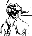
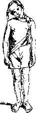
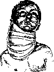
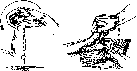
contractures, infectious contractures in the stage of subsidence of the main process, improper bone fusions in the stage of primary callus, various forms of clubfoot, sometimes also rachitic skeletal curvatures. Contraindicated in persons with status thymico-lympha-ticus, in poliomyelitis patients and rheumatics with多年 contractures of large joints, especially of the hip joint. Great caution is required in the operation during the period of puberty. In R., both the degree of deformation and especially the elastic properties of tissues are taken into account. These properties vary within very wide ranges depending on the type of tissue (skin, fascia, tendon, muscle, ligament, bone), its normal or pathological state (scar degeneration, nutritional atrophy, osteoporosis, etc.), the age of the patient and to some extent his sex. The elasticity of living tissues has considerable adaptability. Thus, if by R. a certain complex of soft tissues is brought to the limit of their extensibility, beyond which the tissues face rupture, and then these tissues are fixed in the achieved position for 1-2 weeks, it turns out that after this period it is possible to subject the tissues to some further stretching without risk of rupture. This is the basis of the method of staged R. (Yu. Vol'f). To a lesser, but still noticeable degree, physiological adaptability of the elastic forces of tissues manifests itself even during one session of the operation, if it is conducted with gradually increasing force and short breaks. This is the basis of the method of modeling R. (A. Lorenz). In performing the operation, the guiding role belongs to the muscular sense of the operator. The technique of R. Firmly grasping with both hands the part of the body being formed, produce cautious, short pushes in the direction opposite to the curvature. The force of the pushes gradually increases to significant sizes. Having achieved some correction, the operator holds the tissues for a short time in a state of correction and then releases them. After a half-minute 'rest', beneficial to the tissues and the operator himself, the manipulations are resumed. With each subsequent moment of violence, the tissues become increasingly flaccid, pliable, plastic, so that in the end they retain the shape given to them. The result is fixed with an immovable, most often plaster cast, for a period of 2 to 8 weeks. If complete correction cannot be achieved in one session,
Figure 6. Redressation according to Lorenz.
Figure 7. Redressation by bandaging according to Fink.
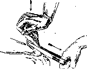
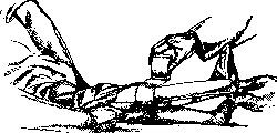
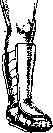
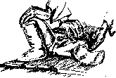
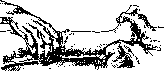
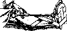
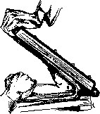
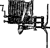
then fixation is performed in a partially corrected position for a period of 5 to 10 days, after which the cast is removed and R. is repeated. The method of staged R. can achieve correction of very large deformations, in which a single act of violence would lead to dangerous complications. The methods of grasping various parts of the body being redressed are different. Thus, in contracture of a limb, the latter is grasped with hands immediately above and below the contracted joint. Sometimes it is more convenient to apply pressure with hands not directly at the site of curvature, for example in contractures of the knee joint, in correcting diaphyseal deformations at the site of bone callus. In R. of the shoulder and hip joints, it is important to firmly fix the shoulder and pelvic girdle. For this, in the first case, the patient is placed somewhat on his side so that the corresponding scapula is firmly pressed to the surface of the table by the patient's own weight. In the second case, the operator's assistant presses the patient to the table, leaning with hands on the crests of the iliac bones; it is even better to bend the healthy leg at this time and press its thigh to the patient's abdomen. The technique of redressation in wryneck is easily understood from figures 1-3. Figures 4-10 show manual redressation of equinus in clubfoot with application of a cast. When operating on adult individuals, as well as in very rigid forms of curvatures, especially if the operator does not possess great physical strength and the area of force application is small (foot), use is made of special devices and instruments. These include rubber traction (fig. 11), König's wedge, Schultze's board (fig. 12), then apparatuses built on the type of a lever or screw: Thomas-wrench, osteoclast-brace of Bradford, Redar, Lorenz (fig. 13), etc. During R., various complications may occur: rupture of large vessels and nerves, constriction of the vascular bed, overstretching of large nerves (more or less permanent paralysis), fracture of healthy parts of the skeleton, reflex convulsions, acute dilation of the stomach and fat embolism. To avoid these dangerous disorders, it is necessary to perform the entire redressation procedure with great attention, to constantly consider the degree of tissue tension, to fix in plaster tissues only with their elastic forces completely suppressed, not leaving to the cast an additional corrective function, and finally, one must not operate on patients with the presence of the contraindications listed above.
Mi Fridland. REDUCTION DIVISION (maturation division), the cell division of developing sexual elements (future eggs and spermatozoa), as a result of which in the 'mature' sexual cell-egg and spermatide-the number of chromosomes is reduced by half compared to the normal number (see Haploid). The morphological details of the process under consideration are not completely clarified, however, the research of a number of authors over the last several decades (Hertwig, Haecker, Gregoire and others) allow to draw the following schematic picture. The development process of sexual elements is divided into periods: 1) multiplication, 2) growth, and 3) maturation. At the end of the growth period, the chromosomes take on a special arrangement within the nucleus, concentrating in one part of it and forming there a compact tangle (stage of synapsis, meiosis of some English and American authors). The processes occurring here are mainly reduced to conjugation
/Щ (Ш: > «Ъ* <Я/ Jк Figure 1. Phenomena of reduction during spermatogenesis in Tomopteris onisciformis: A-shortly after the last mitosis of spermatogonia; B-D- stages of the bouquet [B-parallel conjugation (leptotene stage), C-conjugation almost completed, only at the bends of the chromosomes both partners are not yet connected, D-pachytene stage]; E and F- strepsitene stage; G-diakinesis; H-metaphase of reduction division, the equational furrow is visible; I-early anaphase of reduction division; J- the same, from the pole; K-metaphase; L-anaphase of equational division. (According to A. and K. E. Shreiner.) (connection) of corresponding (allelomorphic) chromosomes, i.e., chromosomes of paternal and maternal origin. As a result, the chromosomes of sexual cells are found to have split and connected together in pairs. The resulting quadruple groups, each consisting of 4 chromosomes, are called tetrads; their number is equal to half the normal number for a given species. Thus, even before the onset of the R. d. itself, there is an apparent decrease in the number of chromosomes (pseudoreduction), associated or with the parallel application of corresponding chromo
Figure 2. Diagram showing the essential facts in the reduction of chromosomes during the development of spermatozoa in males of Ascaris megalocephala. The number of chromosomes in germ cells is assumed to be four. A and B-division of spermatogonia with the full number (four) of chromosomes; C-primary spermatocyte; chromatin forms two tetrads; D, E and F-first division into secondary spermatocytes, each of which contains two diads; G and H-division of the secondary spermatocyte into four spermatids; each spermatid contains two chromosomes and one centrosome, which remains in the middle part of the spermatozoon. (According to Wilson.)
Figure

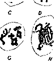
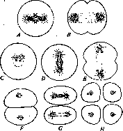
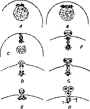
depict phenomena of conjugation (parasynapsis, parallel conjugation) or with the attachment of paired corresponding chromosomes at their ends (metasynapsis, conjugation at the ends). The resulting tetrads can be considered as bivalent chromosomes (see), which in a latent form contain two sets of chromosomes. The tetrads pass directly into the metaphase of the first maturation division, as a result of which each of them divides into two double groups (dyads). Immediately after this, usually without a resting stage, the second division occurs, in which the components of the dyad diverge into the daughter cells, each of which thus receives a number of chromosomes reduced by half. On a concrete example of the roundworm Ascaris megalocephala bivalens, these phenomena appear as follows (figs. 2 and 3): in the somatic cells there are 4 chromosomes, from which 2 tetrads, i.e., 8 chromosomes, are formed; as a result of two successive maturation divisions, two chromosomes, i.e., their haploid number, pass into each of the daughter sex cells. Of the two maturation divisions, one is normal, i.e., associated with the divergence of homonymous chromosomes (equational division), the other with the divergence of heteronymous conjugated chromosomes (proper R. d.). Depending on which of these divisions is first, the following are distinguished: 1) post-reduction division, which for the O (paternal) and M (maternal) chromosomes can be expressed by the following scheme: the tetrad at the first R. d. breaks down into two O M, and at the second R. d. into four cells. Diagram of the essential phenomena observed during the maturation of the egg of Ascaris megalocephala. The number of chromosomes is taken as four. A-initial phase; two tetrads have formed in the germinal vesicle; B-tetrads have arranged themselves near the spindle and formed the equatorial plate of the first division for the formation of the polar body; C-mitotic figure has turned; D-formation of the first polar body; each tetrad divides into two dyads; E-the first polar body is ready; it and the egg each contain two dyads; F-beginning of the second division; G-formation of the second polar body and division of the first; each dyad divides into two separate chromosomes; H-final result: three polar bodies and an egg nucleus (oocyte); each contains separate chromosomes (half of those contained in the cells); 1-egg centrosome, which should disappear or degenerate. (After Wilson.) The dyads I- O M. The reduction division is pre-reductional-first, and 2) pre-reductional division, proceeding according to the scheme where the reduction division is first in time. The latter case is apparently most common. In the male and female sex, R. d. proceed fundamentally the same, with the difference that in spermatogenesis, four equivalent cells-spermatids-are obtained from the primary seminal cell (spermatogonium), while in oogenesis, R. d. occurs on the periphery of the oogonium (primary egg cell), and its result is the formation of one full-fledged egg cell and three directing, polar, or reduction bodies, which are abortive egg cells with a minimal amount of protoplasm. Half of the chromosomes pass into the formed directing body (the first directing body, which usually divides in half again); the chromatin apparatus of the egg, without passing through a resting stage, divides again, as a result of which the second directing body is formed. Polar bodies in the overwhelming majority of cases die off shortly after being extruded. In some cases (mollusks), the reverse resorption of the extruded directing body by the egg has been described. R. d. have also been described for plants. In recent years, the research of Belar, Dobell, and Reichenow [Belar (1923-24), Dobell (1915), Reichenow (1921)] has shown the presence of R. d. (sometimes occurring in a peculiar way) also in the simplest organisms. Interesting are the R. d. in parthenogenesis. In diploid parthenogenesis (proceeding with a double set of chromosomes), reduction in its final form is absent, and the only division observed is equational (although sometimes a short-term conjugation of chromosomes is observed). In cases of haploid parthenogenesis (with a reduced set of chromosomes, for example in male bees, mites, etc.), both R. d. occur, and the egg receives a reduced number of chromosomes. Parthenogenetic haploid eggs produce males, in whose spermatogenesis R. d. begins but then breaks off, so that the original half number of chromosomes is preserved.
The theoretical significance of the phenomena of maturation is very great. According to Weismann's (1881) reduction hypothesis and Montgomery's (1901) hypothesis, the conjugation of chromosomes and R. d. ensure some 'revival' of the germ plasm, the appearance of new sets of hereditary factors, which should lead to the possibility of the appearance of the most favorable combinations, which are then fixed by selection. The exchange of homologous hereditary factors received from both parents should occur, according to the currently prevailing views, during the conjugation of chromosomes. The concepts of conjugation and reduction of chromosomes are in good agreement with the basic laws of modern genetics (Mendel's laws, Morgan's theory).
s. zapis'.
Related articles
Mentioned in
Cite this page
“Regression.” Soviet Medical Encyclopedia. English translation of Bolshaya Meditsinskaya Entsiklopediya, 1st ed. (Moscow, 1928–1936), ed. N. A. Semashko. https://sovietmedicalencyclopedia.pages.dev/article/regression/