Hemiazygos Vein
Historical document, translated for reference. It reflects medical knowledge of the 1920s–30s and is not medical advice.
Summary
This article from the 1928-1936 Great Medical Encyclopedia describes the anatomy, anatomical variations, and embryological development of the hemiazygos vein, detailing its connections, tributaries, and relationship with adjacent venous systems.
Encyclopedia article (1928–1936)
HEMIAZYGOS VENA, the hemiazygos vein. It lies on the left side of the spinal column and represents a common collector for the intercostal and part of the lumbar veins of the left side. The mode of union of the intercostal veins is subject to various variations. The most common type is the combination of them into two or three trunks (see Figure 1). Most
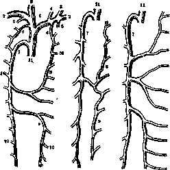
Figure 1. Diagram of the variations in the relationship of the vv. azygos, hemiazygos, and intercostalium: 1-v. anonyma dextra et sin.; 2-v. subclavia dextra et sin.; 3-v. jugularis dextra et sin.; 4-v. thyreoidea inf.; 5-v. mammaria int.; 6-v. intercostal. suprema; 7-v. azygos; 8-v. hemiazygos accessoria; 9-v. hemiazygos; 10-vv. intercostales; 11-v. cava superior (after Henle).
often, however, the hemiazygos vein is composed of two branches—the upper, sometimes called the accessory hemiazygos vein and receiving blood from the upper seven intercostal veins, and the lower branch, receiving blood from the five lower intercostal veins. Located behind the thoracic aorta, both branches run, one descending and the other ascending, along the bodies of the thoracic vertebrae
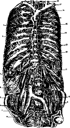
Figure 2. Vena hemiazygos and vena azygos with their anastomoses: 1-v. bronchialis post.; 2-v. azygos; 3-crura medialia diaphragmatis; 4-v. cava inf.; 5-v. iliaca communis; 6-v. iliaca ext.; 7-v. hypogastrica; 8-pl. sacralis; 9-v. sacralis med.; 10-v. ilio-lumbalis; 11-v. lumbalis ascendens; 12-v. oesophagea; 13-vv. intercostales VIII, IX; 14-v. hemiazygos; 15-v. hemiazygos accessoria; 16-v. anonyma sin.; 17-v. subclavia; 18-v. jugularis interna.
and receive the veins of the esophagus and posterior mediastinum. At the level of one of the lower or middle thoracic vertebrae (most often the VIII), they pass to the right anterior to the vertebral body, cross behind the thoracic duct, and empty into the azygos vein (v. azygos) either separately each, or having previously formed a single common trunk, or even an entire venous plexus. The initial part of the lower branch of the hemiazygos vein (lower end) is located between the inner and middle crura of the diaphragm on the left and is in close connection with the left ascending lumbar vein, sometimes being its direct continuation. At this same level, one most frequently encounters constantly existing, but variously expressed anastomoses with the renal vein and the portal system. The initial part of the upper branch of the hemiazygos vein is connected with the left brachiocephalic vein and with the vertebral vein system (see Figure 2). The complexity of the structure of the hemiazygos vein has its explanation in its embryological development. After the connection of the cardinal veins with the inferior vena cava, the right section becomes predominant, while the left lags in growth. The section of the left cardinal vein lying between the renal vein and the pelvis ceases to function and disappears with the regression of the primary kidney. In the thoracic part of the body, the initial relationships of blood circulation change due to the regression of the superior vena cava, as a result of which direct blood outflow from the left cardinal vein into the atrium becomes difficult and finally stops altogether. At the same time, an anastomosis forms between the right and left cardinal veins, through which blood from the left side begins to pass to the right. Thus, the thoracic part of the left cardinal vein with its anastomoses turns into the left hemiazygos vein. (Pathology of the hemiazygos vein—see Retroperitoneal space, Mediastinum.)
Related articles
Mentioned in
Cite this page
“Hemiazygos Vein.” Soviet Medical Encyclopedia. English translation of Bolshaya Meditsinskaya Entsiklopediya, 1st ed. (Moscow, 1928–1936), ed. N. A. Semashko. https://sovietmedicalencyclopedia.pages.dev/article/hemiazygos-vein/