Thoracic Duct
Historical document, translated for reference. It reflects medical knowledge of the 1920s–30s and is not medical advice.
Summary
This article from the 1928–1936 Soviet Medical Encyclopedia details the anatomy, development, histology, pathology, and surgical significance of the thoracic duct. It describes the origin from the cisterna chyli, its course through the thoracic cavity, its termination in the venous angle, and various clinical conditions such as obstruction and traumatic injuries.
Encyclopedia article (1928–1936)
THORACIC DUCT (ductus thoracicus), emerges at the level of the first or second lumbar vertebra from the cisterna chyli, into which the lumbar lymphatic trunks and intestinal trunk, as well as two descending trunks receiving lymph from the six or seven lower intercostal lymph nodes, empty from both sides of the spine. The lymphatic vessels from the upper surface of the liver and upper intercostal nodes on the left also empty into the thoracic duct, whereas a special trunk, the right lymphatic trunk, is formed from the upper intercostal nodes on the right and empties into the right venous angle. The thoracic duct, having emerged from the cisterna chyli, penetrates into the thoracic cavity through the aortic hiatus, locating to the right of the aorta and behind it (see figure). In the thoracic cavity, it approaches the median line of the spine and runs between the aorta (on the left), the azygos vein (on the right), and the esophagus (in front). However, these relationships are variable, and according to Minkin's studies, the thoracic duct may also be located at the left edge of the esophagus and at some distance from it. At the level of the third to fifth thoracic vertebrae, the thoracic duct passes to the left side of the spine and departs from it ventrally in a forward direction. Having reached the upper aperture of the chest, it arches over the top
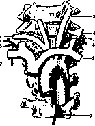
1-right subclavian artery; 2-right bronchomediastinal trunk; 3-left common carotid artery; 4-right and left jugular trunks; 5-right and left subclavian trunks; 6-left subclavian vein; 7-thoracic duct. (After Schultze.)
left subclavian artery, and having passed between it and the common carotid artery and previously received the jugular lymphatic trunk, subclavian trunk, and mammary lymphatic trunk, it empties into the left venous angle. In reality, significant deviations from the described scheme are observed both regarding the origin of the thoracic duct and the manner and site of its termination. The following is of practical importance: passing behind the common carotid artery, the thoracic duct in 50% of cases divides into two trunks of equal caliber or into a larger number of trunks (up to four and even up to six) of unequal caliber; in the latter case, the larger branch is located higher; in cases of multiple endings, the thoracic duct may either, without reaching the vein or within the thickness of its wall, reunite into a single trunk, but in the majority of cases it has several orifices (Wendel, Parson, and Sargent). At the site of entry, there is usually a dilatation of the lumen resembling an ampulla, and the opening itself is most often located in the lowest division of the internal jugular vein, protected by a valve present here in the lumen of the vein. Less frequently, the duct opens into the venous angle and even less frequently into the left subclavian vein. The practical value of multiple endings lies in the possibility of restoring the movement of chyle in cases of compression or wounding of the thoracic duct. In the same sense, the presence of connections with veins—the azygos vein, renal veins, intercostal veins, and veins of the right side of the neck—is of significance (Wendel, Sappey, Beneteau). The level of the arch of the thoracic duct in the neck varies greatly, rising up to the fifth cervical vertebra. Lecène, depending on this, distinguishes a 'high-position arch' and a 'low-position arch.' The lymphatic system develops from a source common with the circulatory system and, with the exception of mammals, is laid down earlier than the latter (Huntington). The thoracic duct in the form of a true lymphatic duct (paired in the anterior part of the body) appears only in birds. In amphibians and fish, it appears as a lymphatic reservoir located peri-aortically (around the aorta) and communicating with the subclavian and jugular veins and the veins of the posterior part of the body in the former, with the caudal vein in the second, and with the anonymous vein, sciatic veins, and renal veins in Sauropsida. Histologically, the wall of the thoracic duct consists of: 1) tunica intima (endothelial cells and longitudinally arranged thin elastic fibers); 2) tunica media (a small amount of elastic fibers and transversely arranged smooth muscle fibers); and 3) tunica externa (longitudinally arranged connective tissue, elastic, and muscle fibers). Obstruction of the thoracic duct can be caused by microfilariae (Filaria sanguinis) penetrating into its lumen, which, however, is not encountered in the USSR. Diseases of the thoracic duct are usually secondary, resulting from involvement in the process of compression or growth by malignant neoplasms, or inflammatory processes (especially tuberculosis) originating from an adjacent organ. Usually, such involvement of the thoracic duct is accompanied by phenomena of dissemination and generalization. Injuries of the thoracic duct are relatively rare and, due to its deep position in the thoracic cavity, are possible only with significant chest injuries. Wounding of it in the lower cervical region is more frequent, especially during operations for tuberculous or cancerous lesions of the lymph glands (high position of the steeply curved arch). Accidental wounding during surgery can in most cases be noticed by the outflow of a characteristic milky-white fluid flowing out synchronously with respiratory movements. However, it is possible that within a few hours after the injury and even days (up to 15 days, Halsted), the injury will remain unrecognized. This applies especially to wounds of the thoracic and abdominal sections, when an effusion of chyle into the thoracic and abdominal cavities (chylothorax, chyloperitoneum) or into the subcutaneous tissue is possible. Profuse loss of chyle upon wounding entails rapid exhaustion and death. The simplest and most effective means of combating the outflow of chyle is (in view of the presence of anastomoses of the thoracic duct) ligation of the affected trunk or tight packing; when possible, suturing.
Related articles
Mentioned in
Cite this page
“Thoracic Duct.” Soviet Medical Encyclopedia. English translation of Bolshaya Meditsinskaya Entsiklopediya, 1st ed. (Moscow, 1928–1936), ed. N. A. Semashko. https://sovietmedicalencyclopedia.pages.dev/article/thoracic-duct/