Bronchial Glands
Historical document, translated for reference. It reflects medical knowledge of the 1920s–30s and is not medical advice.
Summary
This article from the 1928–1936 Soviet medical encyclopedia details the anatomy, classification, and clinical significance of bronchial glands, which are regional lymph nodes for the lungs, bronchi, and thoracic trachea. It describes their four main groups, their lymphatic drainage pathways, and how their enlargement can cause serious complications.
Encyclopedia article (1928–1936)
Bronchial Glands (lymphoglandulae bronchiales sive broncho-pulmonales), in anatomical terms, represent lymph nodes that are regional for the lungs, bronchi, and the intrathoracic part of the trachea. Therefore, the term B. g. should apply only to the glands adjacent to the respiratory tube and accompanying it, starting from the small branches of bronchioles and ending with the thoracic part of the trachea at the level of the thoracic inlet. According to the "most widespread classification at present, proposed by V. Sukennikov, B. g. are divided into 4 main groups: 1. Lgl. pulmonales, situated in the lung tissue, in the angles of converging bronchi. 2. Lgl. broncho-pulmonales, located in the area of the hilum (root of the lung) in the angles of branching of bronchi of the 1st and 2nd order. 3. Lgl. tracheo-bronchiales, adjacent directly to the bifurcation of the trachea, and 4. Lgl. paratracheales thoracales, located on the anterior-lateral surfaces of the thoracic part of the trachea. In the same sequence in which this division was made, all these groups of glands are connected to each other by an upward flow of lymph vessels, playing the role of nodes and transmitting lymph from the visceral pleura of the entire lung, lung alveoli, bronchi, and interstitial tissue of the lung to the right and left angulus venosus (angle of convergence of the v. jugularis cvv. subclaviae). The trunks draining lymph from this system—the right and left truncus broncho-mediastinalis—infiltrate the blood venous system either directly or, after preliminary inflow, on the left into the ductus thoracicus and on the right into the ductus lymphaticus.
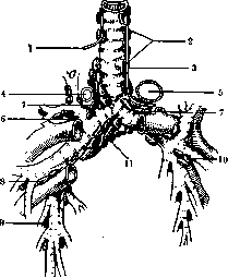
Figure 1. 1-n. recurrens dexter n. vagi; 2-lgl. paratracheales thoracales; 3-n. recurrens sin. n. vagi; 4-v. azygos; 5-aorta; 6-lgl. broncho-pulmonales 1st order dext.; 7-lgl. tracheo-bronchiales super. dext. et sin.; 8-lgl. broncho-pulmonales 1st order dext.; 9-lgl. broncho-pulmonales 2nd order; 10-lgl. broncho-pulmonales sin.; 11-lgl. bifurcationis (according to Sukennikov with modifications) dexter. According to experimental studies by Beitzke (N. Beitzke), verified by the works of other authors confirming his conclusions (Esipov, Abakelia), the bronchial lymph flow constituting in this way has an independent, isolated significance from the cervical flows and does not enter the cervical lymph glands, nor does the reverse B. g. have anatomical connections with the cervical glands and under no conditions receive lymph from them. From the point of view of topographical position and possible influences in pathological cases, the greatest clinical interest is represented by the tracheo-bronchial and paratracheal glands. Lgl. tracheo-bronchiales (syn. interbronchiales) occupy the three angles of the bifurcation of the trachea open to the right, left, and downwards; accordingly, lgl. trach.-bronch. dextrae et sinistrae, or superiores, and lgl. trach.-bronch. inferiores, s. bifurcationis (see Figure 1) are distinguished. All three groups are richly connected to each other by transversely crossing tracheal lymphatic anastomoses, due to which each of them can be involved in the pathological process not only from the homonymous side of the lung, but also from the opposite side and be the first regional stage for any lobe of the lung.
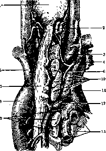
Figure 2. Right tracheobronchial and paratracheal lymph glands, pathologically enlarged (view from behind, esophagus slightly displaced to the left): 1-pharynx; 2-a. thyreoidea inf.; 3-a. anonyma; 4-n. recurrens dext.; 5-a. subclavia; 6-a. vagus dext.; 7-esophagus; 7-aorta; 9-lgl. tracheo-bronchiales dext.; 10-lgl. paratracheales; 11-v. cava superior; 12-v. azygos; 13-lgl. broncho-pulmonales (according to the preparation of K. D. Esipov).
Besides the lungs, B. g. serve the upper thoracic part of the esophagus, since they receive from it the draining lymphatic paths and, through the medium of anastomoses, are functionally connected with the lymph system of the heart and part of the pericardium. In some cases, the clinical significance may have the anastomosis of B. g. (lgl. bifurcationis) with the anterior mediastinal glands of the diaphragm (lying on the surface of its dome, at the place of entry of the n. phrenici). Through this connection, B. g. enter into communication with the lymph circulation of the diaphragm and the organs it serves, in particular, for example, the diaphragmatic peritoneum and the liver. Tracheobronchial glands occupy, to a greater extent, the anterior-lateral surface of the trachea and bronchi; on the posterior side of the latter, only a small number of glands are located. The number of glands varies individually. On the right, the number is smaller (from 3 to 6), at the bottom—of the trachea, during inflammatory enlargement, during periadentitis, suppuration, malignant neoplasms, and other pathological conditions, it can entail serious complications on the part of the trachea, causing narrowing of its lumen, ulcerations upon opening of abscesses through the wall and tumor dissolution, with greater and lesser consequences, etc. Besides this, each of the 3 groups of tracheobronchial glands can exert a certain influence, depending on its anatomical location. Right tracheobronchial lymph glands (see Figure 2), when enlarged, can come into contact in front with the vena cava superior; externally, the mediastinal pleura of the upper lobe of the lung (adhesions, pleuropneumonia, etc.) is adjacent to them; from above—vena azygos (narrowings, thrombophlebitis, perforations, etc.); from inside—the n. vagus dext. at the place of its intersection with the bronchus and transition to the posterior side (see Figure 2 and 3); backwards—the pleura and, when the glands are enlarged, the esophagus (narrowings of the esophagus and perforations). Left tracheobronchial lymphatic glands in the greater part of their mass are hidden behind the arch of the aorta passing over them from front to back (see Figure 1 and 3) and the left pulmonary artery passing in front of them. Besides this, clinical significance has the direct contact of these glands with the n. recurrens sin. s. n. laryngeus sin. inferior passing under the arch of the aorta (influence on phonation!). The left vagus trunk itself is displaced from the tracheobronchial glands by the arch of the aorta and the vessels departing from it to the left (see Figure 3). Lgl. bifurcationis are arranged in a packet in the connective tissue, occupying a triangular-shaped niche under the bifurcation (see Figure 3), the floor and anterior wall of which is formed by the pericardium covering the atria here and above their level forming the posterior wall of the sinus transversi pericardii (in other words, adjacent to the a. pulmonalis and aorta ascendens). Behind, the glands are adjacent to the esophagus and the descending aorta. On the posterior surface of the glands, between them and the esophagus, is located the network of the plexus of branches of the vagus and sympathetic nerves (plexus pulmonalis posterior; see Figure 3). Such intimate neighborhood of the lower tracheobronchial glands, besides the bifurcation of the trachea, with the pericardium, esophagus, aorta, nerve plexus, and trunks of the vagus nerves creates a particularly important significance of this group in the occurrence of corresponding severe complications in all these organs during the involvement of the glands by inflammatory processes with their consequences (purulent dissolution, perforations, narrowings, scars, ossifications, etc.) and during the occurrence of metastatic malignant neoplasms in them.
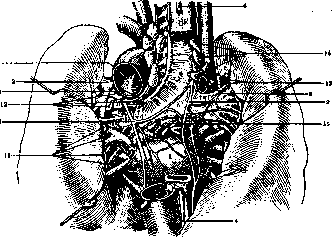
Lgl. paratracheales thoracales (see Figure 1-3) are adjacent to the lateral surface of the trachea and are surrounded externally by the mediastinal pleura. In the closest neighborhood with them on the left is the n. recurrens, a. subclavia; on the right—the right n. vagus crossing the lateral wall of the trachea from front to back. With considerable magnification, both right and left paratracheal glands can to a significant extent come into contact with the esophagus lying behind them (narrowings, ulcerations of the esophagus!). Inflammation of B. g.—see Bronchoadenitis and separate table (st. 287-288), fig. 2-4.
Related articles
Mentioned in
Cite this page
“Bronchial Glands.” Soviet Medical Encyclopedia. English translation of Bolshaya Meditsinskaya Entsiklopediya, 1st ed. (Moscow, 1928–1936), ed. N. A. Semashko. https://sovietmedicalencyclopedia.pages.dev/article/bronchial-glands/