Broncholiths and Bronchomycoses
Historical document, translated for reference. It reflects medical knowledge of the 1920s–30s and is not medical advice.
Summary
This article defines broncholiths as calcified concretions within the bronchi and discusses bronchomycoses, which are respiratory tract infections caused by fungi. It details the formation of these stones, the nature of fungal infections in the lungs, and the secondary role of fungi in pre-existing pulmonary conditions.
Encyclopedia article (1928–1936)
BRONCHOLITHS, broncholithon (from the Greek bronchos—bronchus and lithos—stone), bronchial stones, concretions forming in the lumen of the bronchi. Broncholiths consist of thickened mucus impregnated with calcareous salts. They have a rounded shape and are no larger than a cherry pit (occasionally larger). They are encountered rarely; they form most often in the cavities of saccular bronchiectasis (see). If a broncholith falls from a bronchiectasis into the lumen of the bronchus, symptoms of a foreign body in the respiratory tract may develop. One must distinguish true broncholiths from stony formations that may appear in the bronchial lumen due to the rupture into it of a calcified caseous focus from adjacent lung tissue or a bronchial lymph node. BRONCHOMYCOSES (from the Greek bronchos—bronchus and mykes—fungus), diseases of the respiratory tract caused by fungi—molds and other closely related species. Most often, the causative agents are varieties of so-called ray fungi—actinomycetes (Actinomyces, Streptothrix). These fungi, initially fixing themselves on the mucosa of the small bronchi, usually cause the development of specific granulations there, which quickly spread to the adjacent alveolar parenchyma and propagate widely throughout the organ, due to which both in the clinical and anatomical picture of the disease, the lesion of the lung (pneumomycosis) always comes to the fore. In other, much rarer cases, the disease is limited for a long time to the bronchial system,
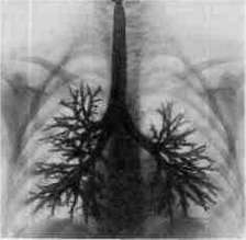
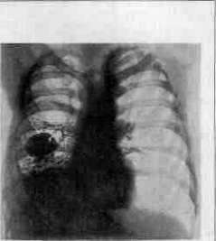
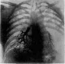
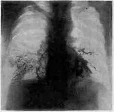
proceeding at first in the form of a protracted bronchial catarrh. However, even here, with the passage of time, on the one hand, a deeper and more significant damage to the bronchial wall occurs (deep and extensive infiltrates, partial necroses and suppurations, development of granulation foci, formation of bronchiectasis, etc.), and on the other, the involvement of the pulmonary parenchyma in the process (interstitial and exudative pneumonias, sometimes with subsequent necrotization or carnification). Thus, phenomena on the part of the lung also significantly prevail here (see Pneumomycoses). Among the lower mold fungi, the causative agents of such diseases can be Aspergillus fumigatus, less often Aspergillus niger, and even more rarely Mucor (hence the name of the disease—mycosis aspergillina s. aspergillosis et mycosis mucorina). Infection of healthy respiratory tracts with the named fungi occurs in exceptionally rare cases. This requires some disease of the airway system or lungs (chronic bronchitis, protracted pneumonia, infarcts, etc.). Fungi fix and multiply especially easily in the stagnant secretion of bronchiectatic cavities and other cavernous formations of the lung (abscesses, old tuberculous cavities, foci of gangrene, etc.), where the fungi, growing, either form a coating along the walls or accumulate in the form of more voluminous, tangled, smeary masses of a dark brown, greenish, yellowish, or gray color. Besides mold fungi, blastomycetes can also occasionally cause the development of granulation or purulent foci in the respiratory organs. The latter, however, never arise primarily, aerogenically, but exclusively by way of hematogenous metastases, being a complication of chronic blastomycosis of the skin. By their nature, the changes observed here already belong entirely to the section of pneumomycoses (see Pneumomycoses; see also literature there).
M. Skvortsov.
Related articles
Cite this page
“Broncholiths and Bronchomycoses.” Soviet Medical Encyclopedia. English translation of Bolshaya Meditsinskaya Entsiklopediya, 1st ed. (Moscow, 1928–1936), ed. N. A. Semashko. https://sovietmedicalencyclopedia.pages.dev/article/broncholiths/